The eruption of wisdom teeth is often accompanied by various complications. This is due to the lack of space in the dentition for the number eight, which erupts last. In dental clinical practice, third molar retention is often encountered - incomplete eruption of the crown above the gum. Removing a wisdom tooth in the gum is a small operation that requires not only extraction of the tooth, but also the creation of access and an incision in the gum. Our dental clinic performs extractions even in the most complex clinical cases. Dentists have extensive experience and high qualifications, and perform procedures painlessly and safely.
A wisdom tooth remains in the gum - is this critical?
Retention or partial eruption is characterized by incomplete emergence of the crown above the gum. Only 1-2 bumps or part of the tooth crown may appear. Retention can be independent or together with dystopia - incorrect positioning of the molar. Symptoms of the disease will depend on the individual clinical situation.
Depending on the location, retention can be vertical, horizontal or angular. With a vertical crown, the tooth crown is located normally in the bone, in accordance with other teeth. Horizontal is characterized by the location of the tooth perpendicular to the others, horizontally to the arch of the jaw. With angular retention, the crown is tilted to the side. According to the depth of its location, the tooth can be covered only by gum, but by a bone plate.
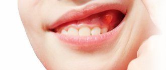
Retention is accompanied by the ingress of food debris and plaque under the gum. This causes inflammation, redness, tissue swelling, and pain. Pericoronaritis may occur - acute inflammation of the gums and mucous hood above the crown of the impacted tooth. The disease can be serous and purulent, causes acute pain, hyperemia, tissue swelling, and makes it difficult to open the mouth.
In most cases, a tooth in the gum is a source of various complications: pericoronitis, the formation of caries and its complications, cysts, stomatitis, periostitis, abscess. Therefore, in case of pathology, dentists recommend removal.
To date, exact methods for preventing retention are unknown. General measures include proper hygienic care of the oral cavity and monitoring the correct development of the child’s jaws and bite. As well as timely and correct orthodontic treatment of malocclusions.
Main diseases characterized by tissue hyperplasia
Fibrous or hypertrophic gingivitis
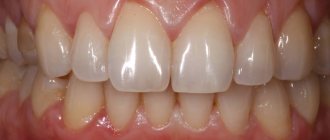
Between the teeth there are gingival papillae, which during the development of fibrous gingivitis begin to gradually increase in size, they become denser and grow. In this case, the mucous membrane remains painless for a long time, and there is no bleeding.
Read more about all types and symptoms of gingivitis in a separate article on the website.
The disease develops against the background of poor oral hygiene and the accumulation of a large amount of plaque; it can appear due to poor-quality prosthetics and constant trauma to the mucous membrane. People who have hormonal imbalances or gastrointestinal diseases also encounter fibrous gingivitis. The disease often occurs in both adults and children.
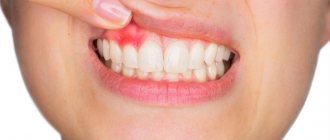
Important! A growth over a tooth is not always associated with hyperplasia; it can be nothing more than a gumboil, abscess, fistula or cyst, indicating advanced dental diseases. Most often, such manifestations are very painful, they are white in color, because... pus accumulates in them.
Fibromatosis
This is a hereditary disease and therefore often manifests itself in childhood. It is characterized by the growth of individual areas of the gums and proceeds very slowly. The color of the affected tissues is usually no different from healthy ones; they are painless.
The disease occurs mainly in the frontal area of the smile, and soft tissues can grow so much that they cover the crowns by more than 2/3. If fibromatosis is not treated, it is fraught with destruction of interdental septa, osteoporosis, and a severe violation of the aesthetics of the smile.
Fibropapilloma
If the gums have grown onto the crown, then a disease such as fibropapilloma may be to blame. This is a benign tumor. The tumor is hard to the touch, but painless. Its appearance in childhood is more dangerous, because can lead to disruption of the formation, growth and eruption of permanent elements.
Epulis
Epulis, like fibropapilloma, is a benign formation. It has a mushroom shape, because the tumor grows on a stalk. Appears as a result of chronic irritation and tissue injury. The most common location is the area of the incisors or canines. If the tumor is not removed, it can lead to pathological mobility and tooth loss.
Indications for removal
Retention can manifest itself as a stage of eruption. Over time, the crown completely appears above the gum and the tooth can function normally. Therefore, the decision on the need for removal should be made by a doctor only after examining and diagnosing the condition of the dental system. Indications for surgery are the following clinical cases:
- Constant pain;
- Complications of eruption;
- Trauma to an adjacent molar;
- Pressure on the dentition and its deformation;
- Horizontal or angular position of the third molar in the jaw;
- Dental crowding, which can be aggravated by wisdom teeth;
- Orthodontic and orthopedic indications, that is, the need for extraction to correct the bite or prosthetics.
How to prevent a relapse?
Any treatment, including surgery, unfortunately, does not exclude recurrences of the disease.
Simple measures taken by the patient himself will help minimize their risk. Necessary:
- Give up some habits. Namely, stop smoking, minimize alcohol, give up soda. Dishes and drinks should be consumed warm. Too hot or cold promotes chemical/thermal damage and usually relapse.
- Brush your teeth with soft bristles (low or medium hardness) and Sensitive toothpastes for painfully sensitive teeth. Initially, after treatment, it is better to use baby hygiene products that are mild and without aggressive components.
- Visit the dental office regularly to monitor the process of regeneration of operated tissues and prevent any unhealthy changes. It is necessary to come to the doctor for examination every 1-1.5 months.
The process of removing a wisdom tooth in the gum
The removal operation consists of the following steps:
- Preparation. To reduce the risk of postoperative deposits, sanitation of the oral cavity and professional hygiene are carried out with the removal of dental deposits. Elimination of pathological microorganisms and foci of infection promotes rapid wound healing without complications.
- Anesthesia. Before the operation, local anesthesia is performed using a carpule syringe and modern painkillers. The anesthetic is selected for each individual, taking into account individual sensitivity, age, and medical history.
- Creating access to the tooth. In case of retention, the dentist must provide good access so that the forceps can be securely fixed. To do this, size the gums and, if necessary, remove the bone and gingival hood.
- Extraction. The difficulty of molar removal depends on the type of retention and the individual situation. The tooth is fixed with forceps and dislocated; if necessary, additional instruments are used.
- Procedures after removal. After extraction, the tooth socket is inspected and bone fragments and protrusions are removed. The doctor applies a gauze pad and, if necessary, medications to stop bleeding. Sutures are placed on the gum to speed up healing.
- After surgery, the patient is given instructions, advice, and recommendations in the postoperative period.
Hypertrophic pulpitis
Overgrowth of gum tissue is a serious pathology that requires immediate attention to a dental clinic.
The gum comes out of the tooth often with advanced hypertrophic pulpitis, which occurs in two possible chronic forms:
- Excessive volume of soft tissue is formed due to the granulation process. The development of the process is preceded by the presence of a huge carious hole over a long period of time. Due to its presence, soft structures are constantly injured, transmitting irritation to the pulp.
- A dense polyp forms in the pulp area due to the growth and layering of flesh on top.
The disease is mostly asymptomatic. And only eating rough food provokes aching pain. The gums in the problem area may also bleed. But the main sign of hypertrophic pulpitis is the appearance of “wild meat” in the hole of the dental unit. Over time, it grows so much that it fills the entire cavity and goes beyond its limits. Pain and discomfort interfere with proper hygienic care. Aggressive microflora contributes to halitosis (bad breath).
Swelling of the mucous membrane due to traumatic or deep carious destruction of the crown
Gum receding occurs when the walls of the tooth are completely or almost completely destroyed. It becomes inflamed due to constant injury and accumulation of food particles. Plaque thickens over time, puts pressure on the gums, and populates it with pathogenic microflora. The proliferation of aggressive microorganisms contributes to the development of swelling and hyperemia. The gums increase in volume and it seems that meat is sticking out of the cavity of the dental unit. The patient experiences pain and severe burning when eating salty and sour foods. The jaw row reacts sharply not only to taste, but also to temperature stimuli. There is discomfort from hot/cold.
Prevention after surgery
The rehabilitation period after removal of an impacted wisdom tooth lasts longer than after a normal one. Healing of the gum socket can take up to 2-3 weeks. During this period, it is important to follow the doctor's recommendations to prevent complications. It is necessary to carefully but delicately carry out hygienic care of the oral cavity. For the first days after the intervention, take painkillers and follow a gentle diet. Too hard, burning foods can damage the gums and cause tissue inflammation.
Dentists also recommend rinsing the mouth after each meal using antiseptic solutions, herbal decoctions, or just water. During the week, you must avoid physical activity, visiting the gym, baths, saunas. If any complications occur, you should not self-medicate; you must immediately seek help from a doctor.
If it hurts, what should you do?
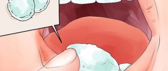
In case of acute pain in the area of the impacted tooth or after extraction, you can take an anesthetic. For this purpose, non-steroidal anti-inflammatory drugs are best suited - Nimessil, Ibuprofen, Paracetamol, Ketanov. At home, you can also carry out hygienic cleaning and rinsing the mouth with antiseptics. For a professional examination and treatment, you must visit a dentist.
How long does the gum “grow” and the stages of development of hyperplasia
Pathology can develop slowly if it is caused by fibromatosis, epulis or fibropapilloma. And it can develop more rapidly if it occurs against the background of gingivitis.
As for the stages of development of hyperplasia, doctors identify the following:
- first stage: the gingival papillae thicken and thicken, while there are no other alarming signs,
- second stage: soft tissues grow and can cover up to half of the crown. Due to the volume, tissues are easily injured during oral hygiene and when chewing food, they can begin to bleed,
- third stage: the most rare stage. The tissues grow so much that they can even cover the cutting area of the crown. Granulations appear on the affected areas.
If you break off a tooth, gum growth may take several years.
Causes
Most often, gum growths appear after some kind of trauma to the gums. This can be facilitated by either external or internal factors. Such cases may be:
- insufficient hygiene;
- pathology of the jaw bones;
- malocclusion (extra or protruding teeth, curvatures);
- removal of a tooth;
- unprofessional dental care (poor quality fillings, infection after minor operations);
- injuries or scratches to the gums;
- periodontitis;
- bad habits (smoking or alcohol);
- diseases of internal organs (usually digestive);
- infection after jaw surgery.
Treatment
Any growths in the mouth should be treated by a doctor.
To determine the type of cyst, an x-ray method is used. If necessary, histology of the cyst tissue is also performed.
Dentists best treat cystic formations in the early stages. The most radical and fastest method is to remove the diseased tooth in the area of the damaged gum. This was the only way to treat cysts until recently. Then the cavity was completely sanitized and cleared of infected tissue.
Currently, there is no need to remove the tooth, since modern methods of washing the resulting cavity through the fistula canal with various antiseptic solutions are used. This course of treatment is very long and includes the use of general anti-inflammatory therapy and new generation antibiotics. The cystic cavity is washed until all microorganisms are removed from it. Throughout the course of conservative treatment, a special paste is injected into the root canal and the resulting cavity in the cyst to help restore bone tissue.
What is meant by a growth on the gum?
When people talk about a growth on the gum, they usually mean a cyst, which often appears for no apparent reason. If the growth does not hurt when pressed, then it is called epulis (or supragingival). If you open it, a loose mass is released from the growth or liquid contents flow out. Without timely therapy, the formation will sooner or later open up on its own, transforming into a small tumor with a hole on the surface, from which the fistulous tract goes into the thickness of the cyst. From the resulting fistula, ichor or pus is periodically released. At the same time, the patient’s general condition also suffers: he is bothered by headaches, loss of vitality, and the lymph nodes near the source of infection (jaw, neck or ear) are usually enlarged and painful.
What does a growth on the gum mean?
The formation of a cyst on the gum occurs at any age. Fortunately, such a neoplasm is not always a sign of a serious pathology.
Most often, an unpleasant growth on the gum appears from a wound formed after an infection enters the oral cavity.
This process can especially often manifest itself in children, since parents can only dream of a child observing strict hygiene. Most often, growths on the surface of the gums appear in children during teething. At this moment, all the main factors are present that contribute to the penetration of infection into the gum cavities: putting dirty hands or objects into the mouth, reduced immunity and wounds in the gum mucosa. By putting various objects into the mouth, the child thereby tries to relieve pain and itching at the site of tooth eruption by massaging the gums.
Why is it important to treat flux
Dentists warn that advanced periostitis is very dangerous. If you ignore its occurrence and do not carry out the necessary treatment, the following consequences may occur:
- accumulation of a large amount of pus, loosening of the tooth;
- destruction of the rudiments of permanent teeth (then after the loss of milk units, the permanent ones will not erupt - the child will face complete or partial adentia);
- abscess (in place of the abscess, the tissues seem to melt, forming a huge cavity filled with pus);
- phlegmon (can lead to blood poisoning and death).