According to antiplagiat.ru, the uniqueness of the text as of October 16, 2018 is 99.8%.
Key words, tags: pulp diseases, periodontitis, tooth extraction, oral hygiene, OPTG image, dental microscope, pulpitis, periodontitis.
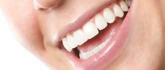
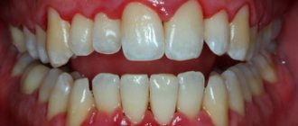
Caries is the most common disease of the human dental system. Today, 98-99% of the adult population of Russia has caries at one stage or another. Its untimely or incorrect treatment leads to complications in the form of pulp and periodontal diseases, the development of purulent-inflammatory processes in the maxillofacial system, and tooth loss. Lack of treatment, as well as the removal of teeth completely affected by caries, cause secondary deformation of the dentition and pathology of the temporomandibular joint. At the same time, timely treatment, regular oral hygiene and other preventive measures help prevent the occurrence and development of caries, and modern dentistry has effective methods for treating caries at any stage.
Historical reference
It is believed that the “history” of caries begins with a change in the human diet, that is, from the time when coarse, unprocessed food began to be replaced by food subject to heat, steam and other processing, as well as from the moment when “sweets” appeared. Judging by archaeological finds, for example, by the teeth of a man who lived 13-14 thousand years ago found near the city of Lucca (Italy, Tuscany region), we can conclude that already then caries was encountered and attempts were made to treat it. In the teeth found near Lucca, cavities were found that reached the pulp, and on the walls of the teeth there were traces of drilling with some stone tools. Moreover, the teeth had fillings made of plant resin with plant fragments. This means that ancient healers tried to protect carious cavities from the penetration of food, and the plants added to the resin composition apparently served as an anesthetic.
The healers of the Mayan civilization, judging by other archaeological finds, made fillings from complex plant compositions with the addition of various minerals. In ancient Rus', caries in the initial stages was treated with honey, and in advanced stages - with propolis, which was applied to the tooth cavity, previously cleaned of decay products and affected tissues; to relieve pain, they applied a cloth soaked in a mixture of vodka and table salt. In ancient Rus', caries in the initial stages was treated with honey, and in advanced stages - with propolis, which was applied to the tooth cavity, previously cleaned of decay products and affected tissues; to relieve pain, they applied a cloth soaked in a mixture of vodka and table salt. In medieval Europe, where the development of medicine was hampered by the dogmas of the church, it was believed that caries developed due to the activity of “tooth worms”, and it was treated with poisons and hot metals introduced without anesthesia into the dental cavity affected by the disease. Moreover, these manipulations were carried out not by doctors, but by barbers. The poor people, having no means for “treatment”, received very strange, unscientific recommendations from the “doctors”: sit under the moonlight with your mouth open, tie a frog to your jaw, apply a mixture of oil and bird droppings, “rub” the affected area with the tooth of a deceased person , etc.
In the 15th century, healers began using metals, including gold, to fill teeth. In 1790, John Greenwood, the personal dentist of the first US President George Washington, came up with a primitive device for drilling teeth, where a foot drive from a spinning wheel was used to rotate the drill. More than 80 years later, in 1871, the American doctor James Morrison patented a dental drill he invented, which gave impetus to the development of therapeutic dentistry. Throughout the 20th century, therapeutic dentistry developed rapidly, adding more and more new techniques, materials and tools for the treatment of dental diseases, including caries. Today, caries treatment is the most common dental service, with high treatment effectiveness at any stage.
Caries and its causes, classification and effect on the body
Caries (lat. caries - “rotting”) is a pathological process in the hard tissues of the tooth, which develops under the long-term complex influence of unfavorable external and internal factors and leads to softening of hard tissues and the formation of defects in the form of cavities. Caries, if left untreated, disrupts the chewing process up to the complete loss of this function, therefore, has an extremely negative effect on digestive function and causes diseases of the gastrointestinal tract. In general, caries has a negative impact on the general condition of the body and on a person’s quality of life, being a constant source of infection and intoxication.1
Caries: its types and classification
Uncomplicated caries:
This is the name of the initial stage of damage to tooth tissue. Caries manifests itself as a small focus of demineralization on the surface of the tooth enamel in the form of a small, light or dark spot. The primary stage of the second degree is characterized by the formation of a carious cavity that does not reach the border between enamel and dentin. Deep tissue damage is manifested by the fact that caries penetrates deeper into the tooth, but without yet affecting the pulp, which is protected by a thin layer of dentin.
Complicated caries:
When caries becomes more severe, it manifests itself as:
- pulpitis – acute inflammation of the neurovascular bundle of the tooth;
- periodontitis is an inflammation of the oral tissues between the alveolar process and the tooth.
Histological classification consists of several points:
- primary damage to tooth enamel;
- damage to the enamel and thin layer of dentin;
- damage to cement:
- dental caries in remission.
Etiology and pathogenesis of caries
The causes of the appearance and development of caries are multifactorial, and the degree and speed of the caries process depends on a number of conditions, the combination of which leads to this disease.
Today, there are about 400 different theories explaining the etiology and pathogenesis of this disease. Among them are physico-chemical, chemical, parasitic, chemical-parasitic, biological, trophoneurotic and others. But there is indisputable evidence that caries is an infectious disease of a bacterial nature that develops as a result of the interaction of the microflora of the oral cavity with resident microorganisms of the dental plaque, forming plaque, and subsequently tartar. It has also been established that dental plaque is not able to form without the presence of a pellicle - an outer compacted layer of cytoplasm on the body surface of many protozoan organisms. The discovery of this fact has recently inclined the scientific community towards the theory of receptor adhesion in the formation of dental plaque. According to this theory, as a result of the vital activity of microorganisms living in dental plaque, organic acids are released that gradually destroy the molecular structure of the tooth. This happens as follows: consumption of carbohydrate foods and poor oral hygiene contributes to the development of pathogenic flora. Due to its constant, long-term impact on the teeth, demineralization occurs, adhesive microdamages are formed on the enamel, and cariogenic microorganisms accumulate in them, forming dental plaque. This process is carried out in several stages: the first - an hour after eating, bacteria adhesion to the pellicle occurs; second – after 24 hours, early (immature) plaque forms; third - after 72 hours, mature plaque forms; fourth – complete maturation of dental plaque and formation of dental plaque on days 3–7.
An important component of dental plaque is sugars (disaccharides, monosaccharides and microorganisms that feed on them) found in the oral cavity due to increased consumption of carbohydrate foods. The fact is that the presence of excess sugars affects the pH level (acid-base balance), causing it to deviate towards acidification or alkalization, which can become one of the factors leading to the formation of dental plaque. The speed and intensity of its development are additionally influenced by such factors as the presence of inflammatory foci in the oral cavity, the viscosity of saliva, and desquamation (flaking) of the epithelium. It is important to understand that the preservation of dental plaque on the surface of the teeth means continued exposure of organic acids to the enamel. This leads to further oxidation of the oral environment, to even greater demineralization, to a qualitative and quantitative increase in defects in the subsurface layers of enamel. But if the organic enamel matrix is preserved during the demineralization stage, caries is quite easy to stop. And then, with careful hygienic care, regular examinations in a dental clinic and remineralizing procedures, the process can be considered reversible. If demineralization leads to the dissolution of a more stable layer of enamel, caries progresses rapidly, has an increasingly destructive effect on the teeth and causes an urgent need for prompt treatment.
Thus, the nature and diet, oral hygiene, oral microflora, the percentage of fluoride in consumed water, the nature of salivation, characteristics of enamel resistance, negative effects on the body in general and the oral cavity in particular - all these factors influence the occurrence of and development of caries.
Diagnosis of hidden caries
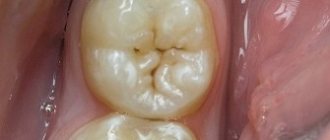
Ordinary caries can be identified through a visual examination and a staining procedure, when a special solution is applied to the enamel, turning the areas of demineralization purple.
With hidden carious lesions, this method may not bring results. In such cases, a dental microscope is used or radiography is prescribed. X-ray allows you to detect caries on any part of the tooth, even at the first stage. Having determined the extent and scale of the disease, the dentist prescribes treatment.
Caries classifications
In dentistry today, thanks to the efforts of scientists and specialists from all over the world, there are several classifications of caries. The basis for diagnosis is the international classification of diseases and related health problems, tenth revision of the World Health Organization (ICD-10). In it, caries is highlighted in a separate section, where it is divided into classes, each of which has a corresponding code:
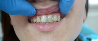
K02.0 Enamel caries. Stage of “white (chalk) spot” (initial caries). This class is characterized by: a change in enamel color (dullness) due to demineralization, and then a change in texture (roughness). In this case, there is no carious cavity, pathological changes do not extend beyond the enamel-dentin border. There is no toothache.
K02.I Dentin caries. This class implies destructive changes not only in enamel, but also in dentin, while the pulp is covered with a larger or smaller layer of preserved dentin. In many cases, at this stage, an uncomfortable reaction of the teeth to hot, cold, sour, and sweet foods may appear.
K02.2 Cement caries. In addition to the signs characteristic of the above classes, it is distinguished by damage to the exposed surface of the tooth root in the cervical region. This stage of caries in most cases is accompanied by sudden pain due to temperature, chemical, mechanical irritation, which disappears after a short period of time. Patients pay attention to the obviously changed aesthetics of the dentition, they may experience pain when eating, and if the chewing teeth are damaged, they may experience inconvenience associated with the chewing function.
K02.3 Suspended caries. It is characterized by the presence of a dark pigmented spot within the enamel as a result of local demineralization of the enamel.
K02.4 Odontoclasia (caries of the roots of primary teeth): childhood melanodentia and melanodontoclasia.
K02.5 Caries with pulp exposure (added: January 2013).
K02.8 Other caries.
K02.9 Unspecified caries. 2
Also, there is a modified classification of carious lesions according to their location - the Black classification, which includes six classes of carious formations:
Class 1 – caries of natural convexities and depressions of the chewing, palatal and buccal surfaces of the teeth.
Class 2 – caries of the contact surfaces of molars and premolars.
Class 3 – caries of the contact surface of incisors and canines.
Class 4 – intense caries of incisors and canines with a violation of the integrity of their cutting edge.
Class 5 – caries of the vestibular surface of all groups of teeth.
Class 6 – caries affecting the cutting edges of the incisors and canines and the cusps of the molars.
In addition, there are classifications of caries according to the depth of the lesion - initial, superficial, medium, deep; according to the presence of complications - uncomplicated (typically occurring superficial, medium or deep caries) or complicated (a carious process that affects the deeper structures of the tooth and the tissues surrounding its root).
At the same time, the Vinogradova classification involves dividing the disease into degrees of activity. Thus, caries can be compensated (sluggish), subcompensated (moderately progressive) and decompensated (progressive or, as it is also called, transient).
The clinical classification of caries distinguishes the disease according to the nature of its course - acute, chronic, acute and recurrent; according to the intensity of the process - single, multiple (generalized) and systemic; according to the localization of the process - fissure, aproximal/interdental caries (or caries of contacting surfaces), cervical, circular and hidden. Thus, caries is a complex and diverse polymorphic pathological process that can stabilize and worsen over a long period, acquire varying degrees of activity with different stages of compensation. This explains the difficulty in its classification. The variety of different classifications suggests that today there is no single system that would 100% satisfy the needs of all doctors and reduce this diversity to a common denominator. However, in global clinical practice this is seen as an obvious advantage, as it allows dentists to approach the issue of diagnosis and treatment more broadly.
In our clinical practice, we work according to international standards and approved clinical protocols. When making a diagnosis, the international classification of diseases and related health problems, the tenth revision of the World Health Organization (ICD-10), is used, which allows for clear and complete documentation of all aspects of the disease. For each specific patient and each dental unit, taking into account the degree of development of the pathology, appropriate treatment methods are selected. This approach to diagnosis and treatment is the most complete and effective.
How to treat caries?
It is worth noting that in order to prevent the development of caries, it is recommended to visit the dentist once every six months. When the first stage of caries is detected, a high-quality filling is installed, and the carious area is removed with a drill. If caries is not treated, it progresses to more complex stages, with a large affected area, and then surgical intervention cannot be avoided. If you notice dark spots on your tooth enamel, contact your dentist immediately.
Medium caries is treated in one visit to the doctor, deep caries in 2 visits, and caries in particularly complex forms requires 3 or more visits to the clinic.
Today, caries can be treated without pain, effectively and comfortably for patients. So there is no need to delay going to the doctor. In addition to the drill, caries treatment methods include the microinvasive method, ozone therapy, air abrasive treatment and laser treatment.
Treatment methods for caries
There are several methods for treating caries, which are divided into two main directions - non-invasive/microinvasive and surgical.
It is possible to determine which method to use in treatment only after a complete history collection. Diagnostics includes subjective and objective research methods, where patient complaints are subjective data, and clinical examination, examination with a dental probe and thermal diagnostics are objective data. To obtain an adequate diagnostic picture of the condition of the oral cavity in general and a specific tooth in particular, additional examination methods are used: an OPTG image (orthopantomogram), targeted X-ray images, which in turn are also necessary to monitor the treatment process at its different stages. Diagnostics allows us to identify the presence of caries, determine the location of carious lesions and the degree of tooth destruction.
Non-invasive or micro-invasive treatment of caries is used at the “white spot” stage, when there are no carious cavities and only a change in the structure and color of the enamel is observed. The treatment prevents further development of demineralization, prevents tooth decay, and preserves its functionality and aesthetics. As a rule, non-invasive treatment is accompanied by hygiene procedures, remineralization therapy and deep fluoridation of teeth.
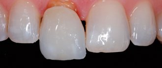
Surgical treatment of caries is the complete removal of tissues altered by caries by preparation with a dental bur and subsequent filling of the cavity. In modern dentistry, there are several main methods of preparation: conventional - preparation is carried out with a bur with a metal or diamond tip; ultrasonic; laser; air-abrasive; chemical. All of the above methods allow you to carefully treat a carious cavity of any location and volume, carefully removing all affected tissue and preserving healthy tooth tissue as much as possible.
The choice of filling technique and the filling materials itself depends not only on the degree of damage to the tooth, the location of the carious cavity and its size, but also on the condition of the dental tissues as a whole, as well as on the general condition of the body at the time of treatment, on the patient’s individual oral hygiene, on his caries susceptibility and caries resistance (caries resistance). To fix the filling, it may be necessary to expand the boundaries of the carious cavity due to the transition of the enamel-dentin contour. When installing a filling, the anatomical features of the tooth shape must be taken into account so that the filling repeats them as accurately as possible. These interventions require painstaking and very careful, concentrated work by the doctor, careful processing so that not a single micron of pathological tissue remains. Of course, such treatment is more effective using a dental microscope. .
At the end of treatment, the dentist-therapist gives the patient recommendations on independent oral hygiene, timing of control examinations, preventive measures to prevent relapse of the disease, and, if necessary, refers to a dental hygienist to obtain and master the skills of independent oral hygiene. Thus, during the treatment of caries, the dentist-therapist simultaneously solves several problems: eliminating local factors that determine the demineralization process; prevention of further development of the pathological carious process; preservation and restoration of the anatomical shape of a tooth affected by caries and its functional ability; prevention of the development of pathological processes and complications.
Stages of treatment of deep caries
Treatment methods for deep caries depend on how much the disease has damaged the tooth tissue and whether it has affected the pulp.
Treatment of deep dental caries without damage to the pulp (deep dentin caries)
- Initial consultation, x-ray, electroodontodiagnosis (EDD for deep caries) to assess the condition of the pulp.
- Anesthesia and preparation of affected tissues.
- In case of deep caries, a therapeutic pad is placed on the bottom of the treated cavity. It contains calcium and has pronounced antimicrobial properties.
- Installation of an insulating lining for deep caries for better fixation of the treatment and isolation of the filling material.
When treating deep caries, pads are necessary before installing a temporary or permanent filling. If the indications are positive (the required dentin thickness and no risk of infection entering the pulp chamber), the doctor can place a permanent filling, and then the entire treatment will take one visit. If a temporary filling is placed, the patient comes to the appointment one more time.
Complications
Untimely or improper treatment of caries leads to complications in the form of diseases such as pulpitis and/or periodontitis, which in turn can lead to the development of purulent-inflammatory processes in the maxillofacial system and complete loss of teeth.
In addition, the lack of treatment, as well as the removal of teeth completely affected by caries, cause secondary deformation of the dentition and pathology of the temporomandibular joint. Indicators of the development of complications of dental caries are very significant: in the age group of 35-44 years, the need for fillings and prosthetics is 48%, for tooth extraction - 24%.3
At the same time, it is worth paying attention to the so-called iatrogenic complications, the occurrence or absence of which depends entirely on the doctor. Since the treatment of dental caries is accompanied by a very large number of medical procedures, the effectiveness and quality of the treatment depends on the level of qualifications, medical skill, quality of the instruments used, materials and other factors. Despite the rapid development of dentistry, today it is often still possible to detect errors and complications that were made during the preparation and filling of a carious cavity during the treatment the patient received earlier. These include: insufficient treatment (preparation) of the carious cavity; overheating and burns of dentin, overheating of the pulp and, as a result, its necrosis; secondary caries; inflammation of the interdental gingival papilla; apical periodontitis – acute and chronic; perforation of the bottom and stenoccarious cavity; damage to adjacent teeth and gingival margins by a dental drill; displacement, fractures or loss of fillings; overbite during filling and/or lack of contact point; applying a single filling to several carious cavities, overhanging edges of the filling; discrepancy between the color of the filling and the color of the tooth/enamel; incorrect choice of filling material and instruments. Most of the listed complications can be eliminated, but, of course, it is better to prevent this by making the right choice of dentist.
Contraindications
Caries is a disease that has no absolute contraindications to treatment. But if certain factors are present, treatment may be delayed or interrupted until they are eliminated. These factors include: the presence of intolerance to drugs and materials that are planned for use in treatment; concomitant diseases aggravating treatment; inadequate psycho-emotional state of the patient before treatment; acute lesions of the oral mucosa and red border of the lips; acute inflammatory diseases of organs and tissues of the mouth; a life-threatening acute condition/disease or exacerbation of a chronic disease (including myocardial infarction, acute cerebrovascular accident), which developed less than 6 months before seeking this dental care; periodontal tissue diseases in the acute stage; unsatisfactory hygienic condition of the oral cavity.
Cost of caries treatment
Treatment of caries is a very individual process, and its cost is determined depending on a particular combination of certain clinical measures. These measures include the diagnostic measures carried out, the selection of a treatment method depending on the established stage of the disease, the methods, technologies, materials, equipment and instruments used in treatment, the qualifications of the attending physician, and consultations with specialists if necessary.
You should not consider caries a “mild” disease and ignore its signs. After all, this is a rather serious pathology that, in the absence of treatment, can leave a person literally “without teeth.” But, fortunately, modern dentistry has studied both caries itself and methods of treating it quite well, and with timely treatment from the patient, this process can be made reversible. Only one thing is required from patients - attention to the health of their teeth, since not a single pathology in the dental system goes away on its own; today there is not a single case of a person self-healing from caries or other diseases. Therefore, visit the dental office regularly to detect and eliminate caries in a timely manner, and then nothing will ruin your healthy and beautiful smile.
According to antiplagiat.ru, the uniqueness of the text as of October 16, 2018 is 99.8%.
Key words, tags: pulp diseases, periodontitis, tooth extraction, oral hygiene, OPTG image, dental microscope, pulpitis, periodontitis.
1 Clinical recommendations (Treatment protocols) for the diagnosis of dental caries. Approved by Resolution No. 15 of the COUNCIL OF THE ASSOCIATION OF PUBLIC ASSOCIATIONS “DENTAL ASSOCIATION OF RUSSIA” DATED SEPTEMBER 30, 2014. 2 https://mkb-10.com 3 Clinical recommendations for the diagnosis of dental caries.
Deep caries after treatment
Considering the serious therapeutic intervention, it is quite normal for a tooth to hurt for 1-2 days after treatment of deep caries (of course, we are not talking about unbearable pain). If the pain continues for a long time, then we are talking about complications, especially when the pulp was initially preserved. In the most difficult cases, therapeutic treatment is not possible, so the only option is to remove the diseased tooth. To avoid such an end, we recommend that you do not delay visits to the doctor: the sooner you come to the dentist’s office, the greater the chance of maintaining your health and money.