In this article
- Who is susceptible to developing interdental caries?
- How to detect caries in interdental spaces?
- Features of the treatment of interdental caries
- Interdental caries of the anterior teeth: what should be done?
- Specifics of treatment of interdental caries of chewing teeth
- How is caries between teeth treated when molars and premolars are affected?
- Prevention of interdental caries
Interdental caries is one of the most “inconvenient” and difficult to treat. In this article we will tell you why it appears, how caries between two teeth is treated, and whether its occurrence can be prevented using preventive measures.
What is interdental caries called and why does it occur? Caries between the lower or upper teeth located next to each other, joint to joint, is called interdental. The denser the arrangement of adjacent teeth in relation to each other, the greater the likelihood of developing carious lesions of the tooth enamel in the area of the joints. When the teeth are tightly packed together, food particles become stuck in the spaces between the teeth, which are difficult to remove with a brush, even if a person regularly brushes their teeth twice a day. To clean the interdental spaces, you need to constantly use dental floss, floss or a special device - an irrigator.
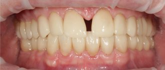
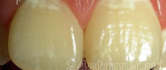
Unfortunately, most people limit themselves to using a toothbrush and toothpaste, as a result of which high-quality cleansing of the interdental spaces does not occur. Accumulated food debris is a favorable environment for the development of microbes and the formation of plaque, which leads to the development of caries between teeth. Due to the location and anatomical features, interdental caries of the anterior teeth most often occurs. Its early detection and timely treatment play an important role from the point of view of not only health, but also aesthetics, because caries between the front teeth, which is located in the most visible place, spoils a person’s smile and appearance. Its specificity is also that the enamel of the front incisors is thinner than the enamel of other teeth, so carious lesions spread deeper and deeper, and treatment presents certain difficulties.
Causes of caries between teeth
The causes of caries are determined by the individual characteristics of tissue structure and certain conditions - previous diseases, diet, location, crowding, hygiene:
- "metabolic explosion"
. Excessive sugar consumption provokes active production and accumulation of acid on the tongue, surfaces and pits. Demineralization occurs, the saturation of saliva with salts decreases, which contributes to the destruction of enamel; - tissue resistance
. The degree of resistance to the action of factors causing pathology depends on heredity, health status, and fluoride content in food/water. Manifests itself in the age-related dynamics of the onset of the lesion, the aggressiveness/prevalence of the process; - ignoring the rules of oral hygiene
.
Diagnostic features
To detect the problem, the doctor examines the patient's mouth and evaluates the condition of the teeth using a special probe. The following techniques help to finally clarify the situation:
- Radiography. On the resulting images, hidden carious cavities are visualized as dark spots.
- Transillumination. Using special dyes, the dentist can identify caries even in the most difficult to reach areas of the mouth.
During the diagnosis, the client’s subjective feelings and complaints about toothache are also taken into account.
Symptoms of interdental caries
The features of the development of interdental caries are not very different from other types of carious lesions. There are 4 stages in total:
- Initial - a small spot forms on the unit, invisible to the naked eye. To identify the initial stage, a hardware method is used.
- Superficial – the disease destroys the enamel. Darkening forms on it, it turns yellow and becomes rough, and sensitivity increases. But bacteria do not penetrate into deeper layers.
- Medium - at this stage the dentin layer is affected, the tooth reacts to irritants: cold, hot, sweet. The patient experiences severe pain.
- Deep - an advanced form of carious damage, pathogenic microorganisms penetrate into the root part. There is a risk of damage to healthy tissues and the development of periodontitis. At a deep stage, filling or restoration is necessary.
How to detect caries in interdental spaces?
One of the specific features of interdental caries is that it is difficult to diagnose. Since enamel damage is located at the junctions of teeth, it can be quite difficult to detect it yourself. Therefore, caries between teeth is rarely diagnosed at an early stage, when it is possible to manage with conservative methods and not prepare the tooth. Much more often it is detected already in the presence of symptoms in the form of pain or a reaction to cold/hot.
The dentist diagnoses interdental caries using several basic methods:
- External visual inspection using a mirror and probe. Only an experienced dentist can detect areas affected by caries and dark spots on the enamel in hard-to-reach areas.
- If the teeth are very closely spaced, a diagnostic examination may be difficult, and then the dentist will order an x-ray, which will show the presence of a carious cavity in the tooth.
- Sometimes, as an alternative to x-rays, a modern sound diagnostic method is used.
After making a diagnosis, the question arises of how caries between teeth is treated.
Diagnostics
- visual examination of the oral cavity
. It is performed using special instruments to assess the condition of the oral cavity. A dental mirror allows the specialist to carefully examine the distal surfaces: to detect the localization of spots, stripes/dots of black, brown color, and a carious lesion in the gap. The probe helps differentiate the depth of the cavity; - percussion method
. Intended for diagnosis and identification of pathological segments by tapping. The diseased unit reacts with a muffled sound, the patient experiences pain and severe discomfort; - radiography
. The x-ray image shows the distance of the lesion from the pulp, the level of prevalence of the process; - transluminescence
. Luminescence (transmission through a filter with an ultraviolet beam) and fiber optic exposure (transmission with a light beam) are practiced. Damaged areas appear darker than healthy areas; - coloring
. Application of staining markers (potassium iodide/methylene blue solution). The technique allows you to diagnose pathology and distinguish it from fluorosis/hypoplasia.
In difficult cases, fissurotomy (opening the grooves on the surface of the molars) and electroodontometry are used to determine the degree of pulp damage.
How to determine the presence of a carious infection
Often this disease is accompanied by such symptoms as:
- pain when cold or hot;
- increased sensitivity of the affected part;
- aching pain that increases at night;
- unpleasant odor from the mouth;
- change in tooth color.
In order to determine the presence of carious bacteria, it is best to contact trusted Dent Prestige dentists. Specialists diagnose the disease through a thorough examination and x-rays.
The presence of professional equipment contributes to the high-quality and quick implementation of the necessary procedures aimed at eliminating the problem.
Treatment of caries between teeth
Treatment of caries between teeth is long and complex, since the doctor has to work with two units - to regenerate their anatomical structure and eliminate the carious lesion. When sanitizing the front teeth, it is also important to achieve an aesthetic result:
- remineralizing therapy
. Remineralization is carried out at the spot stage and involves restoration with the help of fluorine/calcium-containing preparations; - Icon technology
. The modern infiltration method is effective for minor dentin deformations (no more than 1/3): a sealing polymer composition is applied to the damaged area, restoring the density of the enamel; - filling
. The classic method involves installing a filling; in case of serious damage, it may be necessary to install an inlay/pin; - prosthetics
. Restoration with crowns/inlays is practiced for volumetric damage, in the absence of alternative options.
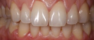
Before and after
Contraindications for treatment:
- First trimester of pregnancy;
- Bleeding gums;
- It is not recommended to carry out treatment during the period of illness with herpes and acute respiratory infections. It is better to postpone a trip to the dentist until you have fully recovered.
Methods for diagnosing contact caries
Visual inspection.
It is carried out during an appointment with the dentist. An old, traditional, cost-free method. But less accurate, because In some places, the dentist may not notice the early stages of caries development.
Diagnodent device
. Detection of lesions occurs by emitting special sound waves, which, reflecting from carious areas, are captured by the device. A very accurate diagnostic method.
X-ray diagnostics.
An excellent diagnostic method, because... allows you to identify carious lesions at an early stage at any location.
Features of the treatment of caries between the front teeth
When treating front teeth, it is important to preserve not only their functionality, but also their aesthetics. Therefore, the dentist is required to have extensive experience and skills in artistic restoration. Treatment of anterior teeth has several nuances:
- Carious lesions develop quickly due to the thin layer of dentin and enamel.
- During sanitation, it is necessary to achieve high aesthetic indicators, which is quite difficult with interdental caries.
The dentist carefully removes the affected tissue from the walls, and then restores the damaged teeth and interdental space. All medical procedures are performed under local anesthesia. There is an opinion that caries between the anterior units of the lower jaw is much easier to treat than on the upper jaw, but this is only a myth. There are no particular differences in the treatment protocol for the lower/upper dentition. To preserve beauty, a durable filling with high aesthetic indicators is installed on the front and side units. The most popular option is artistic restoration using compomer and reflective filling materials that preserve the aesthetics and natural appearance of the tooth.
Locations
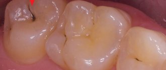
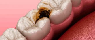
Interdental caries can develop anywhere in the dentition
. Patients turn to dentists both with caries between the front teeth and with damage to the chewing teeth. The photo below shows an example of the development of caries between the chewing tooth and the wisdom tooth.
Also, carious lesions can occur in both the upper and lower teeth. The infection affects not only permanent teeth, but also baby teeth. Therefore, both children and adults are at risk.
Features of the treatment of caries between chewing teeth
Although molars are located in the lateral part of the jaw and are practically invisible when smiling, interdental caries is especially dangerous for them, as it is often diagnosed already at the stage of deep damage, when the dentin layer of one or more chewing units is affected. Very often the disease affects the pulp of two teeth. This situation is the most unfavorable, since endodontic treatment of canals is a long and expensive process and is performed only by an experienced dentist. If you do not start fighting the disease in a timely manner, the patient may encounter complications in the form of periodontitis, pulpitis (nerve inflammation), etc.
When filling molars, special attention is paid to the strength and durability of the filling material. To restore them, high-quality glass ionomer fillings are used.
Treatment of the disease
Caries between teeth is treated in the same way as regular one, with the only difference being that two units have to be “fixed” at once. In some special cases, in order to get to the affected part, the dentist is obliged to get rid of healthy tooth tissue by drilling. First, the specialist must prepare the cavity and then treat it.
It is important to be careful about the process so as not to touch or even damage the nerve. After which the doctor installs a plate that separates the teeth and inserts a filling. At the end of the process, it is put in order by grinding and polishing.
Prevention
Preventing the development of interdental caries is quite simple. To do this, it is enough to follow the recommendations of dentists:
- It is recommended to clean your teeth after each meal, moving the brush up and down to remove as much food debris as possible in hard-to-reach places. For care, use pastes that improve the protective properties of enamel.
- Rinse your mouth several times a day. It is not necessary to use special solutions; boiled water will be enough for cleaning. Additionally, use an irrigator.
- It is better to avoid foods such as chips, chocolate, and crackers, as their particles settle tightly in the interdental space and become a haven for bacteria. As a last resort, carry dental floss with you.
- Visit your dentist's office every six months for a checkup and professional cleaning.
To find out the cost of treating caries between teeth, sign up for a consultation at Dr. Razumenko’s dental clinic in Moscow by phone.
Types of disease
Depending on the location of the disease, the dentist determines treatment methods, as well as the necessary medications.
The main types of interdental caries are:
- Disease of the chewing teeth. Typically, some food particles clog the space between these units. It is difficult for the patient to notice this disease and is often diagnosed when the infection reaches the root of the tooth.
- It is much easier to determine caries if bacteria have formed between the front teeth. It is impossible to hide the damaged appearance of the units; immediate treatment and elimination of the infection should be resorted to.
- Carious bacteria between baby teeth are the most dangerous because they can damage the root of the tooth, which in the future will affect new, permanent teeth.
As mentioned earlier, the bacteria of this disease progress very quickly. If proper measures are not taken in time, the adjacent tooth will also become ill.
In any case, if you detect the slightest signs of interdental caries, immediately seek help from a dentist, who will determine a special course of treatment.
Stages of development of caries between teeth:
- Spot. As you know, enamel loses its natural shine due to insufficient mineralization. Tooth decay begins the moment it loses the required amount of minerals. In the initial stage, carious bacteria take the form of a small matte spot. Modern technologies, together with careful oral hygiene, can eliminate infection in the bud.
- Superficial caries. Unpleasant, painful sensations after contact with sour, sweet, hot, cold. Most likely, carious bacteria are already developing in this oral cavity. The possibility of dentin damage should not be excluded.
- Deep caries. It manifests itself as a powerful, aching pain. It happens when softened dentin fills a carious cavity. It is fraught with the formation of pulpitis.
Prices for treatment of caries between teeth
| Code no. | Name of procedures | Unit of measurement | Cost, rub. |
| 101 | Repeated treatment and diagnostic appointment of the patient | 650,00 | |
| 300 | Application anesthesia | 150,00 | |
| 301 | Infiltration anesthesia, conduction | 550,00 | |
| 303 | Treatment of medium caries (ultrasound, cavity formation) | 650,00 | |
| 307 | Placement of a light-composite filling (Sonicfil, Estelite) | 3 950,00 | |
| 310 | Recovery from significant damage is complex | 1 unit | 5 500,00 |
| 313 | Artistic restoration (front, side teeth) simple | 1 unit | 11 000,00 |
* The prices indicated on the website are not a public offer. The exact cost can only be determined by visiting a doctor.
Prices for treatment in Moscow full price list
Share on social media networks:
Article Expert:
Ustinova Elena Fedorovna
Doctor of the highest category. Root canal treatment (complex, repeated retreatment), microprosthetics with ceramic restorations.
Work experience 15 years
Experts' opinion
The effectiveness of ASEPTA products has been repeatedly clinically proven. For example, according to research conducted at the Ryazan State Medical University named after Academician I.P. Pavlova of the Ministry of Health and Social Development of the Russian Federation (GBOU VPO RyazSMU Ministry of Health and Social Development of Russia), during the use of Asepta line products, a decrease in complaints of discomfort in patients is noted. Upon examination, a decrease in hyperemia and swelling of the gums was noted, but bleeding persisted upon probing.
Sources:
- Clinical experience in using the Asepta series of products Fuchs Elena Ivanovna Assistant of the Department of Therapeutic and Pediatric Dentistry State Budgetary Educational Institution of Higher Professional Education Ryazan State Medical University named after Academician I.P. Pavlova of the Ministry of Health and Social Development of the Russian Federation (GBOU VPO RyazSMU Ministry of Health and Social Development of Russia)
- Report on clinical trials to determine/confirm the preventive properties of commercially produced personal oral hygiene products: mouth rinse "ASEPTA PARODONTAL" - Solution for irrigator." Doctor of Medical Sciences Professor, Honored Doctor of the Russian Federation, Head. Department of Preventive Dentistry S.B. Ulitovsky, doctor-researcher A.A. Leontiev First St. Petersburg State Medical University named after academician I.P. Pavlova, Department of Preventive Dentistry.
- Report on determining/confirming the preventive properties of toothpaste “ASEPTA PLUS” GENTLE WHITENING” Author: doctor-researcher A.A. Leontyev, head Department of Preventive Dentistry, Doctor of Medical Sciences, Professor S.B. Ulitovsky First St. Petersburg State Medical University named after. acad. I.P. Pavlova, Department of Preventive Dentistry
We recommend that you read
Dental treatment during pregnancy
Treatment of anterior teeth
Dental filling
Canal filling
History of dental floss
The word “floss” comes to us from the English language and is literally translated as “dental floss”. It was first mentioned in the book A Practical Guide to Dental Care, written by American dentist Levi Spear Parmley in 1819. He suggested to his patients and readers to use silk dental floss to remove food debris from the interdental spaces.
During World War II, when Japan stopped supplying silk to America and Europe, silk dental floss was replaced by nylon floss, developed by DuPont. Nylon was thinner, stronger and cheaper. It was produced in large quantities, and therefore became very popular in the mass market.
Causes
The main cause of the development of cervical caries is considered to be the bacterium Streptococcus mutans, which lives in dental plaque. Accumulating in gum pockets, it contributes to softening of the enamel and destruction of the tooth at the base.
Other factors can provoke the development of pathology:
- regular consumption of excessively acidic foods and drinks;
- improper brushing of teeth;
- long-term use of certain medications that weaken enamel;
- endocrine diseases;
- deficiency of vitamins and minerals;
- pregnancy.
According to statistics, cervical caries often develops in older patients with a history of periodontitis and bad habits (smoking).
Symptoms
At first, the affected teeth may not show themselves in any way: no pain or destruction of the enamel. Over time, the patient begins to experience discomfort when eating, as well as observe bad breath, darkening of the tooth surface and the formation of a carious cavity. In the last stage, interdental caries can completely change the color of the teeth. If you notice any symptoms, contact your dentist immediately. Timely treatment allows you to avoid tooth loss, as well as save the health of the oral cavity and the whole body.
Impregnations for threads
During the production process, floss manufacturers add various impregnations and flavors to the threads to make the process of brushing teeth more enjoyable and effective. Impregnations can be flavoring, mineralizing and antiseptic:
- Menthol (or fruit) - have a pleasant aroma and freshen your breath.
In addition to menthol, fruit flavors are also used, for example, lemon, cinnamon, strawberry, etc. Curaprox DF834 waxed dental floss has a pleasant mint taste . Thread length 50 m.
Waxed floss Curaprox DF834, 50 m
Waxed thread Splat Dental Floss with strawberries, 30 m
Waxed thread Splat Dental Floss with bergamot and lime, 30 m
Waxed thread Splat Dental Floss with coconut, 30 m
- Fluoride - provide additional strengthening of teeth due to the mineralization of enamel with fluoride ions.
These flosses are recommended for those who have sensitive teeth. Pierrot Dental floss with aloe vera from the Spanish company Fushima is made of nylon . It contains fluoride and aloe vera juice for anti-inflammatory effects. Thread length 50 m.
Waxed dental floss Pierrot Dental floss EXPANDING, 30 m
Waxed dental floss Pierrot Dental floss with aloe vera, 50 m
Waxed dental floss Pierrot Dental Floss with strawberries, 50 m
- Chlorhexidine - have an additional antiseptic effect, reducing the likelihood of developing inflammatory gum diseases.
When choosing a thread with chlorhexidine, we recommend paying attention to the high-quality tape thread Mirafloss chx-Tape. The thread is unwaxed. Length 20 m.
Tape floss with chlorhexidine miradent Mirafloss chx-Tape, 20 m
Floss Curaprox PTFE floss tape DF820 with chlorhexidine, 35 m
Some threads contain combinations of several of the impregnations described above, and some are made without any impregnations.