What to do if caries is found between the teeth, is it treated without depulpation, restoration and other radical methods? This problem is quite common in both children and adults. Due to the peculiarities of the localization of carious darkening, diagnosis and further treatment are difficult. For this reason, it is important to visit your dentist regularly for a thorough examination and adhere to preventative recommendations. If you consult a doctor on time, the disease will be eliminated without consequences; prosthetics and other radical measures will not be required.
Reasons for development
The etiology of damage to hard tissues in this case is similar to the factors causing classic damage. The pathological process develops due to the accumulation of food particles and the active reproduction of pathogenic microorganisms in an environment favorable to them. The disease can spread very quickly, affecting not only the enamel, but also dentin, reaching the roots and causing other diseases. If you do not consult a doctor promptly, you may subsequently need to remove the nerve or install a prosthesis.
Where does interdental caries appear?
It is observed in places of close contact of dental units. It often occurs between the posterior chewing molars, which are actively involved in the process of chewing food. The front incisors and canines are affected much less frequently, since they are used only for biting food.
It is especially unpleasant when the disease affects “eights”. The distant location, as well as uneven roots, complicate filling and depulpation. Most often, experts recommend complete removal without subsequent prosthetics.
Caries between the gum and tooth is not considered interdental. A carious cavity should form precisely in the areas between adjacent units located in the vicinity.
Who gets the disease?
This is one of the most common dental problems. The disease occurs in children of all ages and adults.
It is often found on primary incisors and premolars in children 4 years of age and older. This feature is due to the fact that at this time the child begins to eat all the food from the parent’s table and at the same time needs regular high-quality oral hygiene.
Some parents mistakenly believe that if their son or daughter’s first teeth are damaged, they don’t need to be filled, since they will soon fall out anyway. Caries between teeth is treated both in the presence of temporary units and in the development of permanent ones. The pathological process can spread further, including to the rudiments of the molars and molars. Infection will lead to serious problems later in life.
But you should not be afraid that the child will be in pain during treatment. In the Dentika clinic, in case of deep lesions, the need for depulpation and canal cleaning, both the usual method of pain relief and sedation are used, that is, when performing medical manipulations, the baby sleeps and does not feel pain.
Why does pathology appear?
The main reason for the appearance of caries between teeth in adults and children is the accumulation of food debris in the interdental space and the active proliferation of pathogenic bacteria. With regular brushing, it is almost impossible to completely clean the contact points between the units. It is necessary to use additional devices (irrigator, floss).
The disease develops as follows:
- a person does not eat properly or does not regularly cleanse, or provides poor quality care;
- mucous membranes and enamel are covered with a dense coating;
- biological residues produce acids;
- the resulting substances corrode the outer layer and after some time affect the dentin.
The problem occurs in people with malocclusion, dystopic figure eights and other physiological characteristics. This is due to the difficulty of performing hygienic procedures and detecting carious lesions during a visual examination without x-rays and other precise techniques.
There is a list of factors that can lead to caries between two teeth:
- smoking, alcohol abuse;
- poor food quality;
- unfavorable environmental situation in the locality;
- reduced immunity;
- a history of chronic diseases affecting the condition of hard tissues;
- abuse of sweets and other carbohydrate foods.
Causes of pain in the gums
Unpleasant sensations in soft tissues can be temporary or permanent. Most often pain occurs:
- due to damage to the gums by hard food, bone, toothpick,
- using a toothbrush with stiff bristles,
- untimely removal of soft plaque,
- consuming hot foods and drinks,
- allergies to mouthwash components,
- deficiency of vitamins and minerals in the body.
When the mucous membrane is injured, the pain goes away within 2-4 days. Also, sometimes it is enough to change the brush to a softer one, buy another toothpaste and rinse to get rid of discomfort in the oral cavity.
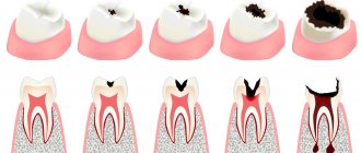
Four stages of development
Pathology goes through several successive stages. At first, this is a small contrasting spot that does not affect deep formations and does not lead to complications with timely treatment. What caries looks like between teeth at different stages of its spread:
- Elementary. Only the upper enamel layer thins. Small white or dark spots are found where surfaces touch.
- Surface. The enamel begins to deteriorate, forming depressions that can be felt by running your tongue over them. Darkening is clearly visible in the interdental space.
- Average. Blackened areas are visible to the naked eye. Symptoms appear, such as sensitivity to cold, hot and sour, and bad breath.
- Deep defeat. The diseased crown begins to collapse. The spaces between adjacent incisors, canines or molars widen. The patient feels severe paroxysmal pain, sometimes there is bleeding after biting food, and the mouth smells rotten. Teeth may become partially chipped or fall out completely.
Recommendations
First of all, take good care of your mouth. When the first signs of food stuck appear, we recommend contacting your dentist for specialist advice. Don't forget to go to the prof 2 times a year. inspections. During a consultation with our specialists, you can find out which teeth should be treated and which fillings need to be replaced with new ones. You also need to come for professional teeth cleaning twice a year.
Secondly, we recommend contacting only trusted clinics with many years of history and experienced doctors. We would like to warn you against the temptation to use free dentistry under compulsory medical insurance; remember, good treatment and prosthetics cannot be free. Don't skimp on your health!
We hope that our article helped you and you found answers to your questions. If you have any further questions or would like to make an appointment, there is an appointment form below.
Why is interdental caries dangerous?
The disease leads to negative consequences, affecting both the front incisors and chewing units. In the first case, the aesthetic component suffers, since the problem is visible to others with the naked eye. A person cannot smile normally while exposing his gums, because people immediately see unattractive black areas. Also, the rows at the front have thinner enamel, so the dark spot grows faster. Dentiki doctors can carry out work of almost any complexity in order to properly heal everything and restore the former beauty of the smile.
If caries between the lower or upper teeth affects the rear molars, this is also dangerous, because the problem may remain unnoticed for a long time, and the process will progress to a more severe stage. Patients often come to the dental clinic with deep lesions, accompanied by complications such as pulpitis or periodontitis. Usually the disease affects chewing units located nearby, so several have to be treated at once.
It also happens that only one crown is affected. This most often occurs at the very beginning or in the presence of prostheses and other artificial devices in the neighborhood. In this case, the pathology is not so dangerous.
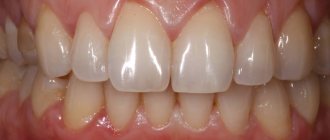
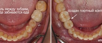
The result of replacing crowns is that food does not get stuck between the teeth
Tight contacts between teeth ensure that food does not get stuck and gums remain healthy.
The cost of ceramic crowns is 55,000 rubles per unit.
Of course, it is possible to make dental crowns cheaper (cheaper material, lack of computer modeling, etc.), but this will invariably affect the quality. The use of computer technology makes it possible to more carefully work out the shape of the crowns, provides additional convenience when taking impressions - all this is reflected in the price, so good dental crowns cannot be cheap. In addition, Dial-Dent uses the highest quality and most modern materials both for the crowns themselves and for their fixation, which increases the service life of dental restorations.
Video about how teeth are scanned with an intraoral scanner:
Symptoms
It is quite difficult to detect the disease in its earliest stages. The first unpleasant signs appear when the integrity of the enamel is damaged. A person begins to react to temperature stimuli, especially food and drink. It is not so easy to examine a carious cavity, since the inner surface of the units may be damaged.
Characteristic symptoms that indicate a problem and require urgent contact with a dentist:
- painful reaction to hot, cold, sour, sweet;
- increased sensitivity when performing routine hygiene procedures;
- foul breath associated with the proliferation of pathogenic bacteria and decay of plaque (experts have not proven whether there can be caries between teeth that are regularly cleaned of particles of eaten food with an irrigator);
- the appearance of white or dark spots on the enamel;
- paroxysmal pain (the larger the damaged area, the more intense the pain);
- the presence of visible carious cavities that can be felt with the tongue (in advanced situations).
It is better not to bring the disease to such an extent that severe pain appears and black spots are visible to the naked eye. With periodic preventive checkups at the dentist, the problem can be identified at the earliest stages.
The first group includes:
Caries
A carious cavity is a depression in the hard tissues of the tooth that appears as a result of their destruction. Food or drinks (most often cold) enter the cavity and cause short-term (several seconds) pain. If food is retained in the carious cavity (especially sweet food), the pain may last longer.
Seal defect
If part of the filling (or part of the hard tooth tissue around the filling) placed previously breaks off under the influence of chewing load, a part of the tooth that is more sensitive to the effects of food is exposed.
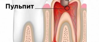
Chronic apical periodontitis in the acute stage
It sounds scary...and in reality it is no less terrible. The mechanism of pain in this case is quite simple. Around the apex of the root, surrounded by a dense bone plate, the inflammatory process is activated. Inflammation in all parts of our body proceeds along approximately the same path and is characterized by an increase in the volume of inflamed tissue (due to blood flow to the affected area). And if this happens, for example, when a hand is bruised, then we will see swelling, that is, an increase in volume. Now let’s imagine the formation of a “swelling” in the area of the root apex surrounded by a dense bone plate... there is no room for the incoming fluid, the nerve endings are compressed, and here you are also trying to chew a delicious steak!
Periodontitis
One of the causes of tooth pain is periodontitis.
The thing is very unpleasant (like most dental diseases). It is characterized by a long-term, most often sluggish inflammatory process of all tissues surrounding the tooth (gums, ligaments, bone surrounding the tooth), leading to their atrophy (reduction). As a result, the tooth becomes mobile and cannot withstand the chewing load, which is indicated by pain.
Functional overload
Each of our teeth, depending on the function it performs, is designed for certain chewing loads. However, if for some reason the number of teeth decreases (extraction, injury, etc.), the remaining ones begin to experience increased chewing pressure (for themselves and for the lost ones). Such excessive load leads to loosening of the tooth and the appearance of the symptom we are discussing. Only high-quality prosthetics will help solve this problem.
Injury to the gingival papilla by hard food
The reasons for this phenomenon may be:
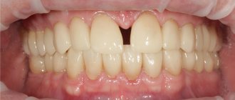
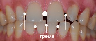
Large distances between teeth (diastema and trema). They can be either physiological in nature (features of the anatomical structure) or pathological (for example, “divergence” of teeth during periodontitis);
Poorly restored contact point
A contact point is a point or area through which adjacent teeth contact each other and through which chewing pressure is distributed. If the carious cavity was located on the side surface of the tooth and after filling it, the contact point was not properly restored, then food will get clogged between the teeth when chewing, injuring the gingival papilla. Hence the pain. This reason relates more to the second group, so let’s move on to it.
Diagnosis of hidden caries between teeth
It is not so easy to detect interdental damage on your own due to the peculiarities of its localization. Patients should first of all pay attention to the appearance of pain, bad breath, dark spots and make an appointment with a doctor. If immediate action is not taken, the pathology may affect neighboring units.
Methods used in the dental clinic:
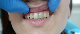
- Visual inspection. The specialist carefully examines the oral cavity using a mirror and probe. A professional can detect darkening even in the most difficult to reach recesses. If the coronal surfaces are located too close to each other and it is impossible to examine everything carefully, the use of other techniques is indicated. You can see what caries looks like between the front teeth in the photo below.
- X-ray. X-rays can reveal areas that cannot be detected externally. The jaws are displayed in white, and the affected areas are highlighted in black and gray.
- Staining and transillumination. Carious structures are detected by applying staining compounds or using fluorescent diagnostics. This way, the presence of shadows in deep, hard-to-reach places is established.
What doctors advise you to do
Some patients notice slight darkening and believe that the damage is not significant enough to contact the dental clinic. Delay in treating caries between the front and back teeth can result in high material costs and complex medical procedures; therapy can take a long time. If the damage is severe, the dentist will have to restore several adjacent incisors, canines or molars at once.
Experienced Dentiki dentists do not begin intervention without a high-quality diagnosis and determination of the stage of the pathological process. First, the specialist will examine the oral cavity with a mirror and probe and only then decide whether additional research is required. A visual examination helps identify the problem in no more than 40% of cases, so it is not surprising that the clinic prescribes x-rays and other procedures.
Doctors note that eliminating deep lesions often requires several visits to the dental office. First, a temporary filling is installed, which is later replaced with a permanent one.
Consequences of regularly getting food stuck between teeth
Some people don’t even think about how to get stuck food out of their teeth. They are guided by the principle, it doesn’t hurt and it’s okay, making a big mistake. Meanwhile, such neglect can lead to dire consequences. For example:
- Gum diseases - food debris is an irritant that damages the mucous membranes, which provokes the development of dental diseases: gingivitis, periodontitis, etc.;
- Unpleasant odor - the food remaining between the teeth begins to decompose, which, in addition to the amber, creates a favorable environment for the life of pathogenic bacteria;
- Root injury – irritation of the gums can cause exposure of the neck of the tooth, which increases sensitivity and causes pain;
- Caries - stuck pieces of food is one of the main causes of carious lesions of the enamel; this disease can only be eliminated through dental intervention;
- Tissue destruction – if you do not clean the interdental space, the teeth begin to gradually deteriorate, which can lead to complete loss.
Treatment of caries between teeth
The therapy is no different from the methods of eliminating ordinary carious cavities, the only difference is that it is necessary to heal several neighboring units at once.
Methods used:
- Remineralization. The enamel is restored with the help of medications with calcium and fluoride. The actions will show a positive effect if the stain stage does not progress to the next one and the integrity of the outer layer is not compromised.
- Filling. This is the method that is used most often. The resulting hole is filled with filling material. How caries is treated between the two front teeth and chewing molars depends on its location and the severity of the pathological process. If the crown is very damaged, it is possible to install pins, inlays, and endodontic treatment.
- Prosthetics. When the tissues are destroyed so much that filling is impossible and the installation of prostheses (for example, inlays) is indicated.
- Icon. At the initial stage of the disease, a special polymer substance is applied to the damaged area. This method will be effective if the dentin is damaged by no more than a third.
Caries between front teeth
If you consult a doctor in a timely manner when the pathology manifests itself in the form of a spot, it is enough to use remineralization and other manipulations to restore the enamel. Physiotherapeutic procedures are important. The treatment course is usually about 5-7 visits to the treatment room. As a result, blood circulation in the tissues significantly improves, and the surface layers are quickly restored. Frequently used methods: laser irradiation, electrical stimulation, magnetic laser therapy.
How to remove caries between teeth with deeper damage:
- injection anesthesia (pain relief) or applications for comfortable treatment;
- cleansing the carious cavity from accumulated deposits;
- isolation of affected areas and mucous membranes using a latex napkin (rubber dam);
- mechanical removal of black deposits with a drill;
- applying a disinfectant and adhesive composition;
- installation of a seal;
- grinding of contact surfaces;
- restoration (use of crowns, veneers, etc.) in case of severe damage.
Caries of chewing teeth
Damage to such units is much more common than to the anterior ones. This is due to the fact that the jaws are involved in the process of chewing hard and soft food, which subsequently accumulates in the interdental spaces. Even small fragments of food are enough for infection to occur and bacteria to multiply.
Sequence of dentist actions:
- Anesthesia. The method of pain relief depends on the depth of the hole. In the early stages, before removing caries between teeth, it is optimal to apply an application; in more serious situations, anesthesia is used.
- Cleaning from plaque and hardened stone.
- Insulation using rubber dam.
- Preparation. In the case of chewing molars, healthy tissue is often removed in order to provide access to the cavity.
- The use of disinfectants and adhesives for better adhesion to the filling material.
- Filling (fillings based on composite substances are most often used).
- Grinding of surfaces after restoring the integrity of teeth.
Dental scanning and dental crown modeling
Before modeling the new crowns, a teeth scan was performed. Dental scanning replaces traditional dental impressions, which many patients cannot tolerate due to an increased gag reflex. When taking digital impressions of teeth, the dentist runs a Sirona scanner along the surfaces of the teeth for several minutes, and a three-dimensional image of both jaws is transferred to the computer - a digital impression of the teeth.
Working with a digital impression in a specialized program, the dentist models crowns with the most tight contacts between the teeth and the correct anatomical chewing surface.
Based on the simulation results, the dental technician produced ceramic crowns from E.max ceramics. Pressed ceramics E.max has high strength and excellent aesthetic properties, therefore it is used for prosthetics of both lateral and anterior teeth.
Prevention of interdental caries
To avoid frequent visits to the dentist with severe pain and other characteristic symptoms, you need to follow simple rules. Hygiene procedures that help prevent disease:
- rinsing and brushing your mouth after every meal;
- regular cleansing with dental floss;
- the use of high-quality pastes with antibacterial components and minerals that help strengthen the enamel (bacteria are the main reason why caries appears between teeth);
- professional treatment for plaque and accumulated stone in the clinic every six months.
Preventive nutritional recommendations:
- give up foods containing large amounts of carbohydrates (sweets, fast food, confectionery, rich bread);
- refrain from eating cold, hot and sour;
- include foods with fluoride and calcium in the menu;
- eat vegetables and fruits more often;
- do not chew nuts, do not bite threads, try not to injure the crown.
Even in the absence of pain and visible darkening, it is necessary to visit the dentist regularly. Only a professional doctor will be able to notice the early symptoms of the pathological process and cure the disease without consequences.
How to understand that caries between teeth has affected the tissue if it is not visible? Pathology occurs quite often, since it is in this place that food accumulates, which cannot be removed with a regular brush. If you do not use an irrigator or thread, a person will most likely encounter this problem. It is important to remember that compliance with preventive measures and regular examinations at the Dentika dental clinic will help prevent the development of the disease and completely cope with it if a stain is detected at an early stage.
Swelling
Swelling may appear after gum injury. In this case, it is localized, extends only to the site of the bruise, may hurt a little, but in general it goes away quickly. If after an injury the swelling persists for a long time, intensifies, the pain becomes acute or throbbing, you need to consult a doctor.
In other cases, swelling of the gums is a symptom of inflammation. It is accompanied by pain, bleeding may appear, the color of the mucous membrane changes (it becomes darker or acquires a bright red tint). Such swelling may accompany gingivitis or periodontitis.