From this article you will learn:
- stages of caries,
- what are the signs of caries?
- how to identify caries yourself.
Caries is the process of destruction of hard tooth tissues with the active participation of cariogenic microorganisms of the oral cavity, such as streptococci, actinomycetes and lactobacilli. The main conditions for its development are irregular or insufficient oral hygiene, which leads to the accumulation of soft microbial plaque on the teeth, as well as food debris between the teeth. Depending on the depth of damage to the hard tissues of the tooth, caries is usually divided into several types, which, in fact, are successive stages of its development.
The initial stage is always spot-shaped caries (foci of demineralization in the form of white spots on the surface of tooth enamel), and if left untreated, it gradually goes through the stages of superficial, medium and deep caries. Symptoms of dental caries, where we include patient complaints and data from a visual examination of the carious cavity, will be slightly different at each stage, and below we will consider them in detail.
Stages of caries: symptoms, diagnosis
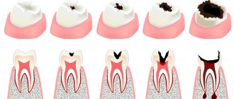
It is customary to distinguish 4 successive stages of caries development. Each stage is characterized by its own symptoms, as well as visual and instrumental examination data, which is important to consider when diagnosing caries.
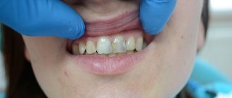
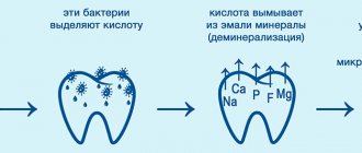
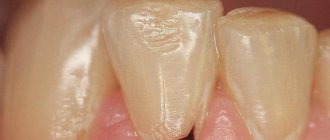
- Caries in the white spot stage (Fig. 1) - manifests itself in the form of white spots on the surface of the enamel, which are nothing more than foci of demineralization. Their formation is associated with cariogenic microorganisms that metabolize food debris in the oral cavity, converting them into organic acids. The latter, upon contact with tooth enamel, begin to dissolve its mineral matrix, which leads to the loss of calcium (hydroxyapatite) by the enamel.
It should be especially noted that at this stage of caries the appearance of the actual carious defect is not yet observed. The changes concern only the appearance of white spots of a chalky or gray hue, the surface of which lacks shine (characteristic of healthy teeth), and roughness of the enamel in the spot area is also observed. If you scratch the enamel with a sharp instrument, it is easily scraped off, leaving a defect. Apart from aesthetic ones, patients at this stage usually do not present any other complaints.
In order to clearly show patients the degree and number of areas of enamel demineralization, after removing plaque, teeth are treated with aniline dyes (1% solution of methylene blue or caries detector). Dyes temporarily stain areas of demineralization dark, clearly showing the extent and extent of the problem. At the same time, healthy dense enamel is not susceptible to such staining with dyes.
→ Treatment of the initial form of caries
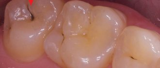
- Superficial form of caries (Fig. 2) - a defect is formed within the layer of tooth enamel, without damaging the dentin underlying the enamel. Typically, superficial caries occurs in areas of demineralization of tooth enamel, i.e. is the next stage in the development of caries in the white spot stage. The formation of a carious defect at the site of a white spot occurs if oral hygiene does not improve, which means that the influence of organic acids and calcium loss continue.
At some point, calcium becomes so low that the enamel framework itself begins to collapse and, accordingly, a superficial carious defect appears. This is especially clearly visible in Fig. 2, where superficial carious defects have formed against the background of foci of demineralization (in the form of white spots). Once again, we draw your attention to the fact that the bottom of the carious defect in superficial caries never extends beyond the enamel-dentin border.
Patients complain of pain from sour, salty, sweet foods, which, however, quickly disappear immediately after the influence of the irritant is eliminated. In addition, if the carious defect is located on the visible surface of the front teeth, patients note a cosmetic defect. An instrumental examination of the carious cavity shows that its edges are soft, the enamel easily chips under mechanical pressure with a sharp instrument (24stoma.ru).
→ Treatment of superficial forms of caries
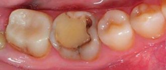
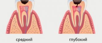
- Average caries (Fig. 3) - in the absence of timely treatment, the carious defect increases, spreading beyond the enamel-dentin border. Thus, the bottom of the carious cavity will be located in the upper or middle layers of dentin (without affecting its deep layers directly surrounding the dental pulp). When examining a carious cavity, it is clear that it is filled with food debris and decay of hard tooth tissue. There are usually no complaints from patients other than aesthetic ones.
→ Treatment of average caries
- Deep form of caries (Fig. 6) - in the absence of timely treatment, the carious process spreads to the deep layers of dentin, so that between the dental pulp (neurovascular bundle) and the bottom of the carious cavity, only a very thin layer of healthy dentin remains. With this form of caries, patients usually complain of pain arising from temperature stimuli (especially cold) and sweets. Moreover, the pain quickly passes when the stimulus stops.
However, if the carious cavity is simply clogged with food debris and decay of hard tooth tissues, the pain syndrome may be weak or absent altogether. In addition, patients may complain of pain when chewing, which is associated with the pressure of hard food on the bottom of the carious cavity while eating. Visually, the carious cavity is usually large in size, but may also have a small entrance hole. There is pain when probing the bottom of the carious cavity and in the area of the enamel-dentin border.→ Treatment of deep caries
Entin's physicochemical theory
Having studied the physicochemical properties of teeth and saliva, Entin decided that dental tissue is a partially permeable membrane that separates two environments - blood and saliva. When the normal pressure of these media on the septum is disrupted (due to an unbalanced diet, a weakened state of the body), problems arise with the nutrition of the enamel. As a result, tooth enamel is attacked by pathogenic microorganisms, contributing to the development of caries.
What does caries look like under a microscope?
Caries looks very interesting under a microscope, especially if you look at it on a tooth cut. Below you can see what superficial, medium and deep forms of caries look like. In addition, in each photograph in the center of the tooth crown you can see the cavity (pulp chamber) in which the neurovascular bundle of the tooth is located.
Influence of the external environment
The likelihood of developing caries is increased by poor ecology, unfavorable climate, constant exposure to sunlight and other environmental factors. People who live in northern regions are more likely to suffer from this disease than residents of southern countries.
In addition, its appearance can be caused by low water hardness, which is determined by the concentration of magnesium, calcium and fluorine. The fewer minerals in the liquid we consume daily, the higher the risk of tooth decay.
The level of air pollution should also be taken into account. In rural areas and in the mountains, where it is clean and fresh, the number of people with dental diseases is 20% lower than in cities.
Complications of caries -
If the deep form of caries is not treated in time, then the infection penetrates into the pulp of the tooth. In this case, inflammation of the nerve in the tooth develops, which is called pulpitis. If, in turn, pulpitis is not treated in a timely manner, then the inflammation extends beyond the boundaries of the tooth - into the tissue surrounding the tips of the roots of the diseased tooth. In this case, granulomas or cysts (purulent sacs) appear at the tops of the roots. This disease is called periodontitis.
Prevention measures
Having summed up all the factors in the development of caries, we can make recommendations that will help prevent the disease and save your teeth:
- Limit your intake of sugar in any form (less candy and baked goods).
- Brush your teeth at least 2 times a day (in the morning and before bed).
- Rinse your mouth with water after every meal.
- To strengthen the enamel, you can use home remineralizing products (ROCS, Elmex, etc.) or undergo fluoridation at the dentist.
- Once every six months, it is useful to do ultrasonic teeth cleaning in the clinic; with the help of this procedure, the doctor removes all bacterial deposits that cannot be removed at home.
- Eat right, eat more raw vegetables and fruits - cabbage, apples, carrots, they contribute to the natural cleansing of enamel. Nuts, fish and dairy products are also good for your teeth.
If you exclude all negative factors, you can forever protect yourself from complications of caries in the form of pulpitis or granuloma.
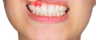
Signs of caries and pulpitis (differences in symptoms) –
Typical signs of caries are the infrequent occurrence of short-term painful attacks under the influence of various irritants:
- thermal irritants (cold water or cold air),
- chemical irritants (for example, sour, salty or sweet foods).
At the same time, caries is characterized by the absence of spontaneous pain, i.e. pain during caries occurs only under the influence of irritants. And as soon as the irritant is eliminated, the pain should immediately disappear. The presence of spontaneous pain not associated with exposure to irritants indicates that the process from caries has already passed into pulpitis.
With deep caries (plus the above), pain may also occur when mechanical pressure is applied to the thinned bottom of the carious cavity. This can occur, for example, while eating - in the process of biting or chewing hard food. In this case, food enters the carious cavity and presses on its bottom, causing compression of the dental pulp (neurovascular bundle inside the tooth). In this case, removing food debris from the carious cavity usually relieves pain.
How to distinguish caries from pulpitis: differences in symptoms
- With caries, pain occurs only in the presence of irritants (thermal, chemical). When the irritant is removed, the pain immediately disappears.
- In acute pulpitis, pain is usually acute, spontaneous and not associated with any irritants.
- But with chronic pulpitis, pain can still be provoked by thermal irritants - cold or hot water. But the difference between caries and chronic pulpitis is that with caries the pain goes away immediately after the irritant disappears, and with chronic pulpitis the pain does not go away immediately, but only after 10-15 or more minutes.
Clinical researches
Asept toothpastes are distinguished by their clinically proven effectiveness - the products have been repeatedly tested. As part of the tests, it was found that:
- regular use of preventive toothpaste ASEPTA ACTIVE for a month can reduce bleeding gums by 60%, improve the overall condition of the oral cavity by 44% and reduce inflammation by 33% (research);
- Regular use of preventive toothpaste ASEPTA SENSITIVE for a month can reduce bleeding gums by 62%, reduce sensitivity of teeth and gums by 48% and reduce inflammation by 66%. (study);
- regular use of professional toothpaste ASEPTA REMINERALIZATION after 4 weeks improved the condition of the enamel by 64% and reduced tooth sensitivity by 66% (study);
- Regular use of professional toothpaste ASEPTA GENTLE WHITENING for a month allows you to lighten tooth enamel by 1.5 tones, increases anti-caries effectiveness by 3.4 times and increases enamel remineralization by 2.6 times (research).
Sources:
- Report on determining/confirming the preventive properties of toothpaste “ASEPTA PLUS” GENTLE WHITENING” Author: doctor-researcher A.A. Leontyev, head Department of Preventive Dentistry, Doctor of Medical Sciences, Professor S.B. Ulitovsky First St. Petersburg State Medical University named after. acad. I.P. Pavlova, Department of Preventive Dentistry
- Clinical and laboratory assessment of the influence of domestic therapeutic and prophylactic toothpaste based on plant extracts on the condition of the oral cavity in patients with simple marginal gingivitis. Doctor of Medical Sciences, Professor Elovikova T.M.1, Candidate of Chemical Sciences, Associate Professor Ermishina E.Yu. 2, Doctor of Technical Sciences Associate Professor Belokonova N.A. 2 Department of Therapeutic Dentistry USMU1, Department of General Chemistry USMU2
- Report on the determination/confirmation of the preventive properties of personal oral hygiene products “ASEPTA PLUS” Remineralization doctor-researcher A.A. Leontyev, head Department of Preventive Dentistry, Doctor of Medical Sciences, Professor S.B. Ulitovsky First St. Petersburg State Medical University named after. acad. I.P. Pavlova, Department of Preventive Dentistry
- Clinical studies of antisensitive toothpaste “Asepta Sensitive” (A.A. Leontyev, O.V. Kalinina, S.B. Ulitovsky) A.A. LEONTIEV, dentist O.V. KALININA, dentist S.B. ULITOVSKY, Doctor of Medical Sciences, Prof. Department of Therapeutic Dentistry, St. Petersburg State Medical University named after. acad. I.P. Pavlova
- The role of anti-inflammatory rinse in the treatment of periodontal diseases (L.Yu. Orekhova, A.A. Leontyev, S.B. Ulitovsky) L.Yu. OREKHOVA, Doctor of Medical Sciences, Prof., Head of Department; A.A. LEONTIEV, dentist; S.B. ULITOVSKY, Doctor of Medical Sciences, Prof. Department of Therapeutic Dentistry of St. Petersburg State Medical University named after. acad. I. P. Pavlova
- Report on determining/confirming the preventive properties of toothpaste “ASEPTA PLUS” COFFEE and TOBACCO Author: doctor-researcher A.A. Leontyev, head Department of Preventive Dentistry, Doctor of Medical Sciences, Professor S.B. Ulitovsky. First St. Petersburg State Medical University named after. acad. I.P. Pavlova, Department of Preventive Dentistry
- Report on determining/confirming the preventive properties of commercially produced personal oral hygiene products: Asepta toothpaste used in combination with Asepta mouthwash and Asepta gum balm Head. Department of PFS Doctor of Medical Sciences Professor S.B. Ulitovsky St. Petersburg State Medical University named after Academician I.P. Pavlova. Faculty of Dentistry. Department of Preventive Dentistry.
How to determine caries at home -
- The presence of pain syndrome, which we wrote about above.
- The cavity may have sharp edges and irregularities, which can be perfectly felt (in some cases) by the tongue.
- Bad breath may indicate the presence of caries. Food debris is retained in the carious cavity, which is difficult to clean; under the influence of the microflora of the oral cavity, they begin to rot, causing an unpleasant odor.
- Inspection of visually accessible surfaces of teeth (for example, in a mirror). At the same time, even if the surface layer of enamel is preserved, darkening may be visible underneath it. Such darkening may indicate caries.
Treatment methods
Modern dentistry offers an alternative to the well-known drill, which often becomes the reason for patients’ fear of dental treatment.
Treatment of dental caries at the Dentpremium center involves the use of innovative techniques and advanced technologies in the field of dentistry.
Patients of our clinic can make a choice of methods for eliminating carious lesions.
However, deep caries, regardless of the localization zone, can only be treated with a drill, since it requires access to dentin.
Types of fillings
The choice of filling material is an important stage in the treatment of caries. The presence or absence of risks of pathological processes occurring in the treated tooth depends on how well the filling is fixed.
Fillings are classified into two main types based on the nature of their installation: temporary and permanent. The former are used when further treatment of the disease is expected, and the latter are installed after completion of all dental procedures.
Depending on the material used to make the fillings, there are:
- cement - the cheapest option, which performs its main function, but has a short service life;
- plastic - a material of an average price category, characterized by increased strength and resistance to thermal and chemical influences, however, it has a toxic effect and deforms over time;
- ceramics are the best option for a high-quality, wear-resistant and durable filling that is not subject to external and structural deformation;
- photo composite is an expensive option that guarantees a long service life, and is characterized by high strength, aesthetics and a wide selection of shades.
When treating caries, the cost of the filling has a significant impact on the price. Photopolymers and ceramics are considered premium materials. These products allow the clinic to guarantee the results of the dentist’s work.