Visually, a fistula is a hole in the gum, directly connected to the source of inflammation located near the upper part of the root of any of the teeth. Typically, the fistula opening is located just above the diseased tooth, which is either filled or has a carious formation. Periodontitis is a disease characterized by the appearance of a fistula.
Development of a fistula on the gum
The process of fistula formation takes a fairly long period of time: for this to happen, inflammatory processes must occur in the tissues of the damaged tooth.
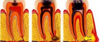
The stages that a tooth usually goes through before a fistula appears are as follows:
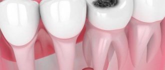
- caries is formed;
- the disease goes into a deep form;
- the development of caries leads to the formation of pulpitis;
- the last stage is periodontitis.
If you ignore periodontitis and do not take measures to treat it, the disease becomes chronic. At the moment of exacerbation, purulent masses come out through the fistulous tract formed in the gum.
Fistula between tooth and gum
A hole between a tooth and periodontal tissue indicates the development of cervical caries or the complications it caused. Cervical caries can be located between the teeth and not be visible to the naked eye. Due to its inaccessible location, such caries often turns into pulpitis.
Pulpitis is an inflammation of the blood vessels and nerve endings located in the pulp chamber of the tooth. The chronic stage of the pathology may be accompanied by the formation of a purulent focus in the soft tissues. If pulpitis progresses to the next stage of periodontitis, several fistulas may appear at once.
Main symptoms of fistula
Before the formation of a fistula, the tooth begins to hurt: when pressing on it and the surrounding area, painful sensations arise, it seems that the tooth is bursting from the inside.
Body temperature may rise and the gums may swell. When the pus comes out, the patient feels relief, thinking that the attack is over. However, this is a misconception: the disease continues to develop, although at this time the person feels much better. The tooth stops hurting, and pus no longer forms.
If no action is taken, the damage will affect the bone tissue, which will require more complex and lengthy treatment.
Prognosis and possible complications
If the fistula is detected and treated in a timely manner, the prognosis is favorable. If you follow all the specialist’s recommendations, within a week the resulting wound will completely heal and heal.
If proper treatment is not carried out, the following consequences are possible:
- abscess formation;
- phlegmon;
- osteomyelitis;
- tooth loss.
Preventive measures
- regular teeth cleaning;
- carrying out professional hygiene in the dental office;
- preventive examinations by a dentist every six months;
- daily consumption of fruits and vegetables.
How to treat a fistula on the gum: diagnosis of the disease
Any treatment is preceded by a diagnosis: the doctor needs to understand what to treat the patient for, as well as decide on the methods.
How to diagnose a fistula? First of all, the patient complains of pain. Often a person may find a small lump in the mouth from which fluid periodically leaks.
A more effective way to establish a fistula is a tomogram or x-ray. Gutta-percha is injected into the fistula tract under anesthesia. An image is taken to determine the location of the inflammatory process.
X-rays performed using modern equipment allow us to establish a complete picture of the disease, which makes it possible to use the necessary treatment methods.
Causes of holes in the gums
In medical terminology, a hole in the gum is called a “fistula”. Such a channel is formed due to the activity of pathogenic microflora located in the area of the alveolar process. The most common causative agents of fistula are coccus bacteria.
Hole in the gum near the tooth
The main reason for the formation of a hole in the mouth near a tooth is the abundance of pathogenic and conditionally pathogenic microflora in the oral cavity. Failure to comply with hygiene measures, the growth of dental plaque, and the formation of mineralized dental plaque provoke the appearance of a hole in the gum. In this case, large dental stones are found, pressing on the edges of the periodontium. In addition, pigmented plaque spoils the aesthetics of a smile.
Another reason for the appearance of a hole near a tooth can be attributed to infectious inflammation. Sore throat, scarlet fever, whooping cough or ARVI reduce local and internal immunity, thereby creating favorable conditions for the proliferation of microorganisms.
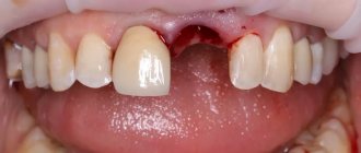
Hole in gum after tooth extraction
The presence of intragingival space after surgical tooth extraction is normal. The location of the hole depends on the location of the extracted tooth. Typically, the resulting hole heals within 2–3 weeks after surgery. If surgery was performed on wisdom teeth, complete retraction of the mucous tissue occurs 6–8 weeks after removal. Impaired healing of the socket is observed when an infection occurs or the wound surface is frequently injured.
If the postoperative hole has healed normally, and a periodontal gap has appeared on the side of the gum, then we can talk about leaving small parts of the tooth in the hole. The same thing occurs when dental instruments and materials are accidentally left in the periodontal space. The foreign body begins to decompose, and pathogenic bacteria enter the external environment, forming a fistula opening.
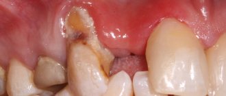
Hole between tooth and gum
A hole in the gum between the tooth and periodontal tissue is formed due to cervical caries and its complications. In this case, caries can be localized between the teeth, affecting both dental surfaces near the gums. Due to their inaccessible location, carious lesions are difficult to detect and most often turn into pulpitis.
Pulpitis is the inflammation of blood vessels and nerve bundles located in the pulp chamber. In the chronic stage of the disease, a purulent focus may appear near the gums.
Ignoring pulpitis leads to periodontitis, which is characterized by the formation of one or more fistulas.
Hole in a child's gum
The appearance of a hole in the gum near a tooth in children occurs for 2 reasons: periodontitis (a complication of caries) and trauma to the mucous membrane.
With periodontitis, bone tissue is affected. A cavity is formed inside the periosteum, filled with purulent exudate. When there is a lot of pus, it breaks into the oral cavity through the fistula canal, causing pain.
With frequent mechanical trauma to the mucous membrane, the surface of the gums is damaged, which is an “open gate” for infection. Pathogenic bacteria are introduced into the periodontium, causing an inflammatory reaction and the formation of a hole.
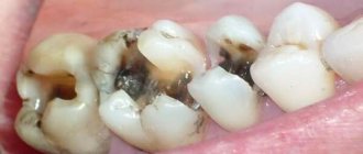
Fistula on the gum with untreated caries
A carious formation requires treatment as quickly as possible: if you delay contacting a doctor, the caries will develop into pulpitis, the infection from which, going beyond the boundaries of the tooth, will cause the formation of a purulent abscess.
When a fistula appears, there is no pain or swelling, since pus can easily come out through the hole. Pain and swelling occur just before the fistula appears. Basically, the tooth in the projection of the fistula will either be affected by caries, or it will have a crown or filling. Quite often, the patient experiences severe pain when biting on the tooth that caused the inflammation.
With inflammation, accompanied by the formation of pus, at the apex of the root, the infection accumulates in the root canals. To eliminate it, it will be necessary to remove the pulp and high-quality filling of the canals. It is extremely important that the dentist seals the canal along its entire length, without leaving a single free millimeter, otherwise the inflammatory process may develop again after some time.
The appearance of a hole in the periodontium in a child
The formation of a hole in the gum near a tooth in a child can be due to reasons such as: periodontitis caused by complicated caries, or injury to the oral mucosa. With periodontitis, the lesion reaches the jaw bone. A cavity appears inside the periosteum, which is filled with pus. When there is an excess of pus, it breaks out through the fistulous tract, thereby bringing severe pain.
Frequent mechanical damage to the mucous membrane and gums causes infection to enter the oral cavity. Bacteria entering the periodontium cause inflammation and fistula formation.
The appearance of a fistula due to unsatisfactory canal filling
Canal filling is a fairly common dental procedure indicated in the treatment of diseases such as periodontitis, pulpitis and in preparing teeth for subsequent prosthetics. However, in more than half of the cases, the canals are not filled as well as required by the rules. The most common mistake is filling not to the very top of the root.
Due to a mistake made by the doctor, an infection will form in the unsealed area of the canal over time, which sooner or later will go beyond the boundaries of the tooth and lead to inflammation. In addition to filling the entire length of the canal, the substance used to fill the canal during the procedure must be packed tightly: the appearance of voids and the formation of pores is unacceptable.
The main symptom indicating problems with the tooth is pain when biting. If you look carefully, in the immediate vicinity of the diseased tooth you can easily find a small hole through which pus will come out.
The appearance of a fistula on the gum, the treatment of which is not recommended to be delayed, indicates that the tooth will not stop hurting on its own. To treat it, the tooth is unfilled, an anti-inflammatory medicine is placed in the canals, which is removed after a certain period of time, and the canals are then filled again.
If a tooth has a crown or a pin, unfilling the canals becomes a real problem for the doctor. It is not always possible to remove the pin, and the crown will have to be re-installed after the procedure is completed, which is fraught with additional financial costs for the patient. Sometimes, if the canal is not sealed at its very end, the doctor decides to resort to a procedure for resection of the upper part of the root.
Resection is far from the most difficult operation: its duration is no more than an hour. The operation consists of several main stages:
- Preparation, during which the canals are carefully sealed a couple of days before the main procedure.
- Anesthesia. The operation is performed under local anesthesia: the patient does not feel pain during the doctor’s manipulations, but discomfort may occur later when the effect of the injection wears off.
- Gaining free access to the upper region of the root. To do this, the dentist cuts the gum, exposing the bone tissue, in which a hole is then cut using a drill.
- Through the resulting hole, the doctor cuts off the tip of the root, to which, as a rule, the cyst is attached, which causes inflammation.
- After removing part of the root, an empty cavity remains in the bone. The doctor can fill the space with synthetic tissue, which speeds up the growth process of natural bone.
- The final stage is suturing the gums. For intensive outflow of ichor, drainage can be installed for several days.
Treatment methods
It is performed under local anesthesia. Therapeutic methods include removal of affected tissue, antiseptic treatment of cavities, and canal treatment. Rinse and use of antiseptic continue until the pus ceases to be released, after which permanent filling of the canals and tooth is carried out. 3-5 visits to the doctor are required.
Surgical methods include:
- tooth extraction, rinsing the cavity with antiseptics, taking antibiotics and anti-inflammatory drugs;
- resection of the root apex, lavage of the cavity and augmentation of synthetic bone tissue;
- hemisection of the tooth with removal of the affected root without cutting the gums or applying sutures.
Fistula due to root perforation
Working with canals requires the dentist to be careful, attentive, and precise, controlled movements. The slightest carelessness can lead to the formation of a non-physiological hole (perforation), which will subsequently cause inflammation and, as a consequence, the appearance of a fistula.
A common mistake that leads to perforation and inflammation is the fixation of the pin when it is installed not along the canal, but its removal into the bone tissue.
With perforation, treatment of the fistula is the most problematic of all cases of its formation. Moreover, the main difficulty is not in medical procedures, but in the behavior of dentists who do not inform patients about the complication that has arisen, so as not to question the professionalism of themselves and their colleagues. As a result, a person who goes to the hospital for dental treatment returns again due to pain and inflammation: often the process becomes so advanced that it is no longer possible to do anything other than remove the tooth.
But still, perforation is not a death sentence, and the tooth can be saved. The sooner the patient applies, the greater the chance that the treatment will be completed successfully. True, this requires expensive materials (Pro Root MTA is often used), with the help of which the perforation area is sealed. Sometimes filling is performed directly through the canals, sometimes a surgical incision is required to gain visual access to the damaged area.
Possible complications if the fistula is not treated
Typically, the appearance of a fistula worries the patient when the gums begin to hurt and swell, making eating and daily oral hygiene difficult. When neglected, a strong bad breath appears, the lymph nodes may become inflamed and the temperature may rise. If treatment is not started in this case, then possible complications include loosening and loss of teeth, sinusitis, the appearance of cysts and even destruction of the jaw.
Of course, it’s better not to let things get to this point, otherwise the treatment will take a long time and cost a significant amount. If you notice a swelling or bump on your gum, don’t hesitate to get it examined!
Fistula on the gum: what to do?
The most important thing that a person who has pain in the gums should understand is that they need to see a doctor as soon as possible, and not wait for the symptoms to disappear and not use various gels and other means. Yes, they can bring relief, but it will be a temporary relief of symptoms, not healing.
There are several treatment options for fistula.
Endodontic method for treating gum fistula
The method is applicable if the following conditions are met:
- the dental canal and its branches are visible;
- it is clearly visible in the tooth canal whether there are cracks or not.
Treatment is carried out under a microscope. The apical part is sealed with modern high-quality materials that are biocompatible with natural tissues. Canal therapy is performed, which allows the fistula to be eliminated.
Mixed treatment
If the lesion is quite severe, the therapist will need the help of a surgeon.
The therapist cleans the canals and seals the apical part of the root. The surgeon's job is to polish this area of the root. Polishing is carried out using ultrasonic nozzles, which allows you to achieve an ideal result.
Surgery
If the inflammatory process has gone too far, the tooth will have to be removed. The source of inflammation is eliminated along with the tooth and root.
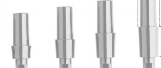
In the future, an implant can be installed in this place at the request of the patient: this is a rather expensive procedure, so it is not recommended to delay the treatment of the fistula. The earlier the patient consults a doctor, the easier and cheaper it will be to cure the disease.
Autotransplantation
This is a type of surgical treatment. In this case, the implant is another tooth of the patient (as a rule, it is a wisdom tooth, which is a kind of reserve, since it is not involved in chewing food).
The advantage of the method is that the person’s own tooth is transplanted, which negates the risk of tissue rejection.
Symptoms
Purulent inflammation is accompanied by:
- discomfort and unpleasant sensations while eating;
- increasing aching or throbbing pain;
- redness and severe swelling of soft tissues;
- itchy sensations;
- mobility of the diseased tooth (not always);
- increased temperature;
- general malaise.
In some cases, due to severe swelling, the entire cheek swells, and the localization of the source of inflammation can only be determined on an x-ray.
After the pus comes out, the pressure on the gum tissue decreases, pain, swelling and other symptoms become less pronounced and then disappear completely.
Regardless of the cause, when purulent inflammation develops, one of two types of formations usually appears on the gums:
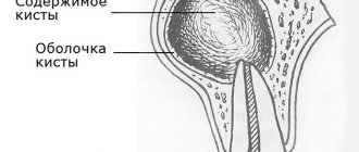
- Cyst. Formed against the background of caries, gingivitis, periodontitis and periostitis, trauma. Inflammation is accompanied by severe swelling and redness of the gums, and the size of the cyst can reach 3 cm.
- Granuloma. A loose formation of small size (no larger than a grain) usually forms in gum pockets and soft tissues. It often develops as a complication of pulpitis and periodontitis.
In both cases, the purulent contents come out over time, leaving a hole in the gum. Such formation constantly oozes and requires competent and timely treatment.
Prevention
To protect yourself from the appearance of a hole in your mouth near a tooth, you need to follow a number of preventive measures:
- Brush your teeth 2-3 times a day using the standard method.
- Use additional oral care items daily: floss, irrigator, tongue scrapers.
- Carry out professional hygiene once every 6 months.
- Visit the dentist annually for the prevention and early detection of dental and periodontal diseases.
- Give up bad habits (smoking tobacco products).
- Reduce the consumption of drinks that contribute to the accumulation of bacterial plaque (soda, strong tea, coffee, alcohol).
- Reduce consumption of high-carbohydrate foods.
- Include vitamin and mineral complexes in your diet.
- Eat as many solid vegetables and fruits as possible.
- Treat somatic diseases (gastritis, gastric and duodenal ulcers, diabetes mellitus) in a timely manner.
Bibliography
- Banchenko G.V. — Combination of diseases of the oral mucosa and internal organs, M.: Medicine, 1979.
- Grigoryan A.S., Grudyanov A.I., Rabukhina N.A., Frolova O.A. — Periodontal diseases, M., 2004.
- Loginova N.K., Volozhin A.I. — Pathophysiology of periodontal disease (methodological manual), M., Medicine, 1993.
- Borisenko A.V. — Secrets of the treatment of caries and dental restoration, M. Book Plus. – 2005.
- Leontyev V.K., Pakhomov G.N. — Prevention of dental diseases, M., 2006.
- L. V. Kharkov, L. N. Yakovenko - Directory of a dental surgeon: diagnosis, clinic, principles of surgical and drug treatment (in children and adults), M.: Book Plus, 2008.
- Afanasyev V.V. - Surgical dentistry: textbook, M.: Geotar-Media, 2011 Tsarev V.N. - Antimicrobial therapy in dentistry, M.: Medical Information Agency, 2006.
Find a clinic