Often young women are perplexed: where did the strange lump on my hand come from?
?
Although painless, but quite large (from 0.5 to 6 cm in diameter), it cannot go unnoticed due to its location. So what is hygroma
- cancer or a purely aesthetic defect?
Although hygroma on the arm is a tumor-like formation, it, contrary to misconceptions, is not an oncological disease
, and also
never degenerates into cancer
. This disease is quite widespread (up to 50% of all neoplasms on the hands) and can be successfully treated. On the other hand, this subcutaneous formation can cause discomfort or even interfere with normal blood circulation in the hands.
Hygroma
(or, as it is also called,
ganglion
) is a capsule-bag that is dense to the touch, filled with a viscous and jelly-like protein liquid mixed with fibrin. The hygroma noticeably protrudes over the ligaments and tendons and practically does not move under the skin. Most often, synovial cysts are observed in women aged 20 to 30 years (⅔ of the total number of patients). Children are least likely to suffer from it.
In what cases should you worry about treating hygroma and can it be prevented?
Hygroma - formation in the form of a tumor
Lump on the leg: causes of the defect
Patients who have a lump under the skin on their leg are often interested in the reasons for the appearance of such an ailment. In this case, protruding formations can also occur in the area of other fingers and do not have a bone origin. According to orthopedists, the main reason why a bunion occurs on the toe is a hereditary predisposition characterized by weakness of the ligamentous apparatus of the foot. As a result, due to the presence of weak connective tissue, the risk of sprains and tears increases, and joint mobility increases.
Pathological processes that can cause foot deformity include:
- A disease accompanied by the deposition of uric acid salts in various tissues (gout);
- The presence of protruding formations in the area of the first metatarsophalangeal joint (bursitis);
- Tendon cyst of the foot;
- Valgus deformity of the fifth metatarsophalangeal joint;
- Having hammertoes.
Among the additional factors that provoke the formation of bumps on the feet on the heels and toes are:
- Endocrine disorders;
- Joint lesions of inflammatory etiology;
- Foot injuries;
- Increased load on the feet during ballet classes;
- Movement disorders in children arising from brain disorders (cerebral palsy);
- Using narrow shoes.
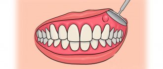
Diagnosis of hygroma
To make an accurate diagnosis, you need to visit a doctor - an orthopedist, surgeon or rheumatologist. Diagnosis of hygroma requires a comprehensive study of the clinical picture, taking into account localization, external signs, and also, if necessary, the results of laboratory or instrumental studies. Typically, a patient examination begins with palpation of the neoplasm, examination and questioning of the patient, as well as checking joint mobility. In some cases, this is enough, and sometimes additional examination of the lump is required to exclude lipoma, fibroma, atheroma or traumatic cyst. If the hygroma is in the palm area, tumor formations in the bone or cartilage tissue should be excluded
To exclude a malignant origin of the formation, the doctor may prescribe a puncture (fluid sampling) from the cyst for histological and/or cytological examination, which is carried out using a syringe.
In the case of an atypical location of the lump, an X-ray examination or ultrasound is performed to make a diagnosis. Ultrasound examination of the cyst helps determine the density of its contents, the presence of veins and other vessels nearby that it can compress, as well as the uniformity of the lump or the presence of several nodules. In some cases, the patient may be recommended an MRI (to determine the structure of the walls, the presence of vessels in the hygroma, and the consistency and composition of the filling of the cyst).
It is important to diagnose hygroma in the early stages
Treatment of bunions on the legs using UVT: features
Shock wave therapy is recognized as the most effective among all existing methods of joint restoration: the procedure allows you to eliminate bumps on the legs in the shortest possible time and with minimal risk of complications for the patient. Through this manipulation, it is possible not only to eliminate the symptoms of the disease, but also to achieve a prolonged therapeutic effect.
Treatment of bumps on the legs using shockwave therapy is characterized by a complex effect on damaged joints, namely:
- It is possible to stop the inflammatory process;
- Provides rapid removal of toxins from the body;
- Regenerative processes in bone, cartilage and soft tissues are enhanced;
- There is a gradual destruction of bone growths;
- It is possible to completely dissolve uric acid salts.
As is known, due to degeneration and inflammatory processes in articular structures, pathological growth of acute bone formations occurs. They can injure soft tissues when changing the position of the elbow or knee joint. The UVT procedure also helps to solve this problem.
The advantages of such acoustic impact are obvious:
- Eliminates the possibility of damage to soft tissues and the integrity of the skin;
- There is no need for general or local anesthesia;
- After the SWT procedure there are no consequences, unlike surgical intervention;
The body accelerates the formation of endorphins, which are responsible for a person’s positive attitude.
Hygroma symptoms
In approximately 35% of cases, a synovial cyst may be asymptomatic
, except for the compaction under the skin.
Therefore, the main symptom of hygroma is considered to be the appearance of a lump
(one or several)
in the area of the wrist joint
,
fingers or hand
;
extremely rarely - on other parts of the body. Typically, such a lump reaches 1, 2 or 3 centimeters in size, but an advanced synovial cyst can be up to 6 cm in diameter. In the scientific literature there are references to even larger seals
! But there is usually no need to worry about its size, if it does not cause severe inconvenience, because the hygroma grows very slowly and does not form a truly large tumor-like body.
Synovial cyst
has a characteristic location: on the back of the wrist or ankle joint, as well as the hand or foot.
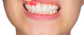
In addition to the elastic dense lump, patients may experience:
- difficulty moving the affected area;
- dull, usually aching, pain in the hand;
- deterioration of sensitivity in the hand;
- weakness of muscles and ligaments;
- discomfort that increases with physical activity on the hands (especially the supporting one);
- crawling sensation, tingling or burning sensation, as well as other symptoms of compression of nerve endings or blood stagnation;
- increased sensitivity of the skin in the area of the lump.
Special mention should be made of hygroma, in which the growing lump is located under the ligament
, and therefore
does not form a distinct protrusion above the surface of the skin
. In this case, patients complain of:
- constant or periodic pain (pressing or dull), which can radiate to the hand, fingers, forearms;
- pain when pressing;
- roughening, peeling of the skin over the formation;
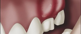
Unlike other similar diseases, hygroma on the arm, as a rule, does not cause pain on palpation (sometimes it can only be slightly painful). It is characteristic that the skin over the lump does not undergo any changes - it remains elastic and retains its normal color, and does not “grow” onto the tumor. The formation, as a rule, can be moved from side to side, but it does not roll under the skin. One of the main differences between a hygroma and a real tumor is that it can shrink and increase in size. As a rule, hygroma loses volume during the rest period and swells after exercise.
Symptoms of hygroma may appear or intensify in women after the birth of a child.
Main periods of disease development
It should be noted that a lump on the foot near the big toe does not appear immediately; this formation is preceded by several stages. Below we will talk about them in more detail.
Stage 1. The first finger is slightly deformed. There is no pain syndrome yet. Only small calluses are noticeable in the area in contact with the shoes.
Stage 2. The process of displacement of the thumb continues, but this does not in any way affect its mobility. During and after exercise, mild pain occurs.
Stage 3. The patient notes constant discomfort and pain. The first and second fingers are deformed. Difficulties arise when choosing shoes. There is a need to reduce the usual loads on the legs.
Stage 4. The thumb joint is subject to significant displacement. There is a pronounced flattening of the transverse arch of the foot. At this stage, calluses may appear in the area of the bone growth and blood flow may be impaired. In addition, the pain is already constant and causes severe discomfort to the patient. Finding comfortable shoes becomes impossible.
Lumps on the legs: treatment methods
So, in order to understand how to get rid of bumps on the legs and draw up an appropriate treatment plan, it is necessary to take into account the stage of this disease. At the initial stages of the development of the disease, eliminating the defect with the help of folk remedies and medications is considered the most effective. In the presence of a more severe stage of the pathology, the UVT technique shows successful results, and surgery is also performed on the bump on the legs.
Both of the above procedures can be performed at the Health Plus clinic. At the same time, you can count on the use of modern equipment, the presence of highly qualified employees and affordable prices.
How to remove bumps on feet using traditional methods?
In folk medicine, there are several remedies that, when used wisely, can quickly get rid of protruding formations. However, if the lump on the toe joint is in an acute stage and causes constant discomfort to the person, it is advisable to use these methods only in combination with other therapeutic measures. So, the best result can be obtained using the following folk remedies:
- Compresses based on bile. A similar composition must be applied to a cotton sponge and applied to the bump. Waterproof paper is placed on top, after which a knitted sock is put on the foot. It is recommended to apply the compress every day for 30 days;
- Baths with the addition of essential oils, sea salt and baking soda. They resort to the help of such procedures in order to reduce fatigue in the legs. To obtain the desired therapeutic effect, add a few drops of lavender oil and 5 tablespoons of salt and sodium bicarbonate to a bowl of warm water;
- Composition with analgesic effect. To prepare it, you need to take 5 tablets of acetylsalicylic acid, crush them, and then add 30 ml of iodine solution. The resulting composition is used to lubricate the bump on the leg, located on the instep or in some other place. Manipulation should be carried out at least 3-4 times a day;
- If the lump on the leg under the skin is hard, using an infusion with lingonberry leaves has a good effect. To obtain it, 2 tbsp. spoons of the collection are necessary, pour 300-400 ml of boiling water and leave for 30-40 minutes. Use the herbal drink for 10 days, 2 times a day, preferably in the morning. With regular use, it is possible to quickly remove uric acid salts;
- Take salt and honey in equal quantities, mix and form small cakes. The resulting paste is applied to the sore spot, covered with cellophane film on top and a warm sock is put on. To achieve the best result, the procedure should be carried out for at least 10-14 days.
How to treat bumps on the legs with medications?
Many patients are interested in the question: “How to treat bumps on the toes with the help of medications?” As a rule, special ointments and creams that have anti-inflammatory, analgesic and distracting effects are used for this purpose. These include:
- Ointment for external use "Indomethacin";
- ValgusStop ointment, based on the use of urea, bile and salicylic acid;
- Ointment with shark oil based on acacia.
All of the above products are gently rubbed into the area of the protruding bone growth.
Physiotherapeutic methods for treating bunions on the legs
When a person has a pain in a lump on his leg, physiotherapeutic procedures provide a good anesthetic effect. Among them, the following have a special positive effect on the body:
- Magnetotherapy . Allows you to reduce pain, relieve swelling and improve blood circulation in the vessels;
- Electrophoresis using novocaine . The procedure helps eliminate pain and relieve swelling, has a powerful anti-inflammatory effect, improves blood flow and restores muscle tone;
- Phonophoresis . This physiotherapeutic method is based on the use of ultrasound. Provides accelerated penetration of drugs into the area of inflammation. Allows you to relieve pain and stop degenerative-dystrophic processes in cartilage tissue;
- Paraffin therapy . Helps relieve inflammation and eliminate muscle spasms;
- Shock wave therapy . It consists of the targeted impact of acoustic waves on connective and bone tissues affected by the disease. Due to this, their cell membranes are stretched, which helps improve blood circulation in the vessels and accelerate metabolic processes. In addition, the ability of damaged joints to regenerate increases, foci of inflammation are eliminated, rapid removal of toxins from tissues is ensured, and calcifications and salts are dissolved. This leads to the gradual removal of bone formations on the toes.
Treatment of hygroma
Although sometimes a hygroma on the arm can be left without treatment, due to the risk of complications and aesthetic discomfort, doctors usually recommend its removal. Sometimes a synovial cyst can resolve spontaneously if the factor that provokes it (for example, occupational stress) is eliminated in a timely manner - then the synovial fluid will simply stop flowing into the capsule.
The indication for treatment of hygroma is its multi-chamber nature, rapid growth of formation, development of the inflammatory process, pain syndrome, and limited mobility in the joint.
The preferred treatment for hygroma is surgical removal.
, since puncture or aspiration of the tumor is usually ineffective or poses a risk of complications.
Thus, conservative treatment of wrist hygroma is characterized by recurrence of the disease in approximately 80-90% of cases
, while for surgical treatment the same figure is only
8-20%
.
Surgery
Doctors do not recommend delaying surgical treatment of hygroma, since a large, albeit benign, formation soon begins to displace blood vessels, muscles, ligaments and nerves. Their non-physiological position significantly complicates the operation and may have consequences for the patient
.
Removal of a hygroma on the arm is usually carried out under local anesthesia with a tourniquet and lasts no more than half an hour. The operation is performed using a scalpel or laser equipment. In advanced cases, large or unusual formations may require general anesthesia. When carrying out the procedure, it is important to remove all degenerative cells without exception (otherwise the altered tissue may again develop into a cyst), wash the capsule, and also bandage the so-called. the mouth of the hygroma, which is associated with the joint and feeds the lump with synovial fluid. The drainage tube for fluid drainage is removed 1-2 days after the procedure, and postoperative sutures after complete healing of the incision are removed on days 7-10. After this, it is recommended to immobilize the operated area with a splint and a pressure bandage for several weeks and avoid serious stress on it for 30-60 days.
.
Recently, surgeons have given preference to endoscopic removal of wrist hygroma, in which the capsule and its contents are removed through a small incision a couple of centimeters in size. Rehabilitation after such an intervention occurs much faster.
Conservative treatment of hygroma
As a conservative treatment for hygroma of the hand, crushing it (without penetration and formation of a wound) is occasionally used, followed by the application of a compressive bandage. This method should never be used on infected synovial cysts due to the risk of sepsis, but it can be used to treat sterile lumps. The main disadvantage of this approach is the rapid recurrence of the disease: as soon as the burst edges of the capsule grow together, it will again begin to fill with serous fluid.
A somewhat more successful technique is aspiration puncture of the formation, in which its contents are sucked out through a small puncture, and glucocorticoid solutions and sclerotization preparations are injected in its place. This allows the capsule to be filled with connective tissue instead of liquid. This method is also indicated for administering antibiotics and treating infected hygroma.
Conservative techniques do not eliminate degenerative cells
, which provoke the formation of hand hygroma and are therefore considered ineffective.
There are several methods for treating hygroma
Physiotherapy
A small cyst that does not grow, does not become inflamed and does not cause pain can often be treated conservatively using physiotherapeutic techniques. They help reduce the volume of formation and ensure the outflow of its contents, slow down cell growth. Although physiotherapy cannot completely cure hygroma, in combination with chondroprotectors
it helps keep the tumor under control without surgery.
Sometimes physiotherapy is the optimal solution for non-standard location of the lump
, in which excision can be a rather traumatic procedure.
This type of treatment helps relieve compression of nerve endings, eliminate pain and inflammation, improve tissue regeneration, and relax the muscles around the tumor.
In modern physiotherapy for hygroma of the wrist and other joints, the following is used:
- electrophoresis
; - ultra high frequency therapy
; - phonophoresis with hydrocortisone
; - magnetotherapy
; - paraffin applications
; - mud baths and wraps
; - UV therapy
.
During the entire course of physiotherapy, and also, as prescribed by the doctor, after it, a tight, compressive bandage is applied to the area affected by hygroma, which prevents further accumulation of fluid in the capsule.
If the tumor is too large or continues to grow, there are signs of inflammation, the attending physician, together with the patient, usually decides on surgical intervention.
Drug treatment
For the medicinal treatment of hygroma, a wide range of anti-inflammatory drugs are used, which are applied to the surface of the cone or injected directly into its cavity.
However, anti-inflammatory therapy provides only a temporary and symptomatic effect
,
and therefore used in combination with other techniques
.
In case of a purulent process inside the cyst, and also as a preventive measure before surgery to remove it, the attending physician selects a specialized antibiotic for the patient - usually in the form of injections.
But the most effective medicine for the treatment of tendon ganglion are chondroprotectors
.
They are helping:
- establish metabolic processes in the tissues adjacent to the hygroma, strengthen muscles and ligaments, prevent the destruction of synovial cartilage;
- improve the quality of synovial fluid - as a result, the joint begins to produce it in smaller volumes, and the nutrition of the hygroma is reduced;
- slow down further growth of tumor formation;
- relieve symptoms of inflammation in the area of hygroma;
- significantly reduce pain (many patients note a reduction in pain by 50-80%);
- cope with discomfort from the tendon ganglion, which cannot be operated on due to its small size.
Chondroprotectors do not allow the joint on which the hygroma occurred to “starve” due to poor supply of nutrients. They also relieve swelling and restore normal mobility in the fingers, hands and wrist joint. In addition, they have a systemic effect on the body, improving the condition of the musculoskeletal system and preventing the appearance of new hygromas, as well as the recurrence of old ones. Due to the almost complete absence of side effects, chondroprotectors can be taken to prevent synovial cysts even before they appear
,
during treatment of a neoplasm
, as well as
after surgery
.
Chondroprotectors stimulate tissue repair after surgery and prevent the re-formation of modified cells. The standard course lasts from 3 to 6 months per year. Compared to other drugs in this series, Artracam
, the course of which lasts 1.5 months, and then can be repeated after 2 months if necessary. It is made exclusively from natural ingredients - seafood.
Drug Artracam
is one of the most effective chondroprotectors for hygroma of the wrist and hand.
Use of orthopedic devices
If you already have a lump on a vein or toe, using orthopedic products will help stop the progression of the disease. In addition, such devices show effectiveness during the rehabilitation period after surgery and prevent the appearance of bunions on the legs. In many cases, the formation of bone growths accompanies flat feet, which determines the need to use products to strengthen the arch.
In some situations, experts recommend using shoes with a special instep support, a small heel, a firm heel, a tight fit to the foot, with liners, etc.
With the help of such products, it is possible to eliminate pain in the legs, prevent the development of pathology, or firmly fix the joint after surgical procedures.
When you shouldn’t delay visiting a doctor
Quite often, bumps on the chin, near the lips, along the spine, or in the groin are mistaken for warts or papillomas. Despite the stereotype that such growths, even on the pubic part, supposedly do not pose a significant danger, this is not the case.
In most cases, the prerequisite for a relatively painless papilloma to form in an unexpected place is a hormonal imbalance or infection. For many people, such growths are only an aesthetic drawback, especially if they form under the eye, on the cheekbone, under the eyebrow, or in the eyelid area.
But in clinical practice, cases are increasingly being recorded where people, ignoring such an anomaly, missed the initial stage of skin cancer. It is impossible to independently distinguish a benign formation, which is simply temporary, from a malignant tumor. Diagnosis should be carried out only by an experienced dermatologist who will examine the entire body, because only a professional can examine any abnormalities on the back of the head, labia, or recognize abnormalities on a healed suture.
Malignant tumors are relatively rare.
They can be divided into three broad categories:
- melanoma, which degenerates from ordinary moles;
- liposarcoma, which affects fat cells;
- lymphoma that occurs at the site of a lymph node.
Liposarcoma mainly affects the lower extremities, forming on the hips and under the knee.
The clinical picture is somewhat similar in all three cases. First, the patient feels a skin lump, which quickly increases in size. An alarming clue is also called the fact that the tumor is immobile or fused to the epidermis.
Another specific type of formation is hernia, which forms on the abdomen after surgery. The catalysts for the occurrence of a hernia are:
- postoperative scars;
- lifting unbearable weights.
Internal organs cannot cope with unnatural loads on them, which leads to their loss. To eliminate the pathology, surgical intervention will be required, healing after which, if medical recommendations are followed, occurs quite quickly.
You should not experiment with the use of traditional medicine recipes if the affected area is the area between the legs. Most often, the provocateurs of many small bumps, or one big one, are:
- viruses;
- bacteria;
- gland blockage;
- blockage of hair follicles.
Immediately after discovering an abnormal tumor on the penis after circumcision, or simply on the penis without external trauma, on a testicle, in the bikini area, or near the anus, you need to consult a doctor. Moreover, it does not matter at all what color the formation is: black with blood on the genitals or round brown on the anus.
Very rarely are cases recorded when a bruise first forms on the forearm, collarbone, foot, heel, and other parts of the body, and then hard edges are felt. This indicates that a foreign body, such as a splinter, got inside and was not pulled out in time.
Features of surgical treatment
If conservative treatment methods do not give the desired result, the patient is prescribed surgery for bunions on the legs. Such surgical interventions can be combined, which involves subsequent restoration of ligaments and bones, and can also be performed in the area of soft and bone tissue.
Based on the specific clinical situation, the following surgical procedures are performed:
- Cutting off the first metatarsal bone;
- Removal of the tendon in the thumb area;
- Excision of the periarticular bursa of the joint affected by the disease;
- Cutting off bone or cartilaginous growth in the metatarsal area. In this case, part of the phalanx of the first finger can be removed;
- Cutting off a fragment of the bone of the thumb.
After the operation on the toe is completed and the lump is removed, experts recommend regularly self-massaging the limb, using special orthoses and performing a set of physical exercises as part of physical therapy. In this case, the load on the legs should be done in doses and gradually. In addition to UVT, honey also practices surgical removal of growths on the legs.
Types of hygroma
As we mentioned above, hygroma can occur in the form of a single formation, delimited from other tissues by a membrane. It is also possible for several cysts to appear, fused or independent of each other.
However, most often the disease is classified according to the location of the lump. So, doctors distinguish the following types of it:
- hygroma of the wrist joint
; - hand hygroma
; - hygroma on fingers
.
Much less common:
- hygroma of the knee joint
(formed in the knee fossa, also known as Becker’s cyst); - hygroma of the ankle joint
; - hygroma of the foot
(located on the dorsal, i.e., upper, side of the foot, usually at its outer edge).
The tendon ganglion, located in the wrist and hands, accounts for up to 88% of the total number of cases of the disease. The ankle joint and foot bothers up to 11% of patients who suffer from hygroma. A synovial cyst in the neck, shoulder, shoulder blade or knee occurs in only 1% of patients. It is extremely rare for patients to see a doctor with formations in the bones, muscles or spine.
Hygroma of the wrist
Hygroma of the wrist joint
- the most common type of synovial cyst. It becomes clearly visible in a bent position of the wrist joint (for visual diagnosis, you need to move the hand forward and back, and then left and right). Often, hygroma of the wrist joint is located on the front or back side of the joint, but it can also grow on the lateral surfaces or at the very base of the palm.
If the lump is located under the ligament, it may not cause any concern and may not even cause any aesthetic discomfort.
As a rule, the consistency of hygroma of the wrist joint is felt as soft or springy, “rubbery”.
Hygroma of the hand
Tendon ganglion in the hand area
(hygroma of the hand) usually occurs on the fingers (from the side of the palm), the palm itself, the back of the hand, or closer to the wrist joint. Typically, hand hygroma does not bother the patient until it reaches a large enough size and does not interfere with everyday activities.
Hygroma on the finger
Synovial cyst on fingers
often occurs on their back side, in the area of the distal phalanx (i.e., the last one on which the nail is located). Often it is located in close proximity to the cuticle or nail fold.
The skin over the hygroma on the finger, as a rule, looks thinned, stretched, and its natural pattern is smoothed out. Under the skin, as a rule, a rounded formation can be easily felt, which does not cause pain, except in case of injury. It is quite dense to the touch, so it can be perceived as a bone or cartilage growth.
Hygromas on the palmar side of the phalanx are usually larger than on the back, and can spread to the entire phalanx and even occupy the adjacent one. They can be quite painful due to compression of the nerves that run along the sides of the fingers. This type of hygroma often prevents patients from performing household chores.
Ganglions also appear at the proximal phalanges
(near the base of the finger). Such cysts are also quite painful, despite their small (about the size of a pea) size. Pain with a hygroma on the finger usually occurs when trying to tightly grasp a hard object (for example, the handle of a shovel, a rolling pin, etc.).
Hygroma of the lower extremities
Synovial cysts of the lower extremities are typically localized on the back of the foot or fingers, as well as on the front of the ankle joint. Such hygroma rarely bothers patients with pain. As a rule, discomfort occurs when the foot is compressed by tight shoes, the foot is rubbed or stuffed, and also when the lump is located in close proximity to the nerve.
Hygroma of the lower extremities
Prevention measures
To prevent the appearance of protruding growths on the feet, you must adhere to the following simple rules:
- Do special strengthening exercises for muscles;
- Buy only comfortable shoes;
- If necessary, use orthopedic products;
- Walk barefoot on rocky surfaces more often;
- Use orthoses if discomfort occurs in the foot;
- Adhere to healthy eating rules and avoid becoming overweight;
- If you have arthrosis and arthritis, consult a doctor in a timely manner and strictly follow all recommendations for the treatment of such ailments.