Problem: parents with a 12-year-old teenager contacted the Dial-Dent orthodontic department. The boy's fangs stick out strongly, and his other teeth grow crooked. The parents wanted advice on how to correct protruding fangs, what the treatment consisted of, and how long it would last.
Solution: orthodontist O.A. Baranova performed treatment with Damon braces, aimed at normalizing the position of the fangs, straightening the teeth and correcting the bite.
Braces for fangs
If we consider all possible anomalies of the dentition, then most often it is the fangs that are crooked. According to statistics, every third orthodontic patient wants to correct the position of the “eye” teeth. Among the possible options for their dystopia:
- noticeable protrusion from the jaws (like a vampire);
- excessive or, conversely, insufficient crown length;
- closure by adjacent units located in front;
- incomplete eruption;
- turning in any direction at the wrong angle.
The “vampire smile” not only looks strange and unaesthetic, but also prevents complete chewing of food, and this negatively affects the functioning of the gastrointestinal tract. Therefore, it is imperative to solve the problem of curvature.
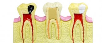
How is canine implantation performed?
- Diagnostic stage – the condition of the bone tissue, the general condition of the body is assessed, and the absence of contraindications is checked.
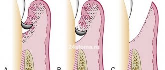
- Implant installation is performed under local anesthesia. The doctor opens the area of mucous membrane above the fang, removes the bone flap, forms a bed and inserts a titanium implant into it.
- The wound is sutured.
- After a week, the stitches are removed.
- Within 3-5 months, the implant takes root without load (a temporary prosthesis is not installed).
- After making sure that the implant has fused well with the bone, the doctor opens the mucosa above the top of the implant (local anesthesia is used).
- An individual abutment is made or a universal adapter is used.
- A gum former is installed, since the canine is located in a visually open area of the row.
- Impressions are taken from the abutment, a crown is made, which will exactly repeat the canine in shape and match the color of the neighboring teeth.
How dentists fight dystopia of “eye” teeth
Determining the curvature of the fangs is quite simple - to do this, the dentist just needs to examine the condition of the client’s oral cavity. When choosing a treatment method, the following criteria are taken into account:
- patient's age;
- features of the existing anomaly;
- degree of neglect of dental pathology.
To remove the curvature and force the canine into the correct position, the doctor may resort to the following treatment options:
- installation of braces on fangs;
- use of mouth guards;
- removal of “eights” (necessary if they are the cause of a dental anomaly);
- transformation of the first molar into a canine if the latter is missing;
- reposition (used exclusively in the most severe cases).
The sooner treatment of the curvature is started, the higher the likelihood of its complete elimination in a short time. If we are talking about providing orthodontic care to children under twelve years of age, then they are advised to wear removable plates. Typically, this method can achieve a beautiful smile in early adolescence in just six months.
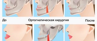
Teenagers fourteen years of age and older can return the canine to its natural place with the help of braces. Adults often have to not only wear braces, but also undergo a simple surgical operation.
Content
- Features of fangs
- What to choose?
- Which implantation to choose?
- How is implantation performed?
- Perform triple implantation
A canine is a single-rooted tooth used for grasping, holding and tearing food. Humans have a pair of fangs on the upper and lower jaws. They are called "triples" because they are considered third after the two incisors. These teeth are tall and cone-shaped.
PROMOTION
Osstem dental implantation turnkey
18,000 rub.
Why canine teeth often grow crooked or do not erupt completely?
For most children, “threes” are the very last to appear - closer to ten or twelve years. Because of this, it may turn out that there is no place for them. Then they begin to grow higher than necessary, behind or in front of their neighbors.
Another common problem with crooked eye units is the discrepancy between the size of the teeth and the width of the jaw. This phenomenon is quite common - a child may inherit a small jaw from one parent, but large, large teeth from the other.
The third cause of dental anomaly is the delayed replacement of milk units with permanent ones. There is no way to influence her. You just have to eliminate the consequences of deviation.
Is it painful to remove molars?
Modern dentistry uses the latest drugs for local anesthesia when removing molars. Therefore, the patient should not be afraid of pain or serious discomfort during the process of tooth extraction. The thinnest needles are used to administer anesthetic injections into the gum area, so the injection is virtually painless.
Installation of braces on fangs
Treatment with orthodontic appliances takes about one to two years. During it, the doctor takes measures to free up space for the fang - widens the jaw or removes low-functioning teeth (for example, “eights”).
If the “twos” and “fours” are too close to each other and do not allow the fang to break through, the “fours” are usually pulled out. If a crooked tooth sticks out against the background of the others, an expansion of the row and a simultaneous movement of the culprit of the problem are indicated. For this purpose, different types of braces are used - metal, ceramic, sapphire, lingual. The first ones are the most popular, as they have a low cost and at the same time guarantee good functionality.
Orthodontic appliances can be fixed exclusively on the canines or on the entire row - it all depends on the degree of curvature. This is what the average pathology correction looks like:
- Carrying out diagnostic measures (making impressions, conducting X-ray diagnostics), treating existing dental diseases.
- Professional oral hygiene designed to remove plaque and tartar.
- If it is necessary to make room in a row, a certain tooth is removed or measures are taken to widen the jaw.
- Fixation of the bracket system. On the appointed day, the doctor installs the leveling device.
- Visiting a doctor to change the degree of tension of the arc, assessing the achieved intermediate results.
- Removing braces and selecting retainers. The latter are needed to ensure that the smile remains aesthetically pleasing forever.
Start of orthodontic treatment
Before installing braces, the patient must undergo professional teeth cleaning by a hygienist. The specialist cleans the teeth, removes all plaque (soft and hard), checks the condition of the gums, polishes the enamel and gives recommendations on how to care for your teeth while wearing braces. Patients who decide to start treatment with braces receive a starter hygiene kit, which includes an orthodontic brush, toothpaste, brushes, dental floss, monobrush, and wax.
Damon metal braces are fixed:
Treatment with braces at this stage is aimed at expanding the dentition in the areas of the 2nd and 4th teeth (special springs are installed on the brace system).
After 5 months, the next stage begins - the return of the fangs to the dentition. The second arch holds the anterior incisors in a stable position.
Stage of wearing orthoelastics (12 months after fixing braces):
Advantages and disadvantages of orthodontic systems
Installing braces on fangs has the following advantages:
- Even the most complex dentofacial curvatures can be cured;
- obtaining positive dynamics after several months of wearing the structure;
- Possibility of use by both children and adults;
- reliable fixation on the teeth (falling off at the most inopportune moment is excluded);
- the ability to use aesthetic devices that are practically invisible to others.
As for the disadvantages, it should be noted here:
- Difficulties in maintaining oral hygiene (cleaning out food debris from under braces is difficult and takes time);
- the need to wear the leveling structure for at least one year;
- change in the appearance of your smile for the worse during treatment;
- high cost of therapy.
Prices for tooth extraction
| Name | Price |
| Consultation | For free |
| Use of bone material | 4,000 rub. |
| Simple tooth extraction | 2,700 rub. |
| Removal of periodontitis tooth | 1,000 rub. |
| Tooth extraction is complicated | 3,200 rub. |
| Removal of a dystopic wisdom tooth | 4,000 rub. |
| Removing a tooth fragment | 500 rub. |
| Removal of exostosis in the area of 1 tooth | 2,500 rub. |
| Removal of cysts and benign formations | 3,500 rub. |
| Treatment of alveolitis | 1,200 rub. |
| Lengthening the clinical crown of a tooth | 5,000 rub. |
| Free flap gum grafting | 8,000 rub. |
| Removing stones from the ducts of the salivary glands | 3,500 rub. |
| Splinting for a jaw fracture | 15,000 rub. |
| Socket curettage | 200 rub. |
| Filling the hole with a medicinal substance (Alvogil, al-vostaz, hemostatic sponge) | 200 rub. |
| Resection of the apex of the tooth root | 4,500 rub. |
| Removing a foreign body from the canal | 1,500 rub. |
Is it possible to improve the bite without installing braces on the canines?
Of course, braces are the most practical, reliable and effective way to straighten teeth today. But it happens that for some reason a person does not want to wear them. This is possible due to the high cost of treatment, its long duration, and the unattractive appearance of the structures.
At the dentist's discretion, such a patient may be offered alternative methods. So, if we are talking about children, then to achieve the goal they can:
- wear custom-made mouth guards at night;
- wear soft removable plates;
- use trainers (these are devices that only affect warped units).
If an adult wants to straighten his fangs without braces, he may be offered:
- Removable mouthguards. They should be worn for about two to three years, but the dentist, as a rule, cannot give a 100% guarantee of a positive result.
- Installation of veneers. They do not remove the existing defect, but mask it perfectly. A thin, high-strength plate is glued onto the pre-ground tooth, which makes the smile much more harmonious and beautiful.
It is important to understand that only an orthodontist can decide whether to replace braces with other alignment options. If the doctor sees that the curvature is pronounced, mouth guards and plates will not help, he warns about this. In this case, there is no point in trying to correct the bite without braces.
Recommendations after removal
Regardless of the method by which the tooth was removed, in the first 2 days the patient must observe a special diet, activity and hygiene. This is necessary to preserve the blood clot in the wound, which helps maintain the sterility of bone tissue and promotes its regeneration.
In the first 48 hours after removal you cannot:
|
Failure to comply with these recommendations most often causes the clot to fall out of the socket and the development of alveolitis or bleeding.
After tooth extraction it is recommended:
- make oral baths from chamomile and sage decoctions (the decoction prepared according to the instructions is taken into the mouth and held there for 10 minutes, without rinsing or moving the tongue) 2-3 times a day;
- take non-steroidal anti-inflammatory drugs (especially after atypical removal, when the tissue is severely damaged and swelling occurs) - nimesulide, diclofenac sodium, ibuprofen, paracetamol, etc. (it is important that the medicine does not reduce blood clotting).
Sometimes patients need preliminary preparation for removal. People taking aspirin and other medications that impair blood clotting should stop taking them a week before the procedure. For those who suffer from increased excitability and anxiety, you can start taking valerian or motherwort 1-2 weeks before removal, having previously agreed with your dentist and your therapist.
Even if the removal was painless and quick, you should not ignore the doctor’s advice. The right approach to rehabilitation will help you avoid complications and recover faster.
Impacted fang - to remove or not
An impacted tooth is a molar or canine that has fully formed, but due to various anomalies, was unable to erupt and fix in its place.
As a rule, this problem arises specifically with “wisdom teeth,” but there are cases when the canine (second lower premolar) is also subject to retention.
If there are no complaints at all about the impacted tooth, and it does not cause any inconvenience (cysts do not form near it, the mucous membranes of the mouth are not injured), then removal may not be necessary. But if the impacted tooth is not removed, then it is necessary to undergo an X-ray examination of the mouth 2 times a year.
- Retention causes constant inflammatory processes in the oral cavity;
- The occurrence of various paradental or follicular carpal formations, abscess, osteomyelitis in the gum tissue;
- Injuries and scratches of the oral mucosa that are caused by retention;
- An impacted molar or canine is also dystopic (improperly positioned in the dental arch) towards the cheeks or tongue.
- The likelihood of injury during surgery,
- The location of the canine or molar and ease of access to them,
- Possibility of complications.
In most cases, the operation is painless and eliminates the possibility of pain even after the anesthesia wears off.
- pain relief by local anesthesia,
- an incision in the gum above the impacted canine and removal of a flap of tissue of the mucous membrane and periosteum,
- sawing out the bone to the dental crown using a drill,
- removal of a fang or molar from the bone using forceps (special biomaterials are left in its place).
- Due to the fact that the roots of the teeth of the upper jaw are sometimes located close to the sinus, their removal must be carried out with the utmost care.
- In the case where the maxillary sinus has nevertheless been opened, medical treatment of the hole is not carried out, and the place where the bone has been broken is immediately sutured.
- Then a biomaterial is placed inside, which will promote rapid and painless overgrowth of bone tissue.
- The previously removed flap of mucous tissue and periosteum is returned to its place and sutures are applied.
- When removing impacted teeth in the lower jaw, the dentist must determine their location as accurately as possible, calculating their proximity to the mandibular canal. The safest way to access such teeth is through the vestibule of the mouth.
Is it worth pulling out children's fangs?
The practice of removing fangs from children to make room for the eruption of molars, adopted in many Western countries, is based on only one study conducted 20 years ago, the conclusions of which were not reliable. This was stated by study leader Nicola Parkin from the Department of Oral Health and Development at the University of Sheffield.
Very often, people's fangs grow in the wrong place. Molars erupt around age 12, and approximately 2-3% of children have these teeth incorrectly positioned. In this case, they can cause damage to neighboring teeth and also move them. Less commonly, cysts form due to improper positioning of teeth.
According to the researchers, the only study that examined this problem was conducted in 1988 in a group of children with abnormal canine growth who had their primary canines removed. The main flaw of the work was the lack of a control group. Since then, two more studies have been attempted with control groups, but their results were discarded due to insufficient reporting.
We suggest you read: Is tooth decay transmitted through a kiss? Is it possible to get infected with it?
“Removing primary canines may help molars grow properly, but we cannot support this with evidence at this time,” Dr. Parkin concluded.
If the presence of fangs confuses you from a visual point of view, you will probably be interested in the fact that special fang extensions are very common among young people. Many consider this feature original and strive to highlight it. So you just have to reconsider your attitude towards this feature - and the intervention of a doctor will not be required at all.
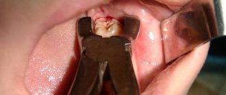
The process of removing the upper canine
Its crown has a spear-shaped (wedge-shaped) shape, a height of 10-12 mm and a width of 7-8 mm. The root is conical, almost oval in cross-section due to its slightly greater flattening in the mediodistal direction than that of the incisors, the diameter at the neck is 7-8.5 mm in the vestibulopalatine and 5-6 mm in the mediodistal directions, the apex is in the thickness of the bone of the canine fossa. The gum in the canine area is thin, the circular ligament is strong.
Removal begins after thorough destruction of the circular ligament of the tooth and detachment of the gums using a trowel or rasp. Extraction is performed using straight or S-shaped forceps for removing premolars, with wide cheeks. The tooth is dislocated using pendulum-like vestibulo-oral and rotational movements. Make the first movement vestibular. With the correct selection of forceps and strict adherence to the removal technique, root fracture or other complications rarely occur.
Reasons for development
The reason for true partial edentia of any teeth is the absence of their rudiment due to a violation of embryogenesis. It can be triggered by various reasons:
- Heredity.
- Deviations in the intrauterine development of the fetus due to a lack of nutrients or metabolic disorders.
- Incorrectly treated baby teeth.
Let's figure out together what a pediatric orthodontist treats and what skills he should have. Come here to determine from a photo what a person’s correct bite looks like.
At this address, evaluate the results of treatment for malocclusion in adults using before and after photos of patients.