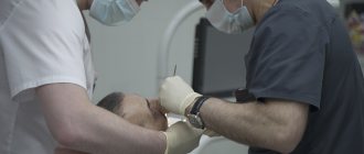
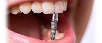
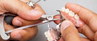
The classic procedure for installing a dental implant involves, although minimally invasive, surgical intervention. A small hole is drilled into the bone, into which the implant is then inserted. Over the next three to six months, the implant fuses with the bone. The final stage of treatment is fixation of the prosthesis. Obviously, the process described above requires special conditions, the absence of contraindications, in particular, the condition of your body must meet the requirements for surgical procedures. Implantation is also problematic for patients with cancer.
Causes
Let's look at the causes of dental cancer
:
- Oral injuries of various origins. Poor-quality dentures rub the gums; the presence of piercings in the tongue or lip can cause inflammation
- Bad habits (smoking, drinking alcohol and drugs).
- Untreated caries.
- Inflammatory diseases of soft tissues.
- Herpes virus, HPV, Bowen's disease.
- Poor oral hygiene.
- Hot or spicy foods that constantly irritate the mucous membranes.
- Work in conditions harmful to health (hot workshop, dusty room).
- A history of cancer of the stomach, kidneys and lungs. In this case dental cancer
is a secondary pathology.
To take care of your health, rule out dental cancer, causes
its development must be monitored. Careful oral care, eradication of bad habits, and regular visits to the dentist will reduce the risk of developing pathology.
Stages of pathology development
Dental cancer
Like any cancer, it occurs in the human body through four stages:
- The first stage is when the tumor is within 10 mm. The place of formation is the mucous membrane or tooth germ.
- The second stage is when the tumor reaches 3 cm. The symptoms of cancer become more obvious. Metastasis is found in one of the regional lymph nodes.
- Stage three – the tumor exceeds 3 cm in size. Metastases are detected in several regional lymph nodes.
- The fourth stage is when the process becomes aggressively malignant, affecting the oral cavity, nose, and also the base of the skull. Metastases may be found in the lungs and liver.
Installation of implants
Implantation for people with diabetes or cancer is carried out in the same way as for ordinary patients. At the same time, the specialist tries to minimally injure the tissue and carefully carry out all procedures. After a thorough examination and preparation, the required tissue area is anaesthetized. Most often, local anesthesia is used, which lasts for several hours. The dentist makes an incision in the gum, accesses the jaw, and a place for the implant is formed in the bone using a special tool. The implant is installed in the prepared bed, fixed and the free space is filled with bone material. Next, the soft tissues, gums and mucous membrane are sutured. All procedures are carried out with periodic cooling of the tissues and washing with an antiseptic solution. After the operation, the implantologist gives advice, prescriptions and recommendations.
Symptoms of tooth cancer
Oncology in the initial stages has hidden symptoms and can develop over years and decades. Gradually the symptoms begin to manifest themselves:
- A formation of soft consistency to the touch appears on the jaw, convex in shape, increasing in size over time.
- The dentition may become deformed and the teeth become mobile.
- Pain sensations increase. It becomes difficult for a person to eat, chew, and swallow.
- Regional lymph nodes increase in size.
- A man loses weight.
- Low-grade body temperature, which periodically rises.
- Fistulas in tissues.
- When pressing on the tumor area, the doctor may feel a crunching sound.
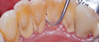
- The enamel becomes fragile and is destroyed very easily.
In the prevention of dental cancer symptoms
may not appear, but it is important to visit your doctor regularly. Identified disease in the early stages provides a better prognosis for treatment. Late stages are dangerous for metastasis to other organs.
Obstacles to implantation in the upper and lower jaws
The main obstacle to implantation is atrophy of the bone of the upper or lower jaw. After the root of a natural tooth is removed, the alveolar process becomes thinner over time. If you install an implant into atrophied bone tissue, over time it will deteriorate even more, and the metal part of the artificial tooth will be exposed. To prevent complications after implantation, jaw bone augmentation is performed.
Problems when installing an implant in the upper jaw are caused by sinusitis - an inflammatory process in the maxillary sinus of the nose. The infection that causes the disease weakens the immune system and complicates the operation. Since the implants are located near the sinuses, pathogenic bacteria can spread to the jaw tissue. The inflammatory process that develops leads to rejection of the metal part of the implants.
Treatment of tooth cancer
When the doctor confirms dental cancer, symptoms
which prompted you to come to the appointment, it is necessary to begin treatment as soon as possible. The most effective method is surgical removal of the tumor within healthy tissue. In the early stages of the process, the tumor is removed along with the teeth and part of the jaw.
In other cases, the jaw is resected completely or partially, and regional lymph nodes are removed. Based on the results of diagnosing the type of tumor and the stage of the process, chemotherapy or laser therapy is prescribed before surgery in order to reduce the tumor in size.
In the first and second stages, the prognosis is favorable: 80% of people survive the 5-year mark. In the third stage of the disease, more than half of people survive five years. In the fourth stage, only 15% reach the five-year survival mark. Regular preventive examinations and adherence to healthy lifestyle standards can reduce the risk of disease.
Dental treatment for oncology
Dental treatment for cancer is associated with difficulties. During chemotherapy and radiation therapy, the body suffers from various side effects. Dental treatment becomes additional stress for a person. The gold standard before starting cancer therapy is oral hygiene, as the body will be less able to resist bacteria during cancer treatment.
Treatment and removal of teeth in cancer patients
In cancer patients, during the treatment of the underlying disease, favorable conditions are created in the oral cavity for the proliferation of pathological microorganisms. Irradiation inhibits cell growth, including in the oral cavity.
The task of a dentist in the treatment of teeth and oncology
the patient is not to harm him. Despite the fact that the body is weakened by a serious illness and no less severe therapy, caries must be treated so that it does not turn into pulpitis. Due to weakened immunity in cancer patients, inflammation progresses rapidly if left untreated. When treating teeth, it is important that the doctor knows the number of leukocytes, blood coagulation status, uses low-traumatic techniques, and coordinates the procedure with the oncologist.
Chemotherapy and tooth extraction
Teeth that cannot be restored are subject to removal. Although patients receiving chemotherapy have a weakened immune system, the consequences of refusing to remove an inflammatory tooth are much more serious than the side effects of anticancer therapy. It is advisable to remove teeth before starting chemotherapy.
in dental treatment
is taken into account, the doctor takes into account the patient’s condition when choosing an intervention technique. Treatment by a dentist during chemotherapy is not advisable, but if there is an emergency and the patient is already taking chemotherapy, the intervention of a doctor will avoid big problems. Removal should be done as atraumatically as possible.
Dental problems and dental treatment for oncology
After chemotherapy, many are faced with the fact that they develop caries, gingivitis, stomatitis, periodontal disease, cysts begin to form, gums become more sensitive and bleed.
This condition creates additional difficulties for the dentist, but does not mean that you need to refuse dental treatment, cancer
not prevent. It is important to tell your dentist about your diagnosis and that you are currently undergoing treatment for cancer. This will allow him to choose the most gentle methods for solving dental problems.
You need to know that when treating teeth, oncology
does not progress and the disease itself is not a contraindication to dental intervention. Dental treatment will remove the source of infection, prevent its further spread, and defeat cancer.
An urgent problem of modern surgical dentistry, maxillofacial surgery and oncology is the treatment of osteonecrosis of the jaw in patients receiving bisphosphonate therapy (BP). The latter are the main treatment for paraneoplastic hypercalcemia, and are also successfully used in conditions with increased resorption of bone tissue by osteoclasts (Paget's disease, osteoporosis) [2]. BFs are part of complex therapy for patients with multiple myeloma, prostate cancer, breast cancer, and osteoporosis. According to statistics on the incidence of malignant neoplasms in the population of Russia in 2007, these malignant neoplasms account for 25% of the overall structure of cancer incidence [1].
Bisphosphonates are a group of drugs that are powerful inhibitors of bone resorption by osteoclasts [3]. By chemical nature, the drugs are analogues of pyrophosphate; the chemical structure of the molecule provides for many possible variants of compounds due to modification of two side chains on the carbon atom or esterification of phosphate groups (Fig. 1).
Figure 1. Chemical structure of BF. The P-S-P fragment determines the affinity of BF for bone tissue. The creation of chemical modifications in positions R1 and R2 of the BP structure led to a progressive increase in the antiresorptive potential and changes in the affinity of BP for bone tissue (Table 1) [4].
Replacing the hydrogen atom with a hydroxyl group at the R1 position increases the affinity for hydroxyapatite by approximately 2 times. Given the obvious differences in the affinity of BP for bone tissue, the R1 fragment is not the only determining factor in this affinity. The introduction of nitrogen components (primary and tertiary, or heterocyclic ring) into the R2 position increased the antiresorptive potential of BF by 3 times compared to BF that did not contain nitrogen. However, not only the presence of nitrogen atoms is important, but also their position in the molecule, since the antiresorptive potential can differ by more than 700 times between isomers of the same BP.
Based on nitrogen substitution in the side chain, BPs can be divided into 4 chemical groups:
— BF without nitrogen substitution (etidronate, clodronate, tiludronate);
- aminobisphosphonates (pamidronate, alendronate, neridronate);
- aminobisphosphonates with replacement of the nitrogen atom (ibandronate);
— BF with basic heterocyclic compounds containing nitrogen [risedronate (contains a pyridine ring), zoledronate (contains an imidazole ring)].
The main effect of BP is inhibition of bone resorption, which is accompanied by a positive calcium balance. BF penetrate into bone tissue, concentrate around osteoclasts, creating a high concentration in resorption lacunae, bind to the mineral matrix of the bone (the selective effect of BF on bone tissue is based on the high affinity for mineralized bone tissue), are captured by osteoclasts, where they disrupt the formation of the cytoskeleton, necessary for the attachment of osteoclast to bone tissue. There is a decrease in the secretion of lysosomal enzymes. BP also causes apoptosis of osteoclasts in the bone resorption zone, inhibiting the synthesis of osteoclast-stimulating factors by osteoclasts [11, 12].
Osteonecrosis of the jaw (Fig. 2)
Figure 2. Clinical picture of osteonecrosis of the jaw during BF therapy. - a serious complication that can occur in patients receiving BF therapy after any dental intervention: professional hygiene, tooth extraction, as a result of chronic trauma to the mucous membrane with dentures, etc. For the development of this complication, the duration of BP therapy (i.e., accumulation of the drug in bone tissue) and the generation of the drug used are also important (with intravenous administration of IV generation BP, the risk of bisphosphonate osteonecrosis of the jaw - ONJ - increases 4.4 times).
Since 2003, when R. Marx first described 36 cases of osteonecrosis of the jaw in patients receiving fourth-generation bisphosphonate therapy, the authors have described more than 500 cases of ONJ in different countries. The incidence of this complication, according to various authors, reaches 12% [5-10].
Over the past 5 years, a large number of publications by foreign authors have been devoted to this problem. The pathogenesis of this disease is still not completely clear and is the subject of research, as evidenced by the growing interest in the problem of specialists: oncologists, maxillofacial surgeons. The literature describes the following pathogenetic factors that determine the development of osteonecrosis of the jaw in patients receiving therapy with nitrogen-containing BP:
- local or general immunosuppressive effect (especially in the case of treatment of cancer patients), the abundance of pathogenic microorganisms in the oral cavity;
- increased production of pro-inflammatory cytokines;
- antiangiogenic effect;
— inhibition of bone tissue remodeling processes [6].
To date, the optimal treatment tactics for patients with osteonecrosis of the jaw receiving BF therapy have not been developed. Consequently, the search for effective methods of treating this category of patients remains an urgent problem of modern medicine.
Material and methods
From September 2009 to the present, at the Department of Hospital Surgical Dentistry and Maxillofacial Surgery of the Central and Maxillofacial Surgery of the Moscow State Medical University, 19 patients with osteonecrosis of the jaw are being monitored and treated during therapy with BF. The patients' age ranged from 55 to 74 years. Patients received treatment with BF as part of complex therapy for breast cancer, prostate cancer, kidney cancer, and multiple myeloma (Table 2).
The diagnosis was made based on:
- the presence of an exposed area of discolored jaw bone tissue, which is determined within 8 weeks or more;
- current or history of therapy for FD;
— lack of radiation therapy in the maxillofacial area.
In the anamnesis, 16 patients noted the removal of one or more teeth due to exacerbation of chronic periodontitis, periodontitis, or for the purpose of sanitation of the oral cavity in preparation for orthopedic treatment. Two patients used irrationally manufactured removable orthopedic structures (clasp dentures).
After tooth extraction, patients noted the presence of long-term non-healing holes, suppuration, pain, exposure of areas of the alveolar bone, and the formation of fistula tracts in the oral cavity or on the skin.
It should be noted that dentists are insufficiently aware of the risk of developing osteonecrosis of the jaw in patients receiving BF therapy after invasive interventions, which leads to errors in diagnosis and treatment. In particular, exposure of bone tissue after removal is regarded as exostosis, a sharp edge of the socket, etc. Attempts to eliminate this using traditional methods of surgical treatment aggravate the course of osteonecrosis, leading to an acceleration of its spread, a deterioration in the patient’s quality of life, and the prognosis of the disease.
We determined the stage of osteonecrosis in patients in accordance with the recommendations of the American Association of Oral and Maxillofacial Surgeons.
Stage 0: there may be no exposure of necrotic bone, but the presence of nonspecific symptoms such as toothache without an odontogenic cause; aching pain in the lower jaw, sometimes radiating to the temporomandibular joint; pain in the maxillary sinus, which may be associated with inflammation and thinning of its wall; mobility of teeth with intact periodontium; fistulous tracts that do not have an odontogenic cause;
Stage 1: exposure of an area of necrotic bone without signs of inflammation and pain;
Stage 2: exposure of an area of necrotic bone, accompanied by pain and inflammation;
Stage 3: exposure of an area of necrotic bone with signs of inflammation, as well as one or more of the following: exposure of necrotic bone extending beyond the alveolar bone (i.e., involvement of the lower edge and ramus of the mandible, maxillary sinus and zygomatic arch with osteonecrosis of the upper jaw); the presence of a pathological fracture; fistulous tract on the skin; presence of oroantral or oronasal communication (Fig. 3).
Figure 3. Scintigram of a patient with osteonecrosis of the mandible.
In our observations, there were no patients with stage 0, stage 1 was observed in 3 (16%) patients, stage 2 in 5 (26%), stage 3 in 11 (58%).
Among patients with the 3rd stage of BONJ, 1 had a fistulous tract on the skin in the submandibular region, in 2 the process spread to the maxillary sinus, in 1 - to the hard palate, in 1 patient - to both maxillary sinuses and the floor of the nasal cavity, in 6 patients - on the body and branch of the lower jaw.
Mandatory additional examination methods are visualization. However, there are certain difficulties in the X-ray diagnosis of BONJ. This is due to the fact that the area of osteonecrosis can be defined as an area of bone tissue destruction or as an area of osteosclerosis, without clear boundaries of transition to intact bone. The periosteal reaction is not typical, however, in a number of cases, with a long course of the process, thickening of the periosteum was detected. Orthopantomography is an insufficiently informative method, while the results of computed tomography can determine the presence of a pathological fracture of the jaw, the spread of the process to the hard palate and maxillary sinuses when the lesion is localized in the upper jaw. However, it is quite difficult to determine the exact size and localization of necrosis zones using scintigraphy. Thus, osteonecrosis is not characterized by complete separation of necrotic areas of bone, although radiographically in many patients they are visualized as sequestration. In this case, intraoperative problems arise, since it is necessary to mechanically separate areas of the jaw affected by osteonecrosis.
Some patients in oncology clinics undergo radioisotope examination of the skeletal system (scintigraphy) to identify metastases in bone tissue. In this study, zones of osteonecrosis are defined as areas of increased accumulation of radiopharmaceuticals (Fig. 3). However, this study is not informative enough to determine the exact size and localization of the osteonecrosis zone.
Treatment of 19 patients varied depending on the stage of osteonecrosis. The patients were divided into two groups.
Group 1 included 11 patients with stage 3 osteonecrosis, as well as 1 patient with stage 2 on the background of a severe course of the underlying disease. Patients of group 1 received symptomatic conservative treatment, which was aimed at controlling the course of the inflammatory process, preventing the spread of inflammation into the surrounding soft tissue, and relieving pain associated with irritation of the inferior alveolar nerve when the process is localized in the lower jaw. For this purpose, antiseptic treatment of the affected area with a 0.05% chlorhexidine solution was regularly carried out, and dynamic observation was carried out. In case of exacerbation of inflammation (increased pain, the appearance of profuse suppuration, swelling of soft tissues), patients were prescribed Tiberal (ornidazole) 500 mg twice a day, Suprastin (chloropyramine) 25 mg 2 times a day for 5 days. Metrogil-denta gel and Levomekol ointment were also used topically. Patients received recommendations on oral care and nutritional recommendations.
Clinical example 1.
Patient Sh.,
Born in 1955. She received therapy with BF (Zometa) as part of complex therapy for breast cancer from June 2008 to 2009.
(11 courses). In April 2009, tooth 4.4 was removed at the local clinic due to exacerbation of chronic periodontitis. The healing of the hole did not occur; an area of exposed bone tissue appeared. The patient has been observed in the outpatient department of the Department of GCS and Maxillary Surgery since 2009 (Fig. 4, a).
Figure 4. Clinical picture at the first visit (a) and during the period of stabilization of the disease (b). The patient received symptomatic conservative treatment. Against the background of stabilization of the general condition, a relative stabilization of the course of the disease occurred (Fig. 4, b). However, subsequently, against the backdrop of an unfavorable course of the underlying disease, osteonecrosis covered the entire body of the lower jaw, and a pathological fracture occurred (Fig. 5, 6).
Figure 5. Extensive area of destruction, pathological fracture of the lower jaw on the left.
Figure 6. Osteonecrosis of the lower jaw, stage 3. BONCH stage: 3.
Treatment of 7 patients of group 2 was surgical. In a hospital setting, laser necrectomy was performed under intravenous and local anesthesia with monitoring by an anesthesiologist, since all patients had concomitant pathology from the cardiovascular or endocrine system. For local anesthesia, articaine anesthetics with a reduced adrenaline content were used. Using an erbium laser in ablation mode with a focused beam of long pulses (long pulse - lp - 700 μs) with an energy of 320 mJ, a frequency of 15 Hz, the edge of the mucous membrane was excised along the perimeter of the osteonecrosis zone, the mucoperiosteal flap was gently mobilized and carefully folded back so that , on the one hand, there was a sufficient overview of the affected area, on the other hand, it was possible to suture the postoperative wound tightly with a tight fit of the flap to the bone surface, but without tension. During the operation, tissue trauma was kept to a minimum, since any invasive action can lead to further development of osteonecrosis. Then, using an erbium laser in ablation mode with very short pulses (vsp - 230 μs) with an energy of 450 mJ, a frequency of 15 Hz, necrectomy was performed to a visually identifiable intact bone. Although on a computed tomogram the necrotic bone was identified as a sequester, during the operation it turned out to be tightly fused with the intact bone, but was different in appearance: it was yellow, rough, but dense. After separating the necrotic bone with a defocused beam of an erbium laser in the mode: energy 320 mJ, frequency 10 Hz, vsp pulses, the surface of the intact bone in the osteotomy area and the inner surface of the mucoperiosteal flaps were treated. The mucoperiosteal flaps were placed in place, the wounds were sutured tightly with Rezopren No. 4 suture material. The suture line was covered with Diplen-Denta L film. In the postoperative period, antiseptic treatment was prescribed with a 0.05% solution of chlorhexidine digluconate, followed by closing the suture line with a film containing an antiseptic and lincomycin. Prescribed: Tiberal (ornidazole) 500 mg 2 times a day, Suprastin (chloropyramine) 25 mg 2 times a day, for 5 days. The sutures were removed no earlier than 10 days later. On the 2nd day after surgery, the patients were discharged from the hospital for outpatient treatment.
Clinical example 2.
Patient B.
Born in 1953. She received therapy with BF (Zometa) as part of complex therapy for multiple myeloma from 2007 to 2009. During this time, sanitation of the oral cavity was carried out in the clinic at the place of residence - removal of teeth of the lower jaw, and a clasp denture was made. Subsequently, two areas of exposed bone tissue appeared on the lower jaw (Fig. 7).
Figure 7. Area of osteonecrosis in the area of the lower jaw on the left (a) and on the right (b).
The patient has been observed in the outpatient department of the Department of GCS and Maxillofacial Surgery since April 2010. Stage of osteonecrosis: 2. According to computed tomography (MSCT), two foci of bone tissue destruction were identified (Fig. 8, 9).
Figure 8. Two zones of destruction in the lower jaw (MSCT).
Figure 9. Zones of osteonecrosis of the lower jaw on an orthopantomogram. In June 2010, laser necrectomy was performed on the lower jaw on the left (Fig. 10-13) in the conditions of the Central Clinic and Maxillofacial Surgery Hospital of the Moscow State Medical University.
Figure 10. Bactericidal treatment with an erbium laser, defocused beam.
Figure 11. Removed sequester during surgery in the lower jaw on the left.
Figure 12. Formation of granulations in the postoperative area on the 10th day.
Figure 13. Epithelization of the postoperative area on the 30th day. in September 2010 - on the right (Fig. 14-18).
Figure 14. Performing laser necrectomy.
Figure 15. Removed sequester during surgery in the lower jaw on the right.
Figure 16. View of the surgical wound after removal of the sequester.
Figure 17. The wound is sutured with Diplen-denta L film.
Figure 18. View of the postoperative area after 2 months (a) and after 5 months (b). Postoperative wounds are completely epithelialized. Currently, the patient is under dynamic observation in the remission stage (Fig. 19).
Figure 19. Current clinical picture.
The use of an erbium laser to prevent the development of osteonecrosis of the jaw after tooth extraction.
We are monitoring 2 patients who underwent tooth extraction due to exacerbation of chronic periodontitis, followed by laser treatment of the bone walls of the sockets of the extracted teeth in ablation mode to prevent the development of osteonecrosis of the jaw. In these cases, healing of the holes occurs without inflammation (Fig. 20),
Figure 20. View of the socket of tooth 3.3 10 days (a) and 30 days (b) after removal. after 1 month, the formation of bone beams in the area of the sockets of the extracted teeth is radiologically determined (Fig. 21).
Figure 21. Intraoral contact radiograph of tooth 3.3. Chronic periodontitis in the acute stage (a); 30 days after tooth extraction (b).
Clinical example 3.
Patient F.,
Born in 1954. Receives therapy with BF (Zometa, Zolerix) as part of complex therapy for breast cancer from 2009 to the present.
On October 25, 2010, teeth 4.5 and 4.7 were removed under local anesthesia with an articaine anesthetic solution with a reduced adrenaline content. Removal was carried out as atraumatically as possible, bone tissue trauma was minimal. After the extraction, a thorough curettage was performed, the bone walls of the sockets of the extracted teeth were scanned with an erbium laser, a defocused beam, in the mode: energy 320 mJ, frequency 10 Hz, vsp pulses. 4-0 Vicryl guide sutures were placed at the orifices of the sockets (Fig. 22).
Figure 22. A guide suture is placed on the socket of the extracted tooth. In the postoperative period, antibacterial therapy was prescribed: amoxiclav 625 mg 3 times a day (5 days), Claritin (loratadine) 10 mg 1 time a day (5 days). The sutures were removed on the 10th day (Fig. 23, 24).
Figure 23. Fibrinous plaque at the mouth of the socket on the 3rd day. The seams are fixed well.
Figure 24. Formation of granulation in the socket on the 16th day.
As a result of the treatment, the sockets of the extracted teeth healed without inflammation. After 1 month, no signs of osteonecrosis were detected (Fig. 25).
Figure 25. Complete epithelization of the socket on the 30th day.
results
The treatment protocol we propose for BONCH patients can improve the quality of life, namely: relieve inflammation and pain, and stabilize the development of osteonecrosis to varying degrees.
In patients of group 2, who underwent surgical treatment using an erbium laser, it was possible to stop the further development of osteonecrosis after complete removal of necrotic bone. In the postoperative period, patients did not complain of pain, deterioration in health, dysfunction of mouth opening, swallowing, or chewing. Locally: the postoperative wound, sutured with Rezopren, had no signs of inflammation, collateral edema was not detected, palpation was painless, the mouth opened fully. After 7-10 days, the postoperative wound was completely epithelialized. At the same time, an improvement in the condition of the mucous membrane was noted. From loose, swollen, thinned, it turned into elastic, normal thickness, tissue with good blood supply, capable of covering and protecting the bone from injury.
With a conservative treatment protocol in the oral cavity, the mucous membrane remained thinned, pale, friable, or hyperemic and swollen with exacerbation of inflammation. Areas of exposed necrotic bone were still identified.
It is necessary to note the wavy course of osteonecrosis, the presence of periods of exacerbation in the autumn-spring period, as well as the deterioration of the process with the progression of the underlying disease and repeated courses of chemotherapy. Thus, among patients with stages 1-2 of necrosis, whose treatment was surgical, 2 patients had a relapse of osteonecrosis of the jaw during repeated courses of chemotherapy, which led to the transition of the process to the 3rd stage.
We also noted a more aggressive, rapid course of osteonecrosis in 5 out of 10 patients with breast cancer who were under our supervision. In these 5 patients, the process of osteonecrosis of the jaw developed to stage 3 within a year.
Conclusion
Currently, the problem of diagnosing and treating osteonecrosis of the jaw during FD therapy is becoming increasingly urgent due to the prevalence of the disease. This dictates the need to search and improve methods for objectively assessing the prevalence of necrosis and determining optimal treatment tactics using high-tech methods. Of great importance is the prevention of the development of osteonecrosis, which consists of sanitation of the oral cavity before the start of BF therapy and treatment of bone alveoli immediately after tooth extraction with erbium laser radiation.
The best results have been achieved in the treatment of patients with the initial stages of osteonecrosis in remission or stable condition of the underlying disease, subject to discontinuation of BP therapy. For patients with stage 1-2 osteonecrosis, it is advisable to use surgical treatment with an erbium laser, which gives encouraging results. However, these patients require lifelong dynamic monitoring, since the risk of relapse remains due to the deterioration of the patient’s general condition or when the oncologist prolongs therapy for BP.
For the treatment of patients with stage 3 osteonecrosis, conservative treatment aimed at controlling inflammation and preventing complications is indicated.
Thus, to this day, research continues to study the pathogenesis, develop optimal etiotropic and pathogenetic treatment of osteonecrosis of the jaw during therapy with FD.