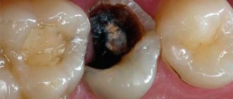
How to understand that there is pulpitis on a tooth?
A person cannot calmly chew food; pain occurs, which is very sharp. In this case, it occurs if a hot or cold liquid gets on the tooth. If we talk about external signs, then usually the gums swell, bleed, and the enamel may darken.
Pulpitis can be acute or chronic. The first option is marked by sharp, sudden pain. Such pulpitis will affect some part of the tooth or the entire area. As for chronic inflammation, the whole process is slow, but the pain will be less. Unpleasant sensations may fade and then return again.
Examples when only the roots of the teeth in the upper jaw remain
Example 1. Restoration from one root of a front tooth
In this example (presented in full here), the situation was created literally by the patient’s hands. She tried to glue part of the tooth with superglue and, under the influence of dangerous substances contained in the glue, the crown of the tooth became completely unusable.
But the root of the tooth under the crown below remained intact and unharmed.
The length of the healthy part of this tooth root removed all doubts about the question - is it necessary to restore the root? Certainly! - it is possible and necessary. The tooth was restored in 1.5 hours using Cerec technology. And here in the photo is the happy owner of a new restored front tooth:
Example 2. Restoring 3 front teeth, of which only the roots remain
Here is a treatment example where I restored the aesthetics of her smile to a 22-year-old girl. The three front teeth on the upper jaw were completely destroyed. From the upper teeth 1.2, 1.1, 2.1 only
the roots
:
These three teeth were actually an aesthetically unsightly frame made from old fillings. Now we will not analyze the whole case in detail, I would like to draw your attention to the following points:
- the shape of the roots of the teeth and their condition made it possible to restore them
- There was no talk at all about any amputation - removal of the roots of the teeth of the upper jaw and implantation.
Our patient's smile was designed in a computer program:
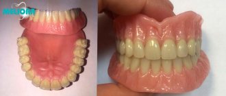
So, in the photo below you see a general set for restoring our patient’s dentition, and among the modules there are 3 with a crown and a root.
The roots of the upper front teeth were quickly and, most importantly, effectively restored:
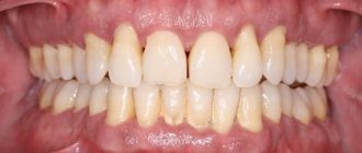
and our patient underwent a beautiful transformation and became the owner of a sweet and chic smile:
This is the enormous potential and aesthetic power of healthy tooth roots! Don't rush to delete them)
Example 3. Complete restoration of the smile zone on the front teeth of the upper jaw
This example is actually very popular on the Internet, since the patient is a TV presenter, and as a result of the treatment she found her new smile. Which is extremely important for her profession. I describe the detailed clinical case itself in another article, but here I would like to note the main points related specifically to the restoration of the roots of the front teeth with crowns.
In the picture below we have removed composite restorations and carious lesions. only the roots remain of the two front incisor teeth.
:
Crowns and “crown + root” modules were prepared in the laboratory:
After installation, the smile began to shine with bright colors:
You can see the entire chronology of our patient’s treatment in one large picture, step by step:
Causes
Pulpitis is inflammation that is caused by infection. The most common causes are lactobacilli, staphylococci, and streptococci.
When these microorganisms get inside the carious cavity, microcracks in the enamel, which leads to destruction of the surface. Their goal is to get to the nerve. Therefore, it is often important to treat the entire body to prevent the infection from spreading further. But treatment of tooth pulpitis should only be prescribed by a doctor.
Examples of canine restoration when only the root remains of the tooth
Example 4. The pin in the root of the lower tooth was removed, and the canine itself was restored
In this clinical case, I was restoring the lower canine, and the task was to save the root of the tooth and restore the coronal part of the canine, which was almost destroyed by caries. The anterior wall of the lower canine was made in the form of a filling, and it fell apart from the cutting edge.
As you can see in the photo, an anchor pin was installed inside the tooth - this is a pin that is screwed into the canal to strengthen the tooth. The tooth under the filling and the root of the lower canine were affected by caries. When the carious tissue was removed, only the root and a small part of the wall
:
You and I remember the main topic of our article, which tells us the following - if the tooth root remains, it can be completely restored. And this is not the first time that the unique Cerec technology has come to our aid. Using it and digital scanning, we restored the missing tooth module, which also included the root part:
Next, we restore the lower front tooth using a CEREC inlay and get an excellent result - the lower canine is completely restored:
Example 5. The root of the tooth under the crown of the upper canine has not rotted, and therefore the tooth is 100% restored
In this case, there was a total restoration of the dentition (veneers and crowns) with the replacement of worn-out crowns on the front teeth. One of the upper canines under the crown was seriously affected by caries, but its root was practically healthy - only a small part of the tooth root was damaged, see the following photo:
The missing “crown + root” module and new crowns were manufactured in a laboratory. In general, everything is in a smile, it looks just great:
And the owner of a new beautiful smile herself cannot hide her admiration:
All the main tasks in this total work were successfully solved, but the main thing you should pay attention to is that we managed to save the tooth root here too.
Symptoms
Different types of pulpitis have their own differences, they develop at different rates, and have different symptoms. If we talk about acute pulpitis, the signs will be as follows:
- unexpected, sharp pain;
- when a person drinks or eats, he can react strongly to high acidity and temperature changes. If you remove the irritant, then this kind of sensation may persist for some time;
- a person cannot independently understand where the pain comes from, because the entire area around the diseased tooth is inflamed and irritated;
- acute pain becomes more frequent when the patient lies down or at night. Sometimes even sleeping pills and painkillers cannot cope with it;
- If not treated on time, the inflammatory process takes on the form of a purulent one. The pain will be constant and more severe;
- Sometimes there is an elevated temperature.
Chronic pulpitis has other symptoms. There are also pain sensations here, but they are not so pronounced. Here the pain becomes aching in nature, in some cases it is not there at all. When the chronic form worsens, symptoms can sometimes resemble the acute form.
When and how can you restore a tooth with Cerec?
A common problem is the development of caries on the frontal units. The result is their complete destruction. The patient sometimes comes too late. Or the previous intervention does not bring the desired result. When several units in the smile area are damaged, and some of them only have tooth root tissue left, you can combine the installation of crowns, veneers and restoration of the upper visible part using special inlays in the root system. Sometimes large-scale intervention is needed - the smile area is corrected, damaged teeth are recreated. You can do a step-by-step restoration of 10 or even 20 teeth above and below.
Using Cerec technology, if there is a healthy root, it is easy to restore almost every tooth within a few hours.
- It will be necessary to determine the condition of the root and undergo a course of treatment.
- Then, after treating the upper roots, it is possible to install a module with the alignment of the tooth crown and the insert into the cavity in the tooth root.
A smile should always be charming. When restored, the new units are ideal, better than natural ones. In addition, natural teeth darken under the influence of pigments or turn yellow with age. Therefore, next to the restored ones they look unaesthetic.
To avoid such consequences, units can be corrected with veneers or crowns (when they are 50% destroyed and there is no stable support). To create an impeccable beauty zone thanks to Cerec, you only need a few visits to the doctor.
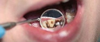
What does pulpitis look like in the picture?
When a patient consults a specialist with acute pain, the doctor examines him and determines the type of disease. Initially, the diseased tooth is examined, tapped, and cold air is directed at it to understand whether there is pain. If there are no visible injuries, the person goes for an x-ray.
The image can only show changes that have occurred in the bone tissue. That is, when there is a radicular cyst, the enamel is deformed and a carious cavity is created. The pulpitis in the picture has an identical appearance to that of a healthy tooth. The only thing is that gangrenous inflammation will be shown. In this case, soft tissues die and toxic substances are released. Because of this, bone tissue slowly dissolves. The result is that the bone becomes less dense, as shown by x-rays.
First aid for pulpitis
Help with the first symptoms of pulpitis should be aimed at reducing pain. To do this you can:
- Use painkillers that are in your home medicine cabinet (analgin, ketanov, ibuprofen, etc.)
- Clean the carious cavity, rinse your mouth with a warm (but not hot) hypertonic solution - dilute 1 teaspoon of salt or soda in a glass of water. Place the prepared solution in your mouth and hold it for a minute.
- Attacks of pain are reduced by rinsing the mouth with decoctions of medicinal plants: chamomile, calendula, valerian, eucalyptus, plantain, sage, St. John's wort.
If the first signs of pulpitis occur, do not delay your visit to the dentist!
Strictly contraindicated:
- Putting something into a tooth cavity.
- Apply warm compresses to the sore spot (they will only increase inflammation).
It is important to understand that the listed methods are temporary and will eliminate only one of the symptoms of dental disease! The cause of pulpitis can only be eliminated by a dentist after conducting a comprehensive diagnosis and performing therapeutic measures.
Complications and consequences
If pulpitis is not treated in time, serious problems may appear. For example, periodontitis may occur. The infection contained in the pulp also spreads to the soft tissues that are around. After this, the jawbone begins to deteriorate. The result is that periodontitis turns into osteomyelitis or purulent periostitis. Also, through the bloodstream, the infection can affect the entire body, and because of this, internal organs can suffer.
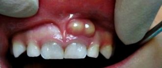
Why do tooth remains appear in the gums?
There are many reasons for this phenomenon:
- Perhaps the tooth has completely crumbled, but the root remains.
- The crown received a strong blow and a fracture occurred.
- An incomplete tooth extraction was performed. The remaining fragments cause inflammatory processes in the soft tissues, causing pain.
- Units with the neurovascular bundle removed are often destroyed. The so-called “dead teeth”. The nutrition of their walls is disrupted, the shell becomes fragile and, due to incorrect loading or due to increased fragility, the natural walls break off “at the root.”
If the process has already started, then urgent treatment of the tooth root is necessary. Otherwise, the remains of the unit will have to be removed to avoid infection of adjacent teeth and surrounding tissues.
The situation cannot be ignored: tooth fragments may not bother you at first, but then turn into a serious problem. When trouble occurs, immediately contact the dental clinic to promptly select treatment and find ways to restore the tooth.
Prevention
Usually, caries leads to the pulp of the tooth. If you do not want to diagnose yourself with pulpitis, then it is worthwhile to prevent this disease. It is important to visit the dentist regularly to treat those teeth that are problematic.
To strengthen the enamel, you can use some products that contain fluoride, calcium, and vitamins. You should not let your sore throat become chronic, as this can also cause dental disease.
If you have an inflammatory process or pain in your teeth, then you should immediately go to a specialist. Until then, to improve the condition, you can rinse the tooth with herbal decoctions and solutions. It could be sage, eucalyptus, chamomile. As for solutions, these are chlorhexidine, miramistin. This will relieve the pain for a while. If there is pus, you can rinse your mouth, but only with liquid at room temperature. This will prevent the spread of the purulent process. And it is important to understand that it is better not to delay treatment, so as not to get more serious problems.
When can a tooth be restored?
When the walls have completely crumbled, but the root of the tooth has not been destroyed, traditional dentistry sometimes suggests removing it. Today, using new protocols, it is possible to preserve the root and create a new stable unit - any of the functional ones, except for the “eights” (wisdom teeth). To do this, you need the root to be healthy:
- Not affected by caries, without fractures or cracks. If such defects exist, then only removal followed by implantation is indicated.
- No cyst. If there is one, then a method of treatment is selected. And only then can you begin to restore.
If a similar problem happens to you, do not rush to the surgeon. New technologies make it possible to accomplish the previously impossible - to preserve the root of a tooth. What to do in a particular case is decided at the appointment, after diagnosis. The vast majority of patients manage to save the tooth after treatment procedures and install a crown on the root of the tooth.
Possible consequences of treating pulpitis
No matter how well the therapy procedure is carried out, there is a chance of developing complications of pulpitis. After treatment, there is a risk of tooth fracture. To avoid this, the patient is given a crown, and if the tooth walls are adequately preserved, restoration is carried out with a photopolymer.
As a complication, in the absence of timely treatment, periodontitis may develop. The disease is accompanied by an acute inflammatory reaction in the tooth and adjacent tissues. The complication has several stages. In the first case, the inflammatory process can be stopped and the tooth can be saved; in the second case, it is removed.
Although x-rays are not able to unambiguously determine the presence of pulpitis, the study remains a key point in diagnosing the pathology. Thanks to an x-ray, the dentist identifies indirect signs of pathology, begins timely treatment and saves the tooth.
X-ray diagnostics
Pulpitis is a study using x-rays passing through the tooth tissue and appearing as a picture on the image. Displayed as dark and light areas.
Classic X-rays are gradually fading into the background. Today, specialized devices are used - visiographs, which project images onto a computer screen.
In fact, x-ray is an additional method for diagnosing pulpitis, since with the help of such a study it is impossible to identify pathology. Based on the results, indirect diagnostics is carried out to determine the depth of the carious lesion.
An exception is the purulent form of pulpitis, which leads to the death of the bone structure and pathological changes. This can be seen clearly in the photographs.
For this reason, x-rays should be considered as an accompanying examination method. A physical examination and EDI (electroodontodiagnosis) are mandatory to assess the condition of the pulp.
X-rays after therapy
X-ray is not only a method for diagnosing pathology, but also acts as a control over the effectiveness of therapy. Through research, the doctor determines:
- Quality of pulp removal. Incomplete removal leads to re-inflammation.
- The nature of the tooth canals, their physiological structure and depth.
- How well were the canals filled using medical cement or gutta-percha? In case of insufficient filling or excess filling material in the canals, the tooth is treated again.
After therapy, control radiography is recommended, usually every six months.
Pulpitis in a child
Pulpitis in a child
It is a mistake to believe that treatment of baby teeth is not necessary. Without adequate treatment, caries of a baby tooth in a child, just like caries in an adult, can very quickly turn into pulpitis with all its complications. There are known cases of death after complications of pulpitis due to the development of sepsis, when no more than two days passed from the first pulpitis pain to the development of facial edema and subsequent death.
To avoid any kind of complications, caries of primary teeth in children must be treated on time. And if the moment is missed and pulpitis has already developed, you need to contact the dentist immediately. A competent doctor will do everything possible to ensure that the treatment of a small patient is as painless, comfortable and, most importantly, effective.
Benefits of Cerec Restoration
The innovation has clear advantages compared to traditional prosthetics:
- The process does not take a week or several days, but only 1.5 hours
per tooth maximum. - Accuracy
. The computer minimizes errors and eliminates errors. - Biocompatibility
. Fabrics do not reject materials.
An inlay in the tooth root is created quickly, then the crown is modeled. The price is fully justified by the reliability, long service life of the structures and their aesthetics.
There is a striking difference between the smiles of patients when they only had the root of the tooth left, and the photo after treatment with Cerec dentures.
X-ray examinations in dental practice
X-ray research methods, due to their informativeness and reliability, occupy a leading position in the diagnosis of diseases of the maxillofacial area. Radiography has found wide application in therapeutic and orthopedic dentistry, maxillofacial surgery, diagnosis of traumatic and inflammatory tooth lesions, cysts, tumors and other pathologies.
Depending on the equipment used and the purpose of the study, modern x-ray diagnostics has the following varieties:
- targeted radiography - an x-ray of one or more teeth;
- orthopantomogram – a panoramic all-round image that shows the entire jaw;
- computed tomography – 3D image of the teeth of the upper and lower jaw
Advantages of X-ray diagnostic methods
X-ray diagnostics is one of the most modern research methods. X-rays provide the dentist with the opportunity to assess the condition of the teeth, the location of the pathology, and the volume of bone tissue. Obtaining a picture of the dentition makes it possible to choose the most correct treatment tactics to achieve a better and long-term result.
In dental practice, equipment with a low degree of radiation is used. Therefore, these diagnostic methods are considered safe and have no contraindications, even during pregnancy.
X-ray examination methods make it possible to determine the condition of the tooth without opening it, to find out the number of root canals for proper treatment and subsequent sealing. The information obtained during x-ray diagnostics during dental implantation is irreplaceable: assessment of the volume and structure of bone tissue is necessary for accurate installation of the implant and its long service life. Dental radiography does not require preliminary preparation of the patient, and the process takes only a few minutes.
Recovery period
Once treatment for pulpitis is completed, the patient will need some more time to fully recover - from one week to a month. During this period, physical therapy is indicated, for example laser therapy using a helium-neon apparatus. This is necessary to speed up the healing process and consolidate the therapeutic effect.
Since pulpitis is an inflammatory disease, after treatment in the clinic it is necessary to take a course of non-steroidal anti-inflammatory drugs at home, such as: Ibuprofen, Ketanov, Naproxen-Acri. However, it should be remembered that all anti-inflammatory and painkillers have their contraindications, so the choice of a specific medication must be agreed with your doctor.