Periodontal diseases are widespread, so it is necessary to use advanced methods to make the most accurate diagnoses and differentiate one pathology from another. For this reason, various periodontal indices have been developed that make it possible to monitor the dynamics of pathology development over a given time period, assess the prevalence and depth of the pathological process, and compare the effectiveness of different treatment methods. In this review we will talk about this method of research as the Schiller-Pisarev test, its advantages, disadvantages and features.
General overview
In dentistry, indices are a quantitative reflection of the progress of the pathological process that has developed in the periodontium.
When compared with clinical diagnostic methods, index assessment is considered relative, since it does not reflect the absolute accuracy of the ongoing process.
But periodontal indexing has some advantages that distinguish it from clinical diagnosis. With its help you can:
- measure the dynamics of tissue infection;
- monitor the spread and development of periodontal diseases;
- monitor the progress of treatment;
- evaluate its success and the results of preventive measures;
- perform epidemiological screening of the population;
- make comparisons of the results of different studies.
There are several dozen assessment indices for studying the condition of periodontium. All of them are divided into 3 groups:
- Reversible . With their help, the dynamics of diseases and the effectiveness of treatment are assessed. Reflect the severity of the inflammatory process, bleeding gums, loose teeth, the size of periodontal and gum pockets.
- Irreversible. They convey the manifestation of such signs as resorption (absorption) of bone tissue in the alveolar process, amyotrophy of the gums.
- Complex. They help to give a comprehensive description of the condition of periodontal tissues, taking into account the existing symptoms and the degree of development of pathological processes.
During one appointment, the dentist may perform several screenings to provide an accurate assessment of tissue health. But all of them, according to doctors, do not provide an individual approach to the patient and cannot completely replace a clinical examination.
What is the Dental Oral Health Index?
The hygiene index is indicators reflecting oral hygiene, the degree of contamination, determining the presence of signs of bacterial infection, indicating the number of teeth that are affected by caries.
The hygiene index allows a specialist to determine the reasons why tooth decay occurs, gum disease occurs, and also prescribe effective preventive measures.
With their help they determine:
- The level of dental health of the patient;
- The severity and stage of caries;
- Number of teeth pulled out;
- Quality of hygiene procedures;
- Presence of malocclusion;
- The degree of effectiveness of therapy.
Important to remember! Each diagnostic criterion for different types of lesions is reflected in an individual index.
Why are hygiene indices needed in dentistry?
The simplest and at the same time effective method of preventing dental diseases is careful adherence to hygiene standards. It is with its help that it is possible to prevent inflammatory and destructive processes in periodontal structures.
During an examination of the patient’s oral cavity, the dentist assigns a hygiene index, which depends on the following criteria:
- quality of teeth cleaning
- oral health
- number of missing teeth, as well as crowns that cannot be restored
- stage of bone tissue destruction
- inflammatory processes and the degree of their neglect
- presence of changes in bite
Hygiene indices in dentistry are used to monitor the effectiveness of treatment. After therapeutic procedures, the indicator should change for the better, which is necessarily recorded in the medical history.
There are different types of hygiene indices, which depend on the parameters being analyzed. In this way, it is possible to note important criteria characteristic of a particular dental disease.
Periodontal index system in general
The following types of periodontal indices in dentistry are distinguished:
- IGs are hygiene indices; they assess enamel contamination and the presence of tartar.
- II - inflammation indices - evaluates inflammatory pathology of the gums, periodontitis and periodontal disease.
- BDI – bone destruction index; combined indices.
All indices are not complicated and do not require special equipment; they are easy to identify. There are a lot of them, the main ones will be discussed below.
What are the subdivisions of indexes?
Periodontal indices are distinguished by reversibility, i.e. subject to regression and not subject to, as well as complex.
Reversible – monitor the dynamics of the pathological process and the effectiveness of treatment. These indices are aimed at the current symptoms of pathologies in their acute period:
- bleeding and inflammation of the gums;
- loose teeth;
- pockets of inflammation – gingival and periodontal.
The most frequently used of these periodontal indices are papillary-alveolar, PI, IG - hygiene indices, of which there are generally more than 15 (Schiller-Pisarev, Pakhomov, Ramfjord, etc.). The data of these indices may change, but the problems respond well to treatment and have a good prognosis, i.e. reversible.
Irreversible indices: gingival recession, X-ray, etc. Irreversible processes are already recorded here when it comes to the consequences and complications of pathologies, such as resorption (resorption) of the bone component of the alveolar processes, recession or amyotrophy of the gums. Treatment is ineffective.
Complex periodontal indices provide a comprehensive assessment of periodontal health. For example, the Komrke index includes a large number of studies: PM index, depth of gum pockets, degree of tissue atrophy, bleeding gums, degree of loose teeth (indicates the degree of inflammation).
Advantages and disadvantages
An important component of implantation at all stages is an accurate index assessment of the condition of peri-implant tissues, implants and supported prostheses. The Schiller-Pisarev test is quite effective and allows you to diagnose a wide range of conditions - this is periodontal destruction, the amount of tartar, plaques, the need for certain therapeutic measures and their volume.
The relationship between the elements of the implant and adjacent tissues and its difference from a natural tooth may make complex periodontal studies impossible.
The Schiller-Pisarev test is quite accurate and objective and has two interpretation options. The first is visual, based on the nature of the gum staining, the second is numerical, that is, index. The main problem of the technique is that dental indices from 30-50 years ago do not meet the current needs of modern implantology.
That is, they can be used, but when interpreting the results, it will be necessary to take into account the full list of current changes and improvements in the field of prosthetics. At the same time, it is the Schiller-Pisarev test that is considered the most informative of all similar diagnostic methods and allows the results to be most successfully adapted to the conditions of endosseous implantation. However, the convention of numerical values still does not disappear anywhere, since diagnostics are carried out using markers, and not high-precision digital equipment. Modern researchers say that the Schiller-Miller test is still relevant, but should be used with certain modifications and clarifications.
Methods used
The objects of index assessment are destructive and inflammatory processes developing in periodontal tissues, the existence of deposits, periodontal pockets, tooth mobility, etc.
There are many research methods, but not all of them receive equal attention from doctors. The most accurate and frequently used research techniques are the following methods.
Papillary-marginal-alveolar index (PMA)
Refers to the basic group of indicators. It was first proposed in 1948 by Massler M. and Schur D. to obtain complete information about gingivitis - to identify its presence, study the severity and depth of penetration. Can be expressed as a percentage (1960 Parma S.) or in absolute numbers.
To detect pathology, the marginal or alveolar gingival zones and interdental papillae are studied.
During the study, the surface facing the lips and cheeks (i.e., vestibular) is covered with a solution with an iodine component. Inflamed areas turn dark brown.
To indicate the presence/absence of a disease, to assess the intensity of the disease, the following encoding is used:
| Code | Result |
| There is no disease. | |
| 1 | Only the gingival papilla is inflamed. |
| 2 | The pathological process occurs on the marginal surface and in the papillae. |
| 3 | The inflammation also spread to the alveolar zone. |
After analysis, the index is calculated using the expression:
RMA = sum of examined elements X per 100: per 3 X per n ( n – number of examined teeth).
The coefficient n is determined by the age of the patient being examined:
- n = 24 – 6-11 years;
- n = 28 – from 12 to 14 years;
- n = there is no exact coefficient, but a number is indicated equal to the number of teeth present in the jaw row.
Explanation of the PMA indicator:
- less than 30% - there is a mild form of inflammation;
- from 31% to 60% - average;
- over 60 – severe.
For the purpose of prevention, this index is used:
- when the disease is detected at an early stage of development;
- to examine the quality of periodontal tissues in dental patients;
- in the treatment of gingivitis/periodontitis in order to assess the severity of the condition and the effectiveness of the therapy.
Measuring periodontal pocket depth
The purpose of the examination is to assess the level of periodontal damage. The presence and parameters of pockets are visualized. The examination is carried out with a probe, a periodontometer and a calibrating stroker with a special scale.
The number of measurements is determined by the group of elements being studied:
2 measurements are taken - for large chewing units from their palatal, lingual and buccal sides and 1 each from the medial and distal sides, 4 measurements - for incisors, small chewing and canines.
Interpretation of pocket measurements:
- Their depth is no more than 2 mm - normal condition.
- This value reaches 3.5 mm - the form of pathology is mild.
- Reaches 4 mm, there is destruction of interdental septa and bone pathology to a moderate degree.
- Exceeds 5 mm – severe form of periodontitis.
This is interesting: What is dangerous about gum burns: causes, symptoms, diagnosis, treatment
Index PI
PI, or Russell's periodontal index, was proposed in 1956 and is intended to establish the stage of development of gingivitis, but also for periodontitis:
- pocket formation, tooth mobility;
- establishes the severity of bone destruction of the tooth, i.e. its loss.
When calculating the periodontal index PI, the index values are summed up and the quotient is obtained taking into account the examined teeth.
The scoring criteria are as follows:
- no signs of pathology – 0 points – no pathological changes, i.e. its intact state;
- 1 - mild gingivitis (the tooth is preserved almost completely, because the inflammation did not cover the perimeter of the tooth);
- 2 - gingivitis has spread circularly, but the tooth-gum connection is not broken;
- 4 – resorption of the tooth septa has begun (this can only be detected on an x-ray);
- 6 - the gum is inflamed, there is a gum pocket, but the tooth is not loose and is fully functional;
- 7 - resorption of the interdental septum has reached the length of the root;
- 8 - periodontal tissues are destructured and the chewing function of the tooth is not performed (the tooth is loose, may be displaced), resorption exceeds the length of the root, and the formation of an intraosseous pocket is possible.
When determining the PI index, all teeth except 8 are examined.
The periodontal index PI determines the degree of plaque on the enamel and refers to periodontitis indices. There are 4 degrees of plaque - from 0 to 3. Zero degree - there is no plaque, the last, third degree - the plaque is pronounced.
The periodontal index PI is obtained by dividing the scores for all teeth by the number of teeth examined. Based on the results of such an examination, we can talk about the degree of gingival inflammation according to an 8-point system, starting from 1.5 points. The last degree is the most difficult.
Complex periodontal index (CPI)
It is used to assess the condition of the periodontium from the initial stage of the disease to its severe manifestations. Designed to determine the health of various age categories of the population:
- 3-5 years;
- 9—14;
- from 15 years old.
All units in the row and their surrounding tissues are checked. The presence of looseness, bleeding, deposits, and the presence of periodontal pockets are observed.
All tissues are examined instrumentally, visually and by palpation. The number and number of elements to be inspected are determined based on the age of the person being examined. So,
- at the age of 3-5 years, numbers are examined: 55, 75, 51, 65, 85;
- 9-14 years –46, 16, 36, 11, 26;
- over 15 years old – 31, 17/16, 11, 47/46, 26/27, 36/
Based on the obtained material, the periodontal condition is coded by the following designations:
| Encoding | Result |
| There is no pathology. | |
| 1 | There is a raid. |
| 2 | Slight bleeding was noted. |
| 3 | A stone has been discovered. |
| 4 | Pockets identified. |
| 5 | The teeth are mobile. |
When calculating KPI, 2 indicators are determined: individual, average:
- KPI in. = sum of codes / per number of units inspected
- KPI environment . = amount of KPI in. /by the number of teeth examined.
Decoding the KPI for different forms of pathologies looks like this:
- up to 2 – the degree of damage is mild;
- up to 4 – average;
- more than 4 – severe.
CPITN Index
The periodontal index CPITN is always considered a marker of the need for treatment of periodontal diseases. It has been used since 1982 and is recommended by WHO. To identify the indicators of this index, the division of the dentition into 3 sextants is used - frontal and 2 lateral. Not all teeth are examined, but only selective ones. It is necessary to examine the tissues around the numbers - 17, 16, 11, 26, 27, 37, 36, 31, 46 and 47. These units, that is, these 10 teeth, give a complete picture of the condition of both jaws. The most periodontally diseased tooth is taken from each sextant. Bleeding gums, the prevalence of tartar and the severity of periodontal pockets are determined.
Research is carried out with a special probe; each tooth is examined for the presence of these disorders. They are registered and analyzed by codes:
- no signs of disease – 1 point;
- if during the examination blood appeared immediately or after 30 seconds. – this is 2 points;
- the presence of tartar (mineralized deposits) - above and below the gum;
- overhanging filling - they delay the plaque that appears - this is 3 points;
- detection of a gum pocket up to 5 mm deep – 4 points;
- if the depth of the periodontal pocket is up to 6 mm or more - 5 points;
X points – there is not a single tooth in the sextant or only 1 (moreover, 8 molars are not included in this calculation).
Next, the amount for each tooth is divided by 6 and the CPITN indicator by code is obtained:
- 0—no treatment required;
- 1 - correction and control of oral hygiene individually for a given patient;
- 2 - professional cleaning and elimination of the above factors delaying plaque on tooth enamel; familiarization with proper oral hygiene;
- 3 — the need for curettage (removal of plaque);
- 4 - comprehensive periodontal treatment.
Pakhomova
Involves applying Lugol's solution to the teeth being examined. The procedure involves 6 frontal teeth of the lower jaw, all 1st molars, 11 and 21 teeth.
The quality of hygiene is assessed according to the degree of staining:
| Grade | Degree of staining |
| 1 | Lack of color upon application |
| 2 | Staining 1/4 crown |
| 3 | Staining 1/2 crown |
| 4 | Staining 3/4 crowns |
| 5 | Staining the entire surface of the tooth |
The overall score is calculated by summing the scores for each tooth examined and dividing by 12.
Periodontal Hygiene Index
1. Periodontal index of oral hygiene Green-Vermillion (Oral Hygiene Index-Simlified, Green-Vermillion, 1964). Allows you to detect not only plaque, but also tartar. Method of determination. The vestibular surface of teeth 16, 11, 26, 31 and the lingual surface of teeth 46, 36 are stained with an iodine-containing solution. On the corresponding surfaces of the teeth being examined, the plaque index (Debris-index) and the tartar index (Calcuius-index) are determined and expressed in points:
Dental plaque (DI): 0—no plaque; 1 - dental plaque covers no more than 1/3 of the surface of the tooth crown; 2 - dental plaque covers from 1/3 to 2/3 of the tooth surface; 3 - plaque covers > 2/3 of the tooth surface.
Dental calculus (CI) : 0 - no dental calculus detected; 1 - supragingival tartar covers less than 1/3 of the tooth crown; 2 - supragingival tartar covers from 1/3 to 2/3 of the tooth crown or there is subgingival tartar in the form of separate lumps; 3 - supragingival tartar covers 2/3 of the tooth crown and/or subgingival tartar surrounds the cervical part of the tooth.
Evaluation of results. Using OHI-S, the level of oral hygiene is determined according to the following criteria: 0 - 0.6 points - good level of hygiene; 0.7 - 1.6 points - satisfactory; 1.7 - 2.5 points - unsatisfactory; more than 2.6 points - bad.
2. Periodontal plaque index Silness-Loe (PI Silness-Löe, 1964). Characterizes the thickness of dental plaque in the gingival area. No special staining is performed to determine it. Method of determination. After thoroughly drying the surface of the teeth, the tip of the probe is passed into the cervical part of the tooth, slightly touching the gingival groove on the buccal and lingual sides. The results are assessed in points: 0 - plaque near the cervix is not detected by the probe; 1 - plaque is not visually detected, only when the tip of the probe moves; 2 - significant accumulation of dental plaque, determined both visually and during probing; 3 - intensive deposition of dental plaque on the surface of the tooth, gum pocket, gingival margin, food residues are determined
Ramfjord's teeth are examined (16, 21, 24, 36, 41, 44). The sum of the indicators is divided by the number of teeth examined.
Silnes Low
A method for assessing oral hygiene without the application of coloring materials.
The dentist examines the oral cavity using a probe to determine the amount of plaque.
Based on the amount of plaque detected, the appropriate assessment is made:
- 0 – no plaque;
- 1 – thin layer of deposits, invisible without the use of a probe;
- 2 – visually noticeable plaques;
- 3 – plaque covers the crown.
Using the Silnes-Low method, the hygiene index of an individual unit, a group of several teeth or the entire oral cavity is calculated.
Gingivitis Index (IG)
Needed to identify the area of formation and the extent of gingivitis. To do this, elements numbered 32, 16, 12, 36, 24,44 are analyzed from four surfaces.
Grades are given based on the following criteria:
| Grade | Result |
| There is no pathology. | |
| 1 | The shade of the gums and its structure are slightly changed, there is no bleeding. |
| 2 | The color is greatly changed, there is swelling and slight bleeding. |
| 3 | Severe inflammation, bleeding with minor gum injury, swelling. |
Based on the data obtained, the average indicator is derived by dividing the sum of assessments for each unit by the number of items examined.
This is interesting: What is tooth depulpation, depulpation (pulp removal) of a tooth during prosthetics
The value of IG is deciphered as follows:
- up to 0.1 – pathology is in the initial stage;
- up to 2 – disease in the middle stage;
- up to 3 – severe form of gingivitis.
CSI
Determining the CSI index allows you to find out the amount of tartar and accumulated plaque in the area where the teeth come into contact with the gums.
The condition of the anterior incisors is analyzed. Each tooth is examined from the lingual, medial and vestibular sides. The examination is carried out using a dental probe.
Each surface is scored by:
- 0 – no deposits;
- 1 – deposits 0.5 mm wide;
- 2 – deposits 1 mm wide;
- 3 – plaque more than 1 mm.
To determine the index, it is necessary to add up the sum of ratings for each examined surface and divide by the number of teeth. The maximum value is considered to be CSI 16.
Simplified Gingival Sulcus Bleeding Index
The technique involves identifying the presence of bleeding only based on the patient’s responses. The manifestation of this symptom cannot be verified by probing or visually.
The patient is asked about the possible appearance of blood in various situations, and based on the answers, a coefficient of the degree of inflammation is set.
Typically, a simplified index is used during treatment to analyze its results. Such an examination provides only a rough estimate of the condition of the gums.
Navi indicator
The method involves examining the anterior incisors from the lips. Before starting the procedure, the patient is required to rinse his mouth with a fuchsin solution. This substance colors soft deposits, allowing you to assess the degree of contamination.
Hygiene rating:
- 0 – no deposits;
- 1 – presence of deposits in the area between the gum and tooth;
- 2 – the presence of a noticeable strip of plaque above the border of the tooth and gum;
- 3 – 1/3 coating;
- 4 – 2/3 coating;
- 5 – the tooth is covered with deposits by more than 2/3.
To give an overall assessment, calculate the arithmetic mean for all examined teeth.
Sulcular bleeding (SBI) according to Muhlemann and Son
SBI - will show the early stages of periodontitis and gingivitis. Externally, the oral mucosa may look healthy, but there may be hidden bleeding. With these pathologies, bleeding is possible even with minor damage.
The dental examination technique is carried out as follows: without pressure, a button probe is passed along a certain gum line and the bleeding reaction is observed.
There are 3 degrees of bleeding severity:
- 0 – bleeding did not appear at all;
- 1 – blood appears only in the second half of a minute;
- 2 – blood appeared immediately or within 30 seconds;
- 3 – blood is visible when brushing teeth and when eating.
Tureski
When calculating the Turesky index, the entire dentition is examined. The procedure involves applying a fuchsin solution, after which the appearance of deposits on the lingual and labial surfaces of the teeth is analyzed.
The score is calculated as follows:
- 0 – no plaque;
- 1 – the appearance of individual deposits in the neck of the tooth;
- 2 – presence of deposits 1 mm thick;
- 3 – deposit of 1 mm but less than 1/3 of the surface;
- 4 – plaque on 2/3 of the tooth;
- 5 – deposits cover from 2/3 to all crowns.
The Turesky Index is calculated by adding the scores for each individual tooth and dividing by the number of teeth examined.
Approximal Plaque Index (API)
The procedure involves applying a dye
The approximal surface is the area of contact of the enamel with the tooth located behind it.
The need to inspect the area presented is due to the fact that it requires careful care, which can be difficult to achieve through routine hygiene procedures.
If the amount of plaque exceeds the permissible level, the patient is prescribed professional cleaning.
The procedure involves applying a dye. After this, it is determined how many teeth the color changes.
The API index rating does not provide a pollution rating. The assessment is the presence of a reaction to the dye or its absence.
To determine the index, it is necessary to divide the number of stained teeth by the number of all teeth in the patient’s oral cavity. The resulting figure is multiplied by 100.
Evaluation of results:
| Grade | Hygiene level |
| Less than 25% | Optimal |
| 26% – 39% | Sufficient |
| 40% – 69% | Moderate |
| 70% – 100% | Unsatisfactory |
Flying Rate by Quigey and Hein
Determining the plaque index involves applying a fuchsin solution to 12 front teeth on both jaws. The survey includes numbers 12, 13, 11, 21, 22, 23, 31, 32, 33, 41, 42, 43.
After applying the solution, the vestibular surface is examined. The plaque index depends on the degree of surface staining.
Results of the procedure:
- 0 – no changes when applying the solution;
- 1 – change in color in the cervical area;
- 2 – color within 1 mm;
- 3 – deposits occupy from 1 mm to 1/3 of the surface;
- 4 – 2/3 plaque;
- 5 – sediments cover more than 2/3.
To calculate the index, the totality of points is summed up, and the resulting number is divided by the number of teeth examined (12).
PHP
Determines the effectiveness of hygiene measures, including the thoroughness of daily cleaning. During the procedure, 6 teeth are examined: 16, 26, 11, 31, 36 and 46. The patient rinses his mouth with a special solution containing a dye.
The rating is based on the presence of a reaction to the solution:
- 0 – no reaction
- 1 – tooth staining
If the indexed tooth is removed, the adjacent tooth is examined.
To calculate the result, the score of all examined teeth is combined, after which it is divided by 6. The code for an individual tooth is the score obtained from the examination of each area (medial, distal, occlusal, central, cervical).
Interpretation:
| Grade | Efficiency of RP hygiene |
| Less than 0.1 | High |
| 0.1 – 0.6 | good |
| 0.7 – 1.6 | Satisfactory |
| More than 1.7 | Unsatisfactory |
Oral Hygiene Performance Index (OHP) Podshadley, Haley, (1968)
Iodine index of enamel remineralization.
The active permeability of iodine in tooth tissue is known. Remineralization index (RI), which characterizes the effectiveness of the remineralization therapy used. It is assessed using a four-point system:
1 point – no staining of the tooth area;
2 points - light yellow coloration of the tooth area;
3 points - light brown or yellow staining of the tooth area;
4 points - dark brown staining of the tooth area.
The calculation is carried out according to the formula:
IR = IRNP x number of teeth with hypersensitivity / n,
where IR is the remineralization index;
RRI—remineralization index of one non-carious lesion;
n is the number of teeth being examined.
Dark brown and light brown staining indicates demineralization of the tooth area with non-carious lesions; light yellow - indicates a certain level of remineralization processes in this area of the tooth, and the absence of staining or its slightly yellow color demonstrates a good level of the remineralization process of a particular non-carious tooth lesion.
Composite Periodontal Index (CPI)
It is used for a comprehensive assessment of the condition of the gums and periodontal canal. The procedure involves performing a standard dental examination using a probe and mirror.
During the examination, the dentist notes the presence of certain symptoms, each of which corresponds to a specific score reflecting the condition of the tissues.
Criteria for evaluation:
- 0 – absence of pathological signs;
- 1 – soft deposits;
- 2 – bleeding;
- 3 – tartar;
- 4 – expansion of the periodontal canal;
- 5 – loosening of the tooth in the affected area.
The KPI index is determined by dividing the sum of indicators by the number of examined teeth. The method of examination depends on the age of the patient.
Interpretation of the CRPD:
- From 0.1 to 1 – potential risk of developing periodontitis;
- From 1.1 to 2 – mild form of periodontitis;
- From 2.1 to 3.5 – moderate severity;
- From 3.6 and above – severe form.
Ramfiord index (dental plaque)
The SP Ramford index (1957) has 2 criteria: the degree of inflamed gums and the depth of periodontal pockets. This is an indicator of periodontal disease. Unlike PI, it not only determines the depth of the pocket from the apex of the papillary triangle, but also takes into account the height of the root exposure due to gum retraction (expansion of the gingival groove with exposure of the neck and part of the root of the tooth).
The distance is measured from the enamel-cementum border to the apex of the papillary triangle. With atrophied gums, these 2 indicators are added; with hypertrophy, the difference between them is taken. The periodontium is examined on 2 surfaces - lingual and vestibular - for the amount of plaque contaminating the enamel, as well as for dental subgingival calculus.
For gingivitis, the indicators will be:
- 0 – no disease;
- 1 – locally the gum is slightly inflamed;
- 2 – noticeable inflammation of a large area of the gum;
- 3 – severe gingivitis.
This is interesting: Treatment, symptoms and prevention of bottle caries in a child
Periodontitis data:
- pocket of acceptable sizes – 0–3;
- 4 – pocket depth 3 mm;
- 5 – depth 6 mm;
- 6 – depth greater than 6 mm.
The sum of the scores obtained is divided by the number of dental units examined.
This index is important for those people who cannot or cannot have an x-ray. In the elderly, it is not practical to identify this index, since there are age-related changes in the periodontium: gum retraction, involution of bone tissue.
Papillary bleeding (PBI) according to Saxer and Miihiemann
The periodontal bpe index (PBI) is needed to determine the degree of gum inflammation. A probe is used to make a groove along the interdental papillae and observe for 30 seconds.
The severity of gingivitis is 4 points:
- 0 – no blood;
- 1 – appearance of only blood spots;
- 2 – pinpoint blood appearances along the line of the furrow;
- 3 – blood fills the triangle between the teeth.
- 4 – intense bleeding.
The study of the papillae - PapillaBleeding - is carried out in the following quadrants: the gums of the 1st and 3rd quadrants from the lingual surface and the 2nd and 4th quadrants - from the vestibular side (the vestibular side is the vertical wall of the tooth from the lips and cheeks). First, each quadrant is counted, then the arithmetic mean is derived.
Gum recession index
It is determined what part of the teeth (%) has an important symptom of periodontal pathology - apical displacement of the periodontal attachment, which clinically looks like exposure of the boundary between the enamel and cementum of the tooth and exposure of the root. Gum recession index = (r/n)*100%,
where r is the number of teeth with gum recession; n is the number of teeth examined.
Gingival attachment loss index
The index is used along with the CPI for epidemiological surveys. The objective of this study is to evaluate the periodontal condition during gingival recession. Using a periodontal button probe, the distance from the enamel-cementum border of the tooth to the periodontal attachment (i.e. to the bottom of the pocket) is measured. Index codes: 0 - the enamel-cementum junction is not visible, the periodontal probe is immersed only to the beginning of the black mark (normal groove depth) or within the black mark (pocket depth no more than 6 mm). Calculation of the distance from the invisible enamel-cementum border to the bottom of the pocket: the pocket depth is less than 6 mm, including 2 mm at the edge of the free gum located above the dentogingival junction - therefore, the distance from the bottom of the pocket to the enamel-cementum border does not exceed 4 mm . 1 - a) the enamel-cement border is not visible, the entire black mark of the probe is immersed; b) the enamel-cement border is visible, from it to the bottom of the pocket 4-5 mm. 2 - the enamel-cement border is visible and, upon probing, is located at the level between the upper border of the black mark and the first ring of the probe - the displacement of the attachment is 6-8 mm; 3 - the visible enamel-cement border is located between the first and second rings of the probe - the displacement is 9-11 mm; 4 - the visible enamel-cement border is located above the second ring of the probe - the displacement is more than 12 mm. The selection of key teeth and sextant evaluation and analysis follow the same rules as when working with CPI.
PFRI
The indicator reflects the rate of plaque formation. Allows you to evaluate the conditions and factors that influence the formation of soft deposits. The diagnostic value of the method lies in the fact that it allows you to assess the risk of caries.
The rate of plaque formation is influenced by the following factors:
- Bacterial composition of the oral microflora;
- Total number of bacteria;
- Composition of salivary fluid and its quantity;
- Condition of enamel, gums, fillings;
- Quality of denture care.
Before assessing the rate of plaque formation, professional cleaning is performed.
The diagnostic procedure is performed 24 hours after cleaning. To do this, a coloring solution is applied.
The following surfaces are examined:
- Buccal;
- Lingual;
- Mesio-buccal;
- Mesio-lingual;
- Distobuccal;
- Distal-lingual.
The appearance of color is assessed as 1 point, while the absence of reaction to the solution is 0 points.
To calculate the PFRI, the total score should be divided by the number of teeth and multiplied by 100. The PFRI results are expressed as a percentage.
Ratings:
- From 0 to 10% – very low;
- From 10% to 20% – low;
- From 21% to 30% – average;
- From 31% to 40% – high;
- Over 40% is very high.
Interpretation:
Periodontium is a complex of tissues that have genetic and functional similarities: periodontium, alveolar bone, gums with periosteum and dental tissue (cement, enamel).
The depth of the periodontal sulcus is usually from 0.5 to 2.0 mm. Its base is located at the site of the intact junction of the epithelium with the tooth. Clinically, the gingival sulcus is a gap between healthy gums and the surface of the tooth; it is revealed by careful probing.
Periodontitis is an inflammation of periodontal tissues, characterized by progressive destruction of periodontal tissue and bone. The etiological factor is dental plaque. Local causes of artificial origin: - crown. Deeply advanced under the gum or incorrectly manufactured. Protruding edges of fillings.
Classifications of tooth mobility
1. According to Evdokimov. 1st degree – the tooth moves in the vestibo-oral direction by no more than 1 mm. in relation to the crown of the adjacent tooth; 2nd degree – the tooth moves in the vestibulo-oral and mesiodistal direction by no more than 1 mm. in relation to the crown of the adjacent tooth; 3rd degree – mobility is added in the vertical direction;
2. According to Entin. 1st degree – displacement of the tooth in the vetibulo-oral direction; 2nd degree - the tooth moves in the vestibulo-oral and mesiodistal direction; 3rd degree - mobility is added in the vertical direction; 4th degree – mobility in three directions and tooth rotation.
3. According to the Miller-Flesar scale (1980) . 0 – the tooth is stable, there is only physiological mobility; 1 – tooth displacement along the vertical axis is no more than 1 mm. along the vertical axis by no more than 1 mm. 2 – the tooth moves by 1-2 mm. in the vestibulo-oral direction, the function is not impaired; 3 – mobility is pronounced, the tooth moves in the vestibulo-oral direction and along the vertical axis, the function is impaired.
Plaque indicators
In dental practice, 0.75% and 6% solutions of basic fuchsin, 4-5% alcohol solution of erythrosine, erythrosine in tablets (6-10 mg each), Schiller-Pisarev solution, 2% aqueous solution of methylene are used as diagnostic stains for dental plaque. blue.
Fuchsin (Fuchsini) is a basic solution of fuchsin. Colors dental plaque crimson. The drug is used for rinsing.
Rp.: Fuchsini bas. 1.5 Spiritus aethylici 75% 25 ml DS 15 drops per 1/2 glass of water (for rinsing the mouth for 20 s)
Erythrosin is a red dye of low toxicity. Contains iodine. Available in the form of a 4-5% alcohol solution and tablets (Mentadent C-Plague, Oga] In Einfarb Plagueindikator, Plague-Farbetabletten, etc.).
Rp.: Sol. Erythrosini 5% 15 ml DS Apply with a cotton swab to the surface of the teeth
Rp.: Tab. Erythrosini 0.006 N. 30 DS Chew 1 tablet for 1 minute
Fluorescein is a plaque dye that does not contain iodine, so it can be used in patients who are sensitized to iodine. Fluorescein-stained plaque is visible only under ultraviolet light. Available under the names “Plak-Lite” (“Blendax”), “Fluorescein” 0.75%.
Schiller-Pisarev solution stains dental plaque yellow-brown. The drug is applied to the surface of the teeth using a cotton swab.
Rp.: lodi 1.0 Kalii iodidi 2.0 Aq. destill. 40 ml MDS Schiller-Pisarev solution. Apply with a cotton swab to the surface of the teeth
Methylene blue (Methylenum coeruleum) is used to identify dental plaque: a 1-2% aqueous solution of methylene blue is applied to the surface of the teeth using a cotton swab.
Rp.: Methyleni coerulei 2.0 Aq. dcstiil. 100 ml MDS To lubricate the surface of teeth
Lugol's solution: Composition: KI - 2.0 g I crystalline - 1.0 g distilled water - 17 ml Method and mechanism are the same as in the previous dye
Disclosing Solution (60ml) —
Directions for use: Prepare a solution: 10 drops of indicator per 30 ml of water. Rinse your mouth. Do not swallow! Colored areas indicate the presence of bacterial plaque. Carry out additional cleaning of problem areas.
Katerol -
Plaque indicator, liquid for removing plaque and cleaning teeth.
KATEROL is an indicator of dental plaque (dental plaque), a drug for removing dental plaque and cleaning teeth.
Ingredients: hydrochloric acid, iodine, acetone, excipients. Properties: Caterol softens calculus deposits and destroys supragingival plaque, facilitating subsequent mechanical removal using instruments.
Caterol also helps detect plaque and tartar by turning it yellow.
Contraindications: allergy to iodine or its derivatives.
Instructions for use.
Using a thick cotton ball soaked in Caterol and wrung out well, thoroughly rub the tartar deposits, trying to avoid contact with the gum as much as possible.
Wait a few seconds for the stone to soften before mechanical.
Curaprox tablets -
It colors old plaque blue and new plaque red. Ideal for home use. Each tablet is individually packaged.
Liquid Curadent
Curadent for plaque detection is a two-color liquid that detects plaque. Old plaque turns blue, and fresh plaque turns red. (60 ml - 1,615 rubles).
Plaque Agent (
Docdont) - mouth rinse to detect plaque (500 ml - 400 rubles)
The mouthwash is suitable for both adults and children.
The mouthwash does not contain erythrosine, so it is successfully used by both adults and children. This method of identifying plaque is especially suitable for children, since they cannot always assess whether they have carried out the process of cleaning their teeth correctly. And if you rinse your mouth with this solution before doing this, you can monitor the quality of cleaning.
Recommendations: Use by children under 12 years of age should be under adult supervision. Pour 10 ml of mouthwash into a measuring cup, rinse your mouth for 30 seconds, and spit it out. Clean blue areas thoroughly with a toothbrush and toothpaste.
Plaquetest tablets for detecting dental plaque
Plaquetest tablets for detecting dental plaque. Old dental plaque is painted dark blue, new plaque is purple-red.
To carry out the test:
Place the tablet on your tongue (half is enough for children), chew it and spread it over the entire dentition with your tongue. Spit it out - it's done! The paint is easily removed with regular tooth brushing.
Ingredients: lactose, magnesium stearate, silicon dioxide, meadow mint, CI 42090 (food coloring), CI 45410 (food coloring).
Classification of root furcation lesions
It is determined using a special curved Naber probe or a regular blunt probe. Furcation lesions are determined in both horizontal and vertical positions.
In the horizontal direction: I – a slight depression is visible in the mucous membrane on the buccal surface, but the probe does not enter the area of depression or penetrates the defect to a depth of 1-2 mm. II – the probe enters the bifurcation by more than 1-2 mm, but the connection between one furcation and another is not determined III – the place of the furcation is visible, with the help of the probe you can pass from one furcation to another.
In the vertical direction (Tarnow, Fletcher, 1984): 1 – vertical loss of bone from 1 to 3 mm; 2 - vertical loss of bone in the furcation area up to 4-6 mm; 3 - vertical loss of bone 7 mm. and more.
How is the procedure performed?
The essence of the Schiller-Pisarev test is to lubricate the gums with a solution of iodine and potassium. As a result, areas with deep connective tissue lesions become stained - this is due to the accumulation of large volumes of glycogen in areas of inflammation. The tests are repeated from time to time - if the treatment is carried out correctly, the condition of the gums will improve, and the inflammation will subside or disappear altogether. That is, if the therapy is correct, then repeated tests should be weakly positive or negative.
Gum staining is due to the high amount of glycogen. When the inflammation passes, there is less glycogen, and the tissues cease to be intensely stained. In this way, the intensity and degree of development of the disease is determined.
Composition of the solution
To take Schiller-Pisarev samples, the composition of the solution is used in the following proportions:
- crystalline iodine – 1.0;
- potassium iodide – 2.0;
- distilled water – 40.0.
Before using therapeutic and prophylactic toothpaste (, Parodontol), the gum mucosa is lubricated with a special solution, then the degree of staining is determined, the data obtained is recorded in the history of the disease. Control – after 1, 2, 3, 6 and 12 months.
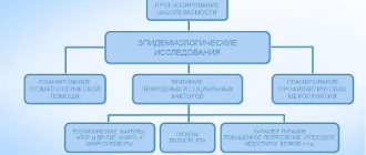
WHO Epidemiological Survey
Epidemiology is a branch of medicine that studies the nature and form of the spread of pathologies.
Epidemiological screening includes 3 stages:
- Preparatory. Planning is carried out for the upcoming survey, indicating its methods, deadlines and tasks.
A working group is created from nurses and trained doctors. For the survey, the population composition differs in terms of living conditions and populations. The number of people depends on the tasks and the required level of viewing accuracy. - Examination. To record the data received, a registration card is used (it has a simplified form for children).
It is prohibited to make additions or corrections to it. All records are coded (each code indicates a specific feature or its absence). - Evaluation of results . The information is calculated according to the required indicators. The result is displayed as a percentage or digital notation.
Such screening helps to assess the dental situation in a certain area, determine the relationship between oral health and social and environmental living conditions, and monitor age-related changes in teeth and gum tissue.
It is essential to determine the most common diseases and the intensity of their manifestation in different areas and age categories.
After analyzing the materials obtained during the study, preventive measures are developed to treat dangerous conditions and teach hygiene rules.
conclusions
The listed periodontal indices contain information about the hygienic situation in the oral cavity, the intensity of the spread of inflammation and the destructive process in the periodontium.
The examinations are simple to perform, do not cause pain to the person, and do not require the doctor to undergo special training.
They help to assess the general condition of periodontal tissues and formulate conclusions about the effectiveness of therapeutic and preventive measures.
Sources:
- https://zubovv.ru/lechenie/desnyi/parodontoz/indeksyi-i-ih-rol.html
- https://dentoland.com/gigiena/indexes-polosti-rta.html
- https://periocenter.com.ua/articles/indeksy-v-stomatologii/
- https://FB.ru/article/439178/parodontalnyie-indeksyi-v-stomatologii
- https://parodont.pro/parodontalnyi-indeks.html
- https://ALVIstom.com/publ/profilaktika/stomatologicheskie_indeksy_gigieny/1-1-0-11
- https://medbe.ru/materials/profilaktika-v-stomatologii/metody-klinicheskoy-i-epidemiologicheskoy-otsenki-kompleksnyy-periodontalnyy-indeks-kpi/