From this article you will learn:
- structural components of enamel,
- the structure of its organic and inorganic matrix,
- electron microscopy images.
Tooth enamel (enamelum) is the outer shell of the crown of the tooth, representing the hardest tissue in the human body. This hardness is explained by the fact that 95-97% of tooth enamel consists of mineral components (mainly calcium phosphate in the form of hydroxyapatite crystals). Organic matter accounts for only 1-2%, plus about 2-3% water. The strongest are the surface layers of enamel - especially on the occlusal surfaces of the teeth, and towards the enamel-dentin border, as well as when approaching the neck of the tooth - its hardness decreases.
The hardness of enamel is 397.6 kg/mm², which is comparable to quartz. Such hardness allows it to withstand extreme mechanical loads, but on the other hand, it makes it very fragile. The enamel of human teeth does not crack or chip only due to the presence under the enamel layer - a layer of dentin, which has a moderate elasticity coefficient. However, despite its hardness, enamel has good permeability to calcium and fluoride ions contained in saliva (or toothpastes and mouthwashes), as well as to pigments contained in food and drinks.
Structure and development of tooth enamel –
Tooth enamel can have different shades - from yellow to various shades of gray and white, which depends on its transparency coefficient. The more transparent the enamel, the more the underlying layer of dentin, which is physiologically yellow, will be visible through it. In addition, the enamel may have a blue tint - at the cutting edges of the incisors (where there is no underlying dentin layer), as well as at baby teeth. The transparency of enamel depends on the degree of its mineralization and homogeneity, which is associated with the ratio of its organic and inorganic matrices. Transparency also depends on the thickness of the enamel layer.
The thickness of the enamel will vary on different surfaces of the tooth crown. For example, in permanent teeth, the thickness of the enamel ranges from 1.7 to 2.5 mm in the area of the chewing cusps of the molars, and to only 0.01 mm in the cervical part of the tooth (where the enamel borders the root cement). Thus, the closer to the neck of the tooth, the less its thickness will decrease. In baby teeth, the thickness of the enamel will be two times less than in permanent teeth, not exceeding 0.8-1.0 mm.
Preface
The goal of root canal therapy is to remove the contents of the root canal space and then fill it. Proper treatment requires knowledge of both the external and internal anatomy of the tooth to reduce the risk of failure and the possibility of iatrogenic biological damage.
Understanding the coronal morphology of the tooth allows us to make endodontic access in the most conservative manner; The shape of the access cavity is described for all teeth. Studying the morphology of the root canal system and its several variations makes it obvious why there are operational difficulties during instrumentation, as shown in the iconographic part. From the morphological and histological tables of Hess, the complexity of the entire root canal system is visible, which confirms the difficulty of completely removing pulp tissue from the endodontic space and encourages the search for new methods and technologies in endodontics.
The microscopic anatomy section summarizes the interaction of the root dentin wall structure with mechanical (files, ultrasound), chemical (irrigants) and physical (lasers) factors during therapy. In particular, in order to understand the different effects of lasers on tissue depending on the wavelength, it is very important to carefully study the ultrastructure and histology of dentin in the canal.
Types of damage to tooth enamel
Over the course of life, even if you provide your teeth with proper care and follow the rules of oral hygiene prescribed by dentists, the enamel layer gradually wears out and is destroyed. This contributes to the occurrence of various diseases of the oral cavity; it is influenced by the food a person eats, etc.
Among the main causes of damage and destruction of tooth enamel, dentists identify:
- Erosion is damage to the enamel layer, and then dentin, which is not associated with carious lesions of the teeth . The essence of this pathological process lies in disorders of mineral metabolism. As a result, disturbances occur in the crystalline structure of the enamel, which is manifested by its focal thinning and destruction. Externally, erosions look like local darkening on a round or oval tooth. The occurrence of erosions is provoked by the consumption of foods with high acidity levels, pathologies of the gastrointestinal tract, the use of certain medications, and the use of aggressive tooth powder or paste.
- Excessive sensitivity of tooth enamel - this disorder is especially pronounced in the form of painful sensations when the teeth cold or hot food, drinks, and even as a result of contact with cool air. The sensitivity of tooth enamel develops due to its thinning under the influence of the factors already described above. A thinned enamel layer puts teeth at increased risk of caries and other dental pathologies.
- Necrosis - this term characterizes multiple lesions of the hard tissues of the tooth, especially the enamel layer and dentin. The pathological process is initially expressed in the appearance of small light spots on the surface of the tooth , which subsequently darken and deepen. The progression of pathology threatens tooth destruction and is accompanied by a number of other oral diseases. The main reasons for the development of necrosis include gastrointestinal diseases, hormonal imbalances, metabolic disorders in the body, and work in hazardous industries.
- Caries is a carious lesion that threatens teeth , primarily affecting the enamel layer of the structure , gradually destroying it and spreading to deeper tissues. There are many reasons for the development of caries, from non-compliance with the rules of oral hygiene and irregular tooth brushing, to pathologies of the structures of the oral cavity, diseases of the gastrointestinal tract and metabolic disorders. If you start caries treatment at the stage when the lesions have affected only the enamel layer, you can only get by installing a filling or even restoring the enamel. But progressive caries is dangerous for teeth due to destruction, which may lead to the need for its complete removal.
- Mechanical damage - due to the fact that the main function of the enamel layer is to provide protection to the teeth , it primarily suffers from the effects of external adverse factors. Mechanical damage to the enamel includes cracks and other violations of its integrity due to blows, bruises, eating too hard food, etc. If the enamel of at least one tooth has been subjected to aggressive mechanical action, you should consult a doctor for an examination and, if necessary, subsequent treatment.
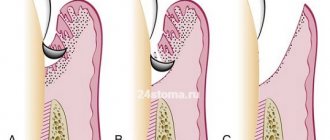
- Wedge-shaped defect – this term characterizes the pathological process in which the area of the dental neck is exposed. In such cases, the thinnest and most vulnerable areas of the enamel layer, located at the base of the teeth, are negatively affected. In addition to visible receding gums, damage to the enamel is indicated by a change in its color, as well as an acute reaction to hot and cold.
Anatomy of primary teeth
The macroscopic anatomy of primary teeth is very similar to that of permanent teeth, with some differences between them outlined in this section.
Ernst Zurcher (1922), Walter Hess school in Zurich, carried out the first scientific work on pulp morphology in primary teeth.
The endodontic morphology of primary teeth is very similar to the endodontic morphology of permanent teeth, but is smaller in size. Primary teeth are usually shorter and smaller than permanent teeth; the roots are narrow, while the roots of permanent teeth are thicker, especially in the cervical third. However, the width of the crowns of primary teeth is more pronounced compared to their height. The roots of temporary molars, in addition to being thinner than permanent roots, diverge to the sides to ensure the eruption of permanent premolars, first of all during their formation, and then during eruption.
Primary upper and lower molars often have a fourth canal in the mesiobuccal root of the upper molar and in the distal root of the lower molar.
As the child grows, the length of the roots of primary teeth decreases due to physiological resorption (exfoliation) (Fig. 1.1 and 1.2). Sometimes resorption of the floor of the pulp chamber at the furcation precedes apical root resorption (Fig. 1.3ad).
Rice. 1.1 Primary upper molar: root resorption starting in the apical region (arrows)
Rice. 1.2. Primary lower molar: root resorption affecting all parts of the root from apex to crown
Rice. 1.3. Primary upper molar: radicular resorption is more obvious at the floor of the pulp chamber in the area of the furcation, which in this case precedes apical resorption; (a) root apices are intact; (b) resorption in the area of the furcation of the pulp chamber; (c) palatal root resorption (arrows); (d) mesial view of furation resorption (arrows)
Unlike permanent teeth, the loss of primary teeth explains why canal filling at the end of endodontic treatment requires the use of resorbable materials that allow gradual root resorption.
Strengthening the enamel of molars
There are more methods to preserve molars and maintain the condition of their enamel layer. Firstly, this is due to fewer contraindications for adults. Secondly, molars require long-term strengthening.
The main methods of strengthening the enamel of permanent teeth include:
- Drug therapy is based on the use of vitamin complexes containing vitamins of groups B6, B12, D. In addition, the patient is selected drugs that promote better absorption of calcium and fluoride by the body.
- Special gels and oral hygiene products – this technique uses specialized toothpastes and gels containing components necessary for teeth to strengthen and maintain the condition of the enamel layer. Also, teeth are subjected to unimaginative cleaning in a dental office.
- Mineralization and preventive cleaning – mineralization is performed using special means to increase the strength of the enamel and reduce its susceptibility to a number of negative factors. As for cleaning, such procedures are performed by dentists in the clinic using special equipment. During cleaning, plaque and tartar are eliminated , pathogenic bacteria and microorganisms that can harm the enamel layer are removed.
- Home prevention - to maintain healthy teeth and enamel, patients are advised to perform a light massage of the gums, enrich the diet with fresh vegetables and fruits rich in vitamins.
Author: Zhukov M.A.
Permanent teeth
Macroscopic anatomy
Human permanent teeth consist of a crown and one or more roots. The endodontic space, created by the dentin of the root and pulp chamber, is very complex and is classified differently depending on the crown-root relationship and canal morphology.
Weine (1996) classified the root canal system into four types when considering the relationships between the pulp chamber, root canals and their apical termination (Fig. 1.4).
Rice. 1.4. Weine's classification of the root canal system takes into account the relationship between the pulp chamber, root canals and their apical termination.
Vertucci (1984) identified eight main types. Later, the Vertucci classification was expanded to include other morphological classifications by Gulabivala et al. (2001 and 2002) and Sert and Bayirili (2004) (Figures 1.5, 1.6 and 1.7).
Rice. 1.5 and 1.6 Graphic display of the morphological classification of Gulabivala endodontic system
Rice. 1.7 Graphic display of the morphological classification of Sert and Bayirili endodontic system
Schneider analyzed single-rooted human teeth and classified them according to the degree of root curvature as straight, with a curvature less than or equal to 5°, with a moderate curvature between 10° and 20°, and with a strong curvature between 25° and 70°. Lautrou [1987] also described and classified various morphologies of root cross sections (Fig. 1.8).
Rice. 1.8 Graphical representation of Lautrou classification of morphology in root cross sections
Zidell (1985) and Ingle and Taintor (1985), in addition to the degree of root and apical curvature, also considered the complexity of the anatomy, including the presence of bifurcations, the presence of accessory canals, and the presence of lateral and accessory canals.
Various studies and texts later described the anatomy and morphology of permanent human teeth and their countless possible shapes.
In paragraphs about a particular tooth, the age of eruption considered is according to Logan and Kronfeld, slightly modified by Schour and included in the text by Ash. The dimensions of human teeth described in this textbook are instead taken from various texts.
International Viola system: a convenient diagram of the arrangement of a person’s teeth by numbers
The described method is very convenient and most common in dentistry. It has received international recognition and has been generally accepted among dentists since 1971, called the two-digit Viola system. The convenience of such a system lies primarily in the fact that there is no need to create a special map of a person’s teeth, the numbering is easily calculated in the mind and information about the condition of certain dental units of the patient can be easily conveyed in an oral conversation, by telephone or by e-mail.
However, many people, having read this far, may say: but our teeth were counted completely differently, and the map contains completely different designations! That’s right, because in addition to the Viola system, there are several other systems that can be used by dentists.
Upper incisors
Central upper incisor
The central upper incisor erupts (one per quadrant) between the ages of six and seven years, and the complete formation of the apical third occurs after 2 or 3 years. The average tooth length is 22-23 mm.
The crown has a triangular shape, about 10.5 mm long, the base extends in the mesial-distal direction, corresponding to the anterior edge of the tooth, up to 9 mm in size, and its buccal-palatal size is 7 mm (Fig. 1.9).
Rice. 1.9 Upper central incisors: palatal view of the crown
The root is usually straight (75%), but according to Ingle, a slight curvature may be present in a small percentage of cases. Lateral canals may be present in more than 20% of cases, and an apical delta is also common (35%).
The coronal pulp space also has a triangular shape, especially in the region of the cervical radicular third, with the base facing the vestibular wall and the apex located palatally, after which it gradually becomes a round canal until the apex.
The access cavity is triangular and follows the shape of the pulp space (Fig. 1.10).
Rice. 1.10 Upper central incisor: graphical representation of the access cavity
Lateral upper incisor
The eruption of the upper lateral incisor (one in the quadrant) occurs 1 year after the central one, and its complete formation takes approximately 3 years.
It is approximately 1 or 2 mm shorter than the central incisor, its mesial-distal size is smaller - about 7 mm, and the vestibular-palatal size is only 5.5-6 mm (Fig. 1.11).
Rice. 1.11 Upper lateral incisor: palatal view of the crown
Normally there is only one root canal, straight in 30% of cases, and often it is curved distally (53%), in a small percentage of cases there is curvature in other directions. The shape of the canal is ovoid in the cervical region, with a tendency to become more rounded in the apical region; the access cavity has a similar structure (Fig. 1.12).
Rice. 1.12 Upper lateral incisor: graphical representation of the access cavity
Bite formation
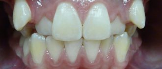
Occlusion is the relationship of the dentition. Normally, the upper arch covers the lower one by a third.
Correct bite:
- Orthognathic bite. Provides uniform distribution of chewing load. Fully performs speech and chewing functions.
- A direct bite occurs when the cutting surfaces of the incisors of two jaws meet.
- A progenic bite is characterized by a slightly protruded jaw.
- Biprognathic. The incisors of both jaws are inclined towards the lips.
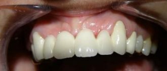
Malocclusion:
- Distal bite. There is underdevelopment of the lower jaw.
- Mesial. The bottom row is strongly pushed forward.
- Open. There is non-closure of the chewing surfaces in the frontal or lateral parts.
- Deep. The upper incisors half cover the lower ones.
- Cross. The intersection of rows in the shape of scissors.
- Reducing. Formed during pathological abrasion of teeth.
The main cause of malocclusion is genetic predisposition. Pathologies during pregnancy, metabolic disorders, anemia, viral infections, and teething of eights also play an important role.
Lower incisors
The lower incisors, two per quadrant, are very similar to each other, with some peculiarity retained.
Lower central incisors
The lower central incisors are the first permanent teeth to emerge in children's mouths, usually between 6 and 7 years of age, and are fully formed by 9 or 10 years of age. The lower central incisor is approximately 21.5 mm long. The apical third of the root is straight in 60% of cases and curves distally in 23% of cases.
The crown has a trapezoidal shape with a large base corresponding to the cutting edge and a smaller base that continues into the cervical third of the root. Its mesio-distal size is 5.5 mm at the greatest width of the incisal edge and gradually decreases to 3 mm at the cervical level (Fig. 1.13).
Rice. 1.13 Lower incisors: lingual view of crowns
The lower central incisor has a buccal inclination. In the root, it is possible to have a second canal located lingually in relation to the main canal in 18-23% of cases (Fig. 1.14).
Fig 1.14 Lower central incisor: graphical representation of the access cavity
Lower lateral incisor
The lower lateral incisor is similar to the central incisor, but slightly larger - about 1 mm, usually erupts 1 year after the lower central incisor and completes its formation after 3 years.
Root examination confirms the presence of two canals in 15% of cases.
The access cavity of the lower incisors is formed in a buccal-lingual direction to search for a second canal located lingually (Fig. 1.15a-c).
Rice. 1.15 (a - c) Lower central incisor: buccal, lingual and proximal views
Fangs
Upper canine
The upper canine, one per quadrant, erupts at approximately 11-12 years of age and completes its formation after 3-4 years.
This is the longest tooth in the arch (about 27 mm or more).
The crown has a diamond shape with a peculiar sharp cusp, which on the vestibular side divides the mesial and distal sides of the crown. The crown is 10 mm high (length) and has a maximum mesial-distal diameter of 7.5 mm at the point of proximal contact and decreases to 5.5 mm cervically. The buccal-palatal diameter is wider (8 mm), and it remains up to 7 mm at the cervical level due to the presence of a prominent marginal ridge (Fig. 1.16).
Rice. 1.16. Upper canine: palatal view
The long root, like the crown, is narrow in the mesial-distal direction and more pronounced in the buccal-palatal direction. There is almost always only one root canal with lateral canals at different levels in 24% of cases. The apical third is straight in 40% of cases, and is often curved distally (32%) and/or vestibularly (13%) (Fig. 1.17).
Rice. 1.17 Upper canine: graphical representation of the access cavity
The pulp space at the level of the cervical third and the root middle third has an oval shape in the buccal-palatal direction; often two canals are present with a tendency to merge into one common round canal in the apical third (Fig. 1.18a, b).
Rice. 1.18 (a, b) Upper canine: buccal and proximal view
The endodontic access cavity is oval-shaped, extending from the cusp to the marginal ridge of the coronal cervical third to gain access to the radicular space without obstruction that could cause instruments to create steps or transposition of the apical foramen (see Fig. 1.19).
Rice. 1.19. Anatomical pictures from Hess and Keller: the endodontic space of the upper canine at the level of the cervical third of the canal is represented by one oval canal, and in the middle third it is divided into two different canals and often merges into one round canal in the apical third
Lower canine
The lower canine, one per quadrant, erupts approximately 8-10 months before the upper one and completes its formation after 3-4 years.
The crown has a length of 11 mm, and its mesiodistal diameter is narrower than the upper one (7 mm), and decreases to 5.5 mm at the cervical level. The maximum buccolingual diameter is 7.5 mm wide and decreases to a minimum at the cervical level. The cervical lingual marginal ridge is less pronounced than that of the upper canine (Fig. 1.20).
Rice. 1.20 Lower canine: lingual view of the crown
The lower canine is about 27 mm long, and has two separate roots in 6% of cases, or two canals in one root, connected by isthmuses, with a common or two separate apexes (Fig. 1.21). In 20% of cases, the apical third has a distal slope (Fig. 1.22).
Rice. 1.21 Lower canine: graphical representation of the access cavity
Rice. 1.22. Anatomical pictures from Hess and Keller: the endodontic space of the lower canine has two separate canals connected by an isthmus in the coronal third; the canals merge into one opening in the apical third
The endodontic space is oval in shape in the bucco-lingual direction for two-thirds of the root, and then gradually becomes rounded.
The endodontic access cavity should be oval in shape in a buccolingual direction, from the cusp to the marginal ridge (Fig. 1.23a, b).
Rice. 1.23 (a, b) Lower canine: buccal and proximal view
Upper premolars
Two per quadrant, premolars erupt between 10 and 12 years of age, and root formation is completed after 3 years. They replace temporary molars.
The first and second premolars have a similar crown but different morphology.
Upper first premolar
The first upper premolar erupts when the child is about 10-11 years old. It has a length of about 21-22 mm, and its buccal-palatal dimensions are 9 mm and mesial-distal dimensions are 7 mm. It has two cusps, buccal and palatine, which are slightly shorter (about 1 mm). The pulp space is determined by the shape and size of the outer crown (Fig. 1.24). In approximately 72% of cases, two roots with two different apical foramina are present (Fig. 1.25). Premolars may also have only one root with two canals (13%) (Fig. 1.26a, b), and in some cases three roots (6%) (Fig. 1.27). In addition, in 37% of cases we found a distal root bend in the apical third.
Rice. 1.24 First upper premolar: occlusal view
Rice. 1.25 Upper premolars: graphical representation of the access cavity; note the mesial position of the second premolar access cavity (right in the figure) to facilitate passage of the distally curved canal
Rice. 1.26 (a, b) Upper single-root premolar: buccal and proximal view
Rice. 1.27 Anatomical pictures from Hess and Keller: complex pulp space of an upper single-rooted premolar; two different channels have multiple messages along the root and two separate outputs
The access cavity to the pulp chamber should have an oval shape in the buccal-palatal direction, from one tubercle to another Fig. 1.28.
Rice. 1.28 Anatomical pictures from Hess and Keller: an upper premolar with three roots, two buccal and one longer palatal
Upper second premolar
The second upper premolar erupts between the ages of 10 and 12 years. It is very similar to the upper first premolar, both in size and crown shape. But there are some main differences between the roots, presented in three versions:
One root with one canal in 75% of cases (Fig. 1.29a, b)
Rice. 1.29 (a, b) Upper first premolar: mesial and distal view
One root with two canals and one or two separate apical foramina (12%) (Fig. 1.30 and 1.31)
Rice. 1.30. Anatomical pictures from Hess and Keller: an upper single-rooted premolar having two canals merging into one exit; lateral canals are also present in the apical and middle third of the canal
Rice. 1.31 Anatomical pictures from Hess and Keller: upper single-rooted premolars have two canals with separate exits, apical and lateral
Two separate roots and canals (12%)
Three separate roots (usually two of them buccal) with three canals (1%)
On the buccal side, the roots have a distal curvature in 27% of cases, and a vestibular curvature in 12% of cases, and in 20% of cases they have two sharp curvatures.
The endodontic access cavity to the pulp chamber has an oval shape in the buccal-palatal direction. If there is significant distal apical curvature, the approach should be moved closer to the mesial marginal ridge, maintaining an oval shape (Fig. 1.28).
What is tooth numbering and why is it used?
If your doctor tells you that there is a problem with one of them, you will most likely want to understand which one. Of course, he can show it to you using a mirror or otherwise explain its location. But entering information related to diagnosis and treatment into the patient’s record requires greater accuracy and a professional approach. The same can be said if a doctor orders an x-ray - the x-ray room worker must know which area needs to be examined. This is what tooth numbers are used for.
Normally, an adult has 32 of them (if there are wisdom teeth), and a child has 20. Each of them takes its place in the jaw, belonging to a specific type. Numbering implies that each has its own serial number, by which a specialist accurately determines its location.
There are various systems, most often used in dentistry, and each of them involves its own algorithm for generating a serial number.
Lower premolars
Two in each quadrant, they erupt between 10 and 12 years of age, with full root formation after about 3 years. They replace primary molars. In contrast, the upper premolars are distinct from each other (Fig. 1.32).
Fig 1.32 Lower premolars: occlusal view
Lower first premolar
The lower premolar erupts sometimes several months before and sometimes after the lower canine and is the smallest of all premolars. The crown has two cusps, one very large and similar to the cusp of a canine tooth. The other is shorter by about 2 mm, similar to the lingual marginal ridge of the canine (Fig. 1.33).
Figure 1.33 Lower premolars: graphical representation of the access cavity
Length is about 21-22 mm, usually has one root with one canal (73-74%). Sometimes one root with two canals is possible (19%) (Fig. 1.34a, b), two roots with two canals (6%) (Fig. 1.35a, b) or three canals (1-2%).
Rice. 1.34 (a, b) First lower premolar: occlusal and proximal view
Rice. 1.35 (a, b) Second lower premolar: (a) coronal view; (b) Proximal view showing the presence of a single root
The root is often distally curved in 35% of cases and with a sharp curve in 7% of cases. The shape of the access cavity to the pulp chamber is oval, extending from the main tubercle to the tip of the minor lingual tubercle (Fig. 1.36).
Figure 1.36 Anatomical pictures from Hess and Keller: a mandibular premolar with a complex canal system that has two main canals connected by several fins and two separate exits
Lower second premolar
The second lower premolar erupts at the age of 11-12 years and is larger than the first premolar by 1 or 2 mm. The crown has two cusps (a larger buccal and a smaller lingual), but the lingual cusp is often divided into two parts (Fig. 1.37).
Rice. 1.37 Anatomical pictures from Hess and Keller: lower premolar with two roots and two canals; several short lateral canals present in the apical third
It has only one root with one canal in 85% of cases, but we can also find two separate canals in one root (11.5%) or two canals that merge into one apical foramen (1.5%) (Fig. 1.37 ). Rarely three channels are possible (0.5-1%). In approximately 40% of cases the root is straight, while in approximately 40% the apical third is distally curved. Severe curvature (7%) and vestibular curvature (10%) are possible.
The access cavity has an oval shape, also in the buccal-lingual direction, located in the center of the occlusal surface (Fig. 1.36).
Upper molars
Three molars in a quadrant without corresponding primary teeth.
Upper first molar
It erupts between 6 and 7 years behind the second primary molar. The formation of the apex is completed between the ages of 9 and 10 years.
The crown has four cusps on the occlusal surface and three roots, one palatal and two vestibular (Fig. 1.38).
Rice. 1.38 First upper molar: occlusal view
The length of the tooth is about 21-22 mm and there is one canal in the palatal and disto-buccal roots
The mesiobuccal root has two separate canals in 60-70% of cases (MB1 and MB2). The second canal in the distobuccal root (DB2) is quite rare (2-3%) (Figs. 1.39, 1.40 and 1.41).
The access cavity to the pulp chamber has a triangular shape with the base facing the buccal side and the apex facing the mesial-palatal tubercle; the cavity is always located mesial (in front) of the transverse ridge, which unites the mesiopalatine tubercle with the distal buccal tubercle. The mouths of the canals are located at the vertices of the triangle, which form the access cavity (Fig. 1.42); if we combine the orifice of the MB1 canal with the palatal orifice, we can find a second mesiobuccal root canal (MB2), which is often closed by calcification (Fig. 1.43).
Rice. 1.39 Anatomical pictures from Hess and Keller: the upper molar has a very complex canal anatomy
Rice. 1.40 Anatomical pictures from Hess and Keller: the upper molar has a very complex canal anatomy; MB1 and MB2 visible in the mesiobuccal root
Rice. 1.41 Anatomical pictures from Hess and Keller: the upper molar has a very complex canal anatomy; MB1 and MB2 visible in the mesiobuccal root
Rice. 1.42 Upper first molar: triangular access cavity with three canals
Rice. 1.43 Upper first molar: access cavity extended to the mesial ridge to locate the MB2 canal on the line connecting the mesiobuccal and palatal canal
Second upper molar
Eruption occurs at the age of 12 years with the completion of the apical third at approximately 14 years.
It is smaller and shorter than the first molar by about 1-2 mm, is about 19-20 mm high and has four occlusal cusps. There are three roots and three canals, just like the first molar, and the MB root also has a second canal (37-42%). The shape of the pulp chamber is rhomboidal rather than triangular (see Figs. 1.42 and 1.43).
Upper third molar
This tooth erupts after age 17. It has a very unique anatomical morphology; the crown has three or five tubercles (Fig. 144). The average height of the third molar is about 18 mm. There may be one root or more often than one - three or four roots, usually curved distally (Figs. 1.45, 1.46, 1.47 and 1.48).
Figure 1.44. The upper third molars have a very distinctive occlusal morphology with three or four cusps
Rice. 1.45. Upper single root third molar
Rice. 1.46. Upper third molar with four distally curved canals
Rice. 1.47. Upper third molar with atypical anatomy
Rice. 1.48. Upper third molar with strong 3D root curvature
Lower molars
Three molars in a quadrant without corresponding primary teeth.
First lower molar
This is the first permanent tooth that erupts at the age of 6 years and completes its formation at approximately 9-10 years. The crown has five cusps, two lingual and three buccal (Fig. 1.49). 22-23 mm long. There are usually two roots, one mesial and one distal (97%), and we rarely find a third distolingual root (3%). In the mesial root we find two canals in 65% of cases: they may have only one apical foramen (40%) or two separate ones (60%). The distal root has one canal (70%) or two canals (30%); there may also be two separate apical foramina (38%) or only one (62%) (Figs. 1.50, 1.51 and 1.52) (Figs. 1.53 and 1.54).
Rice. 1.49. Lower first molar: occlusal view
Rice. 1.50 Anatomical pictures from Hess and Keller: lower molar with very complex canal anatomy
Rice. 1.51 Anatomical pictures from Hess and Keller: lower molar with very complex canal anatomy
Rice. 1.52 Anatomical pictures from Hess and Keller: lower molar with very complex canal anatomy
Rice. 1.53. Micro-computed tomography scan of the roots of lower molars: note the complex morphology of the pulp space
Rice. 1.54. Micro-computed tomography scan of the roots of lower molars: modern digital techniques reproduce the subtle and complex morphology of the pulp space previously illustrated by Hess histological images
All roots on the buccal side have a moderate distal slope. The pulp chamber is triangular or square, depending on the number of canals (3 or 4), located in the center of the crown in the mesial-lingual area. Because of this feature, during the formation of the pulp chamber access cavity, it is important to be careful not to remove precious tooth structure in the distal area.
The shape of the access cavity should be triangular or trapezoidal with the larger base facing the mesial ridge and the smaller (or triangular apex if there is only one distal canal) slightly distal to the central occlusal fossa (Figs. 1.55 and 1.56).
Rice. 1.55 Lower molar: access cavity to three canals
Rice. 1.56 Lower molar: trapezoidal access cavity to four canals
If there is a third root, its mouth can be found along the bottom of the pulp chamber in the corners of the base of the trapezium.
Second lower molar
The tooth erupts at the age of 11 years and is very similar to the lower first molar with some differences.
It is smaller than the first molar by 1-2 mm in each direction. It has four cusps on the occlusal surface and usually two roots, one mesial and one distal. The mesial root has only one canal in 13% of cases; most often there are two channels that end in only one opening (49%), or there are two channels with two independent openings (38%). The distal root has only one canal in 92% of cases. Rarely there are two canals with one apical foramen (5%) or two separate foramina (3%).
The access cavity to the pulp chamber is located in the middle of the mesial-lingual region and has a trapezoidal shape. Due to the size of the pulp chamber and its proximity to the mesial-lingual wall, we advise careful design of the access cavity to avoid unnecessary excision of tooth structure (see Figures 1.55 and 1.56).
Third lower molar
Erupts after 17 years, usually at the age of 25. Also called "wisdom teeth", they have an atypical and varied morphology. It can have from three to five tubercles (Fig. 1.57). Often there is a fusion of roots from two or three roots. And, as a consequence, the anatomy of the canals cannot be schematized, as with the upper molar. Experience in root canal treatment will help the clinician perform the endodontic procedure correctly (Figs. 1.58, 1.59, and 1.60).
Rice. 1.57. The varied occlusal morphology of mandibular third molars may have four to five cusps
Rice. 1.58 (a) Lower third molar with a curved mesial root fused to the distal one. (b) Lower third molar: distal root
Rice. 1.59 (a) Lower third molar with four roots, two mesial and two distal: the mesiobuccal root is a very curved canal in the middle third. (b) Lower third molar: distal view showing graceful curvature of the distolingual root
Rice. 1.60 (a) Lower third molar: on the buccal side, two separate mesial roots are visible with extremely severe curvature, according to Schneider. The distal root has a moderate 3D curvature in the mesial and lingual direction. (b) Lower third molar: from the distal side, a 3D curvature of the distal root is visible in the lingual as well as mesial direction
Igor Lukinykh
Dental formula
At a dentist's appointment, everyone probably heard that teeth are called different numbers. What does this mean and what unit corresponds to a certain number? Numbering is used to enter information about the condition of the teeth and the treatment performed.
The countdown is performed from the middle of the row to the left and right sides. The front incisors are designated 1 and 2, respectively. Canines - 3, premolars - 4, 5, molars - 6, 7, 8 (wisdom teeth).
To determine in which half and on which jaw the required unit is located, the jaw is supposedly divided into 4 segments. The numbering in them starts from the right side of the upper arc. Teeth are written as a two-digit number, where the first is a serial number, and the second is a segment. The units of the upper jaw on the right (I segment) are designated from 11 to 18, the same jaw on the left from 21 to 28. In the lower left segment from 31 to 38, on the right from 41 to 48, respectively. The name of a specific unit uses a serial number and segment number.
In baby teeth, segments are counted from the upper left clockwise. Tens are used: 50, 60, 70, 80.
Dentists use several types of numbering:
The Zsigmondy-Palmer system is also called the square-digital system. Constant units are designated by Arabic numerals, and dairy units by Roman numerals. It is most often used by maxillofacial surgeons and orthodontists.- Haderup system. The symbols “+” and “-” are used to indicate the upper or lower arc. Arabic numbers are used to write teeth. When marking milk units, add 0.
- The Viola system is the designation of teeth in Arabic numerals from 1 to 8 and two-digit numbers of the corresponding segment. This formula is the most convenient and is widely used by dentists all over the world.
- Universal system. Each group of teeth is designated by a specific letter, serial number, or segment number.