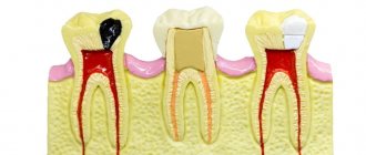
The root system of the tooth and knowledge of its anatomy are an important factor for successful endodontic treatment. The complex anatomy of the canals can significantly complicate treatment.
The main goal of endodontics is complete obturation of the root canal. For successful treatment, it is necessary to clean the canals and tooth cavity from necrotic debris, bacteria and their waste products. We can achieve this through careful mechanical and chemical treatment of the canals.
Persistence of infection due to a missed canal or incomplete treatment of a difficult-to-reach canal can lead to poor results. It is important to study the anatomy of the tooth before treatment to avoid complications.
Anatomy of mandibular molars
In the permanent dentition, the first molars of the mandible have the greatest chewing force and are therefore the most functionally significant. Most mandibular molars are double-rooted with two mesial and one distal canals. But the number of roots and canals may vary.
Additional root
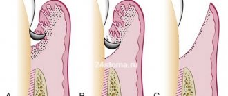
Carabelli was the first to note such a frequent change as an additional root. It is located on the lingual ( radix entomolaris) or buccal ( radix paramolaris ) sides.
Three-rooted mandibular first molars require special attention during endodontics because the accessory root is usually smaller and thinner than the others. Or such a root is partially fused with other roots and is strongly curved.
Identification
To identify additional roots, an x-ray is needed. But sometimes several pictures from different angles are necessary, since one picture may be uninformative.
Rice. 1 - SLOB method allows you to take pictures from different angles.
Rice. 2 – On the radiograph we see the accessory root (Radix Entomolaris) of the first molar of the lower jaw.
Rice. 3 – We see the mouth of the additional canal (Radix Entomolaris), located distally lingually. Asymmetry and deviation from the midline are noticeable.
Central lower incisor
Average age of eruption: 6-7 years
Average age of root formation: 9 years
Average length: 20.7 mm
Narrow and flat in the labial-lingual direction, the lower central incisor is the smallest adult tooth. Radiologically it is visible in only one projection and thus appears more accessible than it actually is. The crown, narrow in lingual projection, has a limited area for access. For gentle access formation, a fissure bur and a spherical bur No. 2 are used. The access cavity should be oval and performed on the lingual side.
The lower central incisor often has two canals. One study reported that 41.4% of mandibular central incisors studied had two separate canals, of which only 1.3% had two separate apical foramina [1]. After completing the access formation, the doctor must examine the tooth cavity to identify an additional canal. Endodontic treatment failures in the lower incisor area are most often associated with an unidentified canal, usually in the lingual location. To create conditions for the straight entry of endodontic instruments into the additional canal, access can be expanded in the incisal direction.
There is a great danger of perforation of the vestibular wall, but it can be avoided if the doctor remembers that it is almost impossible to perforate in the lingual direction, since the bur axis is in contact with the incisal edge. The slit-like lumen of the canal is so common that it can be considered a normal variant, and this requires special attention when cleaning and shaping.
Lateral perforations and pulp anatomy will be discussed in an illustration of the lower second molar.
Actions when changing teeth: how to help your child cope with this process easier
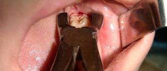
Parents often think that the process of changing baby teeth to permanent teeth is very painful for children. However, this is not the case. If this process is not interfered with, the roots of baby teeth gradually dissolve and the teeth may fall out even without outside help. Or, when it seems that the tooth is completely hanging, it can be easily removed.
In order to disinfect the oral cavity during the period of loss of baby teeth, you need to explain to the child that it is necessary to rinse the mouth. Rinsing can also be done with a special product, chamomile decoction or even plain warm water.
Sometimes it happens that after a tooth falls out, the place in which it was previously located (socket) bleeds. To get rid of this, you need to apply a cotton swab to the hole, or better yet, ask the child to clamp it with his teeth. It is undesirable to eat or drink for 2 hours after tooth loss, provided that the hole is bleeding.
You should immediately consult a doctor only if the loss of baby teeth is accompanied by high fever, swollen gums and severe pain. After all, normally, teeth change occurs almost asymptomatically.
Lateral lower incisor
Average age of teething: 7-8 years
Average age of root formation: 10 years
Average length: 21.1 mm
Very similar to the lower central incisor, so access cavity preparation follows the same principles.
Their similarity can cause rare but serious errors. Hasty installation of a rubber dam, identical fillings and carelessness can lead to preparation of the access cavity on the wrong tooth. This error can be prevented by marking the vestibular surface of the tooth with a felt-tip pen before applying the rubber dam.
Trauma, periodontal disease, carious lesions and malocclusion can lead to obliteration of the canal. When moving apically to identify the orifice, great care must be taken to prevent unnecessary destruction of the crown and root. Labial perforations were discussed in Illustration IX. If the bur is not oriented along the long axis of the tooth, there is a risk of lateral perforation. The situation becomes more complicated with the traumatic loss of an anatomical crown. Without anatomical landmarks, a lateral perforation can be easily made when moving in a coronal direction. To prevent this, access is carried out without a rubber dam so that the root can be palpated.
Lateral perforation with endodontic files and Gates-Glidden burs is facilitated by the presence of slit-shaped canals with a narrow, hourglass-shaped cross-section. To avoid vertical fracture along the approximal root wall, minimal expansion and preparation of the space for the abutment pin is indicated.
Apical curves and accessory canals are common in the lower incisors.
Mandibular canine
Average age of teething: 9-10 years
Average age of root formation: 13 years
Average length: 25.6 mm
The canine of the lower jaw is more powerful and significantly wider than the incisors in the mesial-distal direction. It rarely causes treatment problems. The atypical form with two roots can be problematic, but is rare.
The access cavity is oval and can be expanded in the incisal direction to facilitate vestibular-lingual access. In the cervical region the canal is oval, in the middle third it is rounded. To completely clean its walls, directed instrumental action is necessary.
If there are two roots, one of them will always be easier to instrument. The other canal must also be opened and funnel-shaped in accordance with the first to prevent dentine filings from entering it and impairing access. Pre-bending the instruments during the initial approach will allow the clinician to walk along the walls of the buccal or lingual root until the tip of the instrument enters the orifice. Once a difficult-to-reach canal has been identified, every effort must be made to shape and create a funnel-shaped orifice to keep access open.
First lower premolar
Average age of teething: 10-12 years
Average age of root formation: 12-13 years
Average length: 21.6 mm
Often considered a mystery to endodontists, the mandibular first premolar, with its two canals separating at different levels of the root, can be very difficult to machine.
The crown consists of a well-developed buccal cusp and a small or almost non-existent lingual enamel protrusion. The approach is performed buccally from the central sulcus and directed along the long axis of the root to the central cervical region. The oval-shaped pulp chamber is opened using fissure burs with a cutting apex and elongated spherical burs No. 4 or 6. In teeth with one canal, the pulp cavity in the neck area has an almost circular cross-section, and in teeth with two canals it is oval.
One study reported that “at least 23% of first mandibular premolars have a second or third canal”[17]. The canals can split almost anywhere in the root. Due to the lack of direct access, cleaning, shaping and filling these teeth can be extremely difficult.
In a recent study, Vertucci [13] showed that the first lower premolar has one canal at the apex in 74.0% of cases, two canals in 25.5% and three canals in the remaining 0.5% of cases.
Classification and frequency (%) of canal types in first and second lower premolars
By Vertucci, F. J. Am. Dent. Assoc. 97:47, 1978.
Second lower premolar
Average age of teething: 11-12 years
Average age of root formation: 13-14 years
Average length: 22.3 mm
The second lower premolar, which is very similar in crown shape to the first premolar, has a less complex root.
Its crown has a well-developed buccal cusp and a much better formed lingual cusp than on the first premolar. The access is made slightly oval, wider in the mesial-distal direction. They begin to form access in the central sulcus with a fissure bur with a cutting apex, and then expand and form the contour of the burr hole with spherical burs No. 4 and 6.
Researchers reported that only 12% of mandibular second premolars studied had a second or third canal [17]. Vertucci [13] also showed that second premolars had one apical foramen in 97.5%, while only 2.5% of the teeth examined had two foramina.
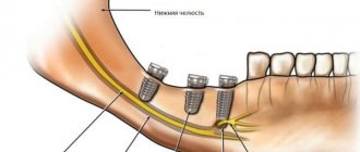
An important circumstance that should not be forgotten is the anatomical location of the mental foramen and the vessels and nerves passing through it. Due to the proximity of these structures, an acute inflammatory process in the area of the lower premolars can cause temporary paresthesia. The exacerbation of the pathological process in this area is more severe and resistant to conservative treatment than in other areas.
Differences in children
Milk and permanent kits are very different from each other. This applies to the structure of the enamel, the quantity, and the presence of individual groups as such. For example, there are simply no premolars in the milk set, so parents who are trying to control the eruption of young children according to the scheme of the adult dental apparatus may be very surprised. The number of teeth in the primary and permanent sets is different precisely because of the premolars.
They are absent throughout early childhood and erupt only after the first molar falls out, closer to 10-11 years of age. By this point, there is more space on the jaw, so all elements of the dentition can be arranged normally.
First lower molar
Average age of teething: 6 years
Average age of root formation: 9-10 years
Average length: 21.0mm
The mandibular first molar erupts earlier than other permanent teeth and most often requires endodontic treatment. It usually has two roots, but sometimes three roots are found, with two canals in the mesial root and one or two canals in the distal root.
The distal root is easily accessible for endodontic cavity preparation and mechanical treatment.
The doctor can directly see the opening(s) of the canal. The distal root canals are wider than those of the mesial root. Sometimes the mouth is wider in the buccolingual direction. This indicates the presence of two channels or a slit-like channel with a complex network-like configuration that can complicate cleaning and shaping.
The mesial roots are usually curved, with the greatest curve in the mesiobuccal canal. The orifices of the canals at the bottom of the pulp chamber are usually clearly separated from each other and located buccally and lingually relative to the upper tubercles.
The tooth often undergoes extensive filling. It almost always experiences a strong chewing load, so the coronal pulp cavity can be obliterated. It is easiest to identify the mouths of the distal canals.
Then the mouths of the mesial canals are found, which will be located in the above locations in the same horizontal plane.
Since the mouths of the mesial canals lie under the mesial tubercles, they may not be detected during normal cavity preparation. In this case, to determine their location, it is necessary to remove the hard tissue of the tubercle or filling. During access preparation, overhanging molar cusps need to be ground down [15]. Remember that this tooth, like all other lateral teeth, after endodontic treatment requires complete restoration of the entire occlusal surface area. Therefore, to identify anatomical landmarks and orifices, it is better to make a wider access cavity than to skip one or more channels for the sake of “sparing” preparation, which may cause failure.
Skidmore and Bjoradal [11] found that approximately one third of the mandibular first molars examined had four root canals. If there are two canals, “they either remain separate with separate apical foramina, or unite and form a common apical foramen, or communicate with one another by transverse anastomoses partially or completely... If the tooth, instead of the usual triangular shape, had a more rectangular shape, this would allow better vision and explore a possible fourth canal in the distal root.”
In the area of bifurcation of the roots of the lower molars there are several orifices of additional canals [9]. They are usually impossible to clean and shape, and are rarely visible except incidentally on radiographs when they are filled with root cement or heated gutta-percha during treatment. It would be correct to assume that if irrigation solutions tend to clean the canal from protein decomposition products, then the area of root bifurcation in the pulp chamber must be thoroughly cleaned (remove denticles, etc.) so that the solutions can reach the small mouths of the canals.
All infected dentin, leaking fillings, and pulp denticles must be removed before endodontic treatment begins. It is recommended to completely cover the cusps with a restorative structure after endodontic treatment.
What are the symptoms of eruption of molars?
When teething, your baby may experience:
- swelling and redness of the gums
- increase in body temperature
- change in stool
- increased sensitivity and irritability
- painful sensations arise
The child becomes more restless and puts his fingers or nearby objects into his mouth. You can alleviate the child’s condition by letting him chew solid food (a piece of apple, carrot, crackers). You can also purchase special teethers and gels that have an analgesic and cooling effect.
Find out about the benefits of the Medissa Art clinic here>>>
Second lower molar
Average age of teething: 11-13 years
Average age of root formation: 14-15 years
Average length: 19.8 mm
The crown is somewhat smaller in size than the first molar, and the more symmetrical second lower molar is characterized by a close arrangement of roots. The roots gradually converge distally and have closely spaced apices.
Exposure is made at the mesial portion of the crown with access extending only slightly distal to the central sulcus. After trephination of the cavity with a fissure bur with a cutting apex, an elongated ball-shaped bur is used to expand it until free access is achieved. The distal root angles often allow for a smaller opening than the lower first molar.
Particular attention should be paid to the shape of the mouth of the distal canal. A narrow oval orifice indicates the presence of a slit-like lumen in the distal canal, which requires more careful treatment.
All carious tissue, leaking fillings and denticles should be removed and replaced with a suitable temporary filling prior to endodontic treatment.
The lower second molar is most susceptible to vertical fractures. After preparing the access, but before starting endodontic treatment, the clinician should examine the floor of the pulp cavity using fiber optics. For mesial-distal crown or root fractures, the prognosis is extremely unfavorable.
To avoid vertical fractures after endodontic treatment, it is necessary to completely cover the cusps with a restorative structure.
Can milk root remain in the gums?
If you carefully examine fallen children's teeth, you will not be able to see any semblance of roots. Some mothers unknowingly begin to panic - it seems to them that a significant part of the unit remains in the deep tissues of the gums.
There is no need to worry - this is how it should be. The absence of roots is the result of their gradual resorption. This process starts long before the day of loss. That is why during a natural change (when a tooth falls out without outside help), the child does not experience pain.