CHEWING MUSCLES
- a group of muscles, the contraction of which moves the lower jaw in directions that ensure chewing.
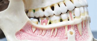
Topographically, this group of muscles includes some muscles of the head (masseter proper, temporalis, lateral and medial pterygoid muscles - Fig. 1) and neck muscles located above the hyoid bone (mylohyoid, geniohyoid and digastric muscles). Rice. 1. Chewing muscles: a - temporal and chewing muscles; b - pterygoid muscles; 1 - temporal muscle; 2 - the masticatory muscle itself: 3 - the lateral pterygoid muscle; 4 - medial pterygoid muscle; 5 - pterygomandibular suture; 6 - buccal muscle.
Purpose of the dental system
The biological mechanics of the dental system and its understanding lead to timely recognition of pathologies of teeth and soft tissues of the oral cavity. The normal functioning of the temporomandibular joint largely depends on the structure and normal functioning of the periodontium - the soft tissues surrounding the tooth cavity.
The anatomy and physiology of the structure of the periodontium determines the biomechanics of the structure and the characteristics of the periodontium. In addition, periodontal biomechanics helps in the functioning of other organs.
Knowledge of the laws of biological mechanics of the jaw and joints helps to design and use features, as fundamental knowledge on the topic of prosthetics and dental implants. Knowledge of the anatomical features of the jaw is necessary in the manufacture of structures and dental materials.
Devices have been developed that almost completely repeat the movements of the jaw and make it possible to reproduce anatomically similar structures to the human jaw. Popular devices include the occluder, facebow, and articulator. The latter design plays an important role in the fitting and creation of individual prostheses.
If disorders of the lower jaw occur, disruptions in articulation, speech, nutrition and swallowing occur. Today, a system of articulation of movements has been developed, led by such authors as Ganau, Gisi, Monson. These authors most accurately described the biomechanics of mandibular movements.
The authors claim that dysfunction of the lower jaw, as well as sore neck muscles, necessarily lead to breathing problems. Different angles of inclination of the lower jaw determine human health.
Occlusion is a condition characterized by full contact of both jaws, which ensures complete chewing of food. Therefore, the contact of teeth is a determining factor for the characteristics of chewing mechanisms.
The work and action of the lower dentition during chewing is due to the synchronous work of all muscles and joints. The work occurs under the influence of the central nervous system. In this case, displacements that occur spontaneously occur under the influence of the neuromuscular structure.
Conscious movements of the dentition include the moment food enters the mouth and swallowing food. The remaining movements that occur after eating food are the result of unconscious motor functions of the body.
The lower part of the human masticatory apparatus is the only moving bone in the human body. However, the connecting and muscle tissue, which sets the mechanism in motion, is of great importance in the structure of the masticatory row and in the normal functioning of the muscles.
In the structure of the lower dentition, there are several muscle groups that are responsible for various movements of the jaw.
Treatment
Treatment of patol, conditions of glandular m. consists of treating the underlying disease (infectious disease of the nervous system, injury, tumor); with true hypertrophy of the t. masseter - orthodontic treatment (see Orthodontic methods of treatment) in order to eliminate malocclusions; with pronounced hypertrophy, resulting in facial asymmetry, partial surgical excision of the hypertrophied muscle is possible; if a tumor localized in the area of the gastrointestinal tract is detected, appropriate treatment is given.
Bibliography:
Vorobiev V. and Yasvoin G. Anatomy, histology and embryology of the oral cavity and teeth, p. 119, M., 1936; Gorenshtein Ya. I. On hypertrophy of the masticatory muscles, Dentistry, No. 4, p. 87, 1965; Ivanitsky M. F. Human Anatomy, vol. 1, p. 379, M., 1965; L e r n e r I. O. Hypertrophy of the masticatory muscles, Dentistry, No. 2, p. 40, 1960, bibliogr.; LimbergA. A. Vascular tumor with multiple stones in the thickness of the masticatory muscle, ibid., No. 4, p. 90, 1965; Morphology of the maxillo-mandibular apparatus, Proc. symp. 9th. Int. congr. Anat., Lpz., 1972; S i with her H. Oral anatomy, St Louis, 1965.
HH Mosolov, V. M. Bezrukov.
Directions of movement of the lower jaw
During the movement of the lower jaw, sagittal, vertical and transversal displacement of its heads occurs.
Vertical displacement is necessary to open and close the mouth. In this case, the movement, depending on the force, occurs with varying strength, which determines the degree of movement of the head.
The sagittal axis of displacement is the ability to move the lower jaw back and forth. In this case, the forward movement of the jaw and the biomechanism of the head of the articular part at the moment of movement is called the sagittal path of the joints.
Transverse is the ability of the jaw to move left or right. In this case, certain muscle groups move. For example, the direction of the masticatory apparatus to the right determines the left direction of the lateral muscles, and vice versa.
Without knowledge of how muscles and joints work, the dentist cannot regulate the normal functioning of the masticatory apparatus, which means he will not be able to manufacture and install suitable dentures and implants.
Wearing the device and care
The child can put on and take off the device independently, but this is not only a plus, because the result of the treatment begins to greatly depend on the child’s discipline.
It is easy to care for the device. No special cleaning products are required: regular toothpaste and a brush will do.
A temporary storage container is provided with the Twin block. And of course, the orthodontist provides instructions on how to wear the device, care, and issues a reminder “what to do if.”
TMJ functioning problems
| Click to sign up for a FREE consultation |
Disruption of the work and function of the joints and muscles of the lower jaw is called TMJ dysfunction. The pathology is caused by the occurrence of a defect in the teeth, increased wear and tear and an abnormal bite of a person.
The main clinical manifestation of dysfunction is clicking and clattering of teeth when opening the mouth. In more advanced cases, pain occurs in the ears, jaw, and painful sensations begin during chewing and yawning.
The main method for diagnosing TMJ dysfunction is radiography, which reveals dysfunction of the jaw and joint.
Treatment consists of a comprehensive effect on the condition of the jaw, which the dentist carries out based on knowledge of the biomechanics of its work.
Pathology of the masticatory muscles
Pathology of the masticatory muscles can manifest itself in the form of dysfunction - paresis, paralysis; for example, with damage to the trigeminal nerve or its nucleus, atrophic paralysis of the stomach is observed. With unilateral damage to the trigeminal nerve, chewing, although difficult, is possible due to the healthy side. With bilateral atrophic paralysis of the stomach, chewing is impossible, the lower jaw droops. This picture can be observed in amyotrophic lateral sclerosis, when the pyramidal tracts and nuclei of the motor cranial nerves are affected. The gland can also be affected by tick-borne encephalitis. The function of the stomach m. is sharply disrupted by trismus (see) - a tonic spasm of the stomach m., which can be caused by an inflammatory process in the lower jaw or in the soft tissues adjacent to the area of location or attachment of the stomach m. Spasm of the stomach m. .—a characteristic symptom of tetanus, can be observed with meningitis, in some cases - as a hysterical reaction.
Rice. 2. True bilateral hypertrophy of the masticatory muscles themselves.
Hypertrophy of the m. is rarely observed, and unilateral hypertrophy of the m. is more common. masseter There are so-called true and false hypertrophy of m. masseter False hypertrophy is the development of lymphoid tissue or a vascular tumor in the area of the masticatory muscle. True hypertrophy of the stomach m. has not been sufficiently studied. Occasionally it is observed with malocclusion. Clinically, hypertrophy manifests itself only as a violation of the configuration of the face (Fig. 2); on the side of hypertrophy, the shape of the angle of the lower jaw can also be changed. It is necessary to differentiate true hypertrophy from benign neoplasms in the area of the gastrointestinal tract (lymphoma, lipoma).
G. m. are involved in patol, the process of jaw injuries, facial wounds, specific inflammatory processes (actinomycosis), as well as malignant tumors on the face.
How often should you visit the orthodontist during treatment and then what?
You need to come to the clinic for device correction on average once every 6 weeks. The correction consists of freeing up space in the plates for the eruption of permanent teeth.
After completing treatment, it is recommended to visit the orthodontist for the first time after six months.
Whether the patient will need orthodontic treatment in the future depends on the stability of the result obtained, which is influenced by many factors, one of which is a possible growth spurt in the child.
Our story has come to an end.
Other examples of orthodontic treatment can be found on the personal page of Irina Konstantinovna Shevchenko.
You will get a lot of useful information from the articles:
- Orthodontics. Why, When and How to correct malocclusion
- A child’s beautiful smile is the care of loving parents
- Correction of distal and mesial bite using additional orthodontic equipment
If you decide to consult with an orthodontist, make an appointment with Irina Konstantinovna Shevchenko >>
Manifestations of distal bite in the mouth
Closing of lateral teeth during distal occlusion.
Distal occlusion is manifested in the mouth by the closure of the upper and lower teeth according to the “second class” (violation of the relationship between the projections of the antagonist teeth of the upper and lower jaw).
In the frontal segment (at the level of the front teeth) there are two closure options (the so-called first or second subclasses).
In the first case, a “sagittal gap” is observed, which reflects the discrepancy between the dentition in the sagittal plane (anterior-posterior direction). And the upper incisors are in protrusion (inclined forward). In the second option, the upper front teeth are tilted back (into retrusion). With the formation of a deep bite (when the lower teeth are not visible from under the upper ones).
Sagittal chain with distal occlusion. 2 class 1 subclass.
Distal occlusion. 2nd class 2nd subclass.
Causes of distal bite (distal occlusion)
They are almost always (in the vast majority of cases) skeletal...
- Posterior position of the lower jaw. This is the most common cause of distal occlusion
- Small (short) lower jaw.
Many people write that the cause of a distal bite may be an overdeveloped (excessively large) upper jaw. They say that this is why there is a “gap” between the front upper and lower teeth (sagittal gap)... Many years of observations and our experience have shown that this is not so. With any anomaly of occlusion, there can be no talk of any OVERdevelopment. Everything, on the contrary, is UNDERdeveloped.
The lower jaw ends up in a posterior position (the first reason) only because the lower teeth bump into the upper teeth while the upper jaw is shortened and narrowed (generally underdeveloped). Or, when the upper incisors are “wrapped” back, into retrusion (second subclass). In addition, with distal occlusion, the upper jaw is always narrower than the lower jaw. Which, by the way, is another reason for blocking the lower jaw in the posterior position. And this same “blocking” does not allow the lower jaw, which is caught in the trap, to develop normally. Here is the second reason for distal occlusion - underdevelopment of the lower jaw.
There is also a “dental” reason for distal occlusion. Or more precisely, the dental cause of closure is “second class”. This is when mesial (front) displacement of the upper lateral teeth occurs. And then, even with a normal relationship of the jaws in the sagittal plane, the closure of the lateral teeth (molars) will also be in the second class. True, this is not a true distal occlusion...
A “dental” cause of the second class may complement or may camouflage true distal occlusion. Therefore, it will not be possible to figure out what is the reason for the closure in class 2 visually (by eye). Diagnostics required.