- How the Mark Ross apparatus is installed and works
- Fixed plate installation process
- Advantages and effectiveness of a plate for maxillary expansion in children
- Why is it better to go to the Aesculapius clinic?
Plates for expanding the jaw for children are special dental devices for correcting the bite and straightening the dentition. The device is used in early childhood and is not removed throughout the treatment. Plates are complex orthodontic structures of different appearances, which are made according to individual impressions of the child’s jaw. Treatment with the apparatus of Dr. Marco Ross shows good results and effectively normalizes the shape of the upper jaw.
How to install and operate the Marco Ross apparatus
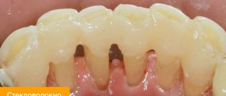
A non-removable plate for expanding the upper jaw for children is a plastic base connected to metal arches of various configurations. A screw with maximum potential for expanding the upper dentition is built into the product. It is this element that is the force that allows the plate to work to expand the child’s upper jaw. A plate is fixed to the upper jaw on the baby’s temporary molars and canines. For the former, with the help of orthodontic rings and metal arches, and for the latter, with dental material.
Expansion of the lower jaw
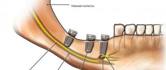
A narrow lower jaw, or microgenia, can be corrected in childhood with special devices - distractors. This is a device that gradually stretches the bones of the lower jaw. New bone and soft tissue grows at the site of the sprain. When installing a distractor, first increase the distance between the canines of the lower jaw. Every day the device allows you to increase the lower jaw by 1 mm. When the jaw is wide enough to accommodate tooth growth, the distractor is placed in place for at least two weeks to stabilize the sprain.
If you have a problem similar to that described in this article, be sure to contact our specialists. Don't diagnose yourself!
Why you should call us now:
- We will answer all your questions in 3 minutes
- Free consultation
- The average work experience of doctors is 12 years
- Convenient location of clinics
Single contact phone number: +7
Make an appointment
In adult patients, the lower jaw bones are fused and cannot be stretched, as in children. Therefore, to enlarge the lower jaw, the surgeon cuts the bone in several places with an ultrasonic knife. On the fourth day after surgery, an expansion device is installed. The first adjustment of the stretching is carried out by an orthodontist, then the patient himself controls the stretching process, which can take up to six months.
After expanding the lower jaw, braces are installed on the lower front teeth to correct the bite. Crowded teeth gradually occupy the resulting space.
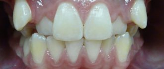
The photo below shows a narrow jaw before and after expansion.
Advantages and effectiveness of a plate for expanding the upper jaw in children
The huge advantage of orthodontic plates is that, despite their simplicity, they have a fairly wide range of uses at a low cost. A jaw expansion plate is recommended in cases such as:
- Narrowed upper teeth.
- There is not enough space for the upper and lower incisors.
- Malocclusion due to narrowing/shortening of the upper jaw.
- Difficulty in nasal breathing and non-closure of lips.
- Problems with diction associated with underdevelopment of the upper dentition.
At the same time, a plate for expanding a child’s jaw has a number of advantages compared to other methods:
- Treatment does not require the child's active cooperation.
- A fixed device cannot be forgotten or lost.
- The space for the eruption of molars is prepared.
- The likelihood that children will need braces in the future is greatly reduced.
Expanding the upper jaw in children is an effective method to take care of your baby’s oral health. Moreover, the procedure is completely safe and reliable, but only if carried out by professionals.
Reasons for the development of micrognathia
Anomalies of jaw development can be congenital or acquired.
Congenital ones include:
- Severe acute illnesses of a woman during pregnancy,
- Anomalies of intrauterine development of the fetus,
- Congenital pathologies of the development of the dental system,
- Genetic predisposition.
An acquired anomaly can result from:
- Maxillofacial trauma,
- Artificial feeding and improper sucking in infancy,
- Diseases of the endocrine system,
- Past infectious diseases and inflammatory processes,
- Early loss of baby teeth,
- Problems with nasal breathing.
Features of orthodontic treatment with the FAGGA device
There are two phases of treatment. The first is the expansion phase of the palate, during which bone growth occurs. The second is treatment with braces to close the gaps between the teeth and create proper interdental contacts. An orthodontist performing treatment with the FAGGA device must have extensive knowledge and experience, and be able to combine various equipment in order to achieve a successful and lasting result of bite correction. Dial-Dent orthodontists are sufficiently qualified to carry out complex treatment and have experience working with various orthodontic equipment.
Installation process
The Marco Rosa apparatus has four elements that are used to fix it to the jaw. Two rings (left and right) that fit on the molars, and two legs (also left and right) that rest on the fangs.
Installation is performed in the following sequence:
- A week before installing the device, separating rubber bands (separation rings) are inserted between the supporting painter and adjacent teeth, which create a gap for installing metal rings on the painters. On the day of installation of the plates, these elements are removed before the start of the procedure.
- All teeth are cleaned with a rotating brush and toothpaste.
- Prepared cement is applied to the rings of the apparatus.
- The device is inserted into the mouth, the rings are put on the molars and pressed all the way with the instrument. The paws should be located on the occlusal surface of the canines. Excess cement is removed.
- The interdental spaces in the area where the rings are fixed are cleaned with thread.
- A composite is applied to the fangs at the point of their contact with the paws to fix the paws. Excess composite is removed.
- The junction is irradiated with light (to polymerize the composite).
This completes the installation of the plate..
In the video, watch how the Marco Ross apparatus is installed.
Price issue
As a rule, the cost of mandibuloplasty indicated by clinics in their price lists includes all costs associated with the operation:
- initial and subsequent examinations;
- expenses associated with the patient's stay in the hospital (food, provision of a robe, towel, slippers, hygiene kit);
- anesthesia and the operation itself;
- dressings;
- postoperative procedures and rehabilitation monitoring.
The cost of the operation is influenced by the type and complexity of the surgical intervention, the technology used, the general health of the patient, which may require additional procedures, the pricing policy of the clinic and its location.
In general, the entire mandibuloplasty procedure may require the patient to pay from 100 to 250 thousand rubles. In some cases, even more.
The video provides additional information on the topic of the article.
What is the FAGGA orthodontic device
FAGGA (Fixed Anterior Growth Guidance Appliance) is a non-removable orthodontic intraoral appliance that is used for directed anterior development of the upper jaw and the correct formation of the entire facial structure.
The parts of the craniofacial system are interdependent. There is constant mutual influence between them. If there is an imbalance in one of its parts, the system adapts. Since the closure of the teeth is the result of adaptation to the individual structure of the skull, it is impossible to straighten the teeth without “straightening” the skull. Correction of bony facial structures leads to a more stable result of bite correction, extends the service life of orthopedic structures (crowns, veneers), and makes it possible to significantly improve aesthetics (facial symmetry, harmony).
Treatment with the FAGGA device is not a mechanical, but a physiological process, since it uses the body’s natural ability to recover in response to stimulation (when the device is activated) and irritation of soft tissues (through a special design).
The FAGGA device was developed by Dr. Steve Galella in the early 1990s.
The development of the upper jaw creates the correct position and size of the lower jaw, and thus helps the development of the airway. Expanding the airways eliminates sleep apnea and snoring, and improves health by normalizing breathing.
When using the FAGGA device in adults, maxillofacial surgery to change the size of the upper jaw can be avoided. For children, an orthodontist can offer a removable device RAGGA (Removable AGGA), which is essentially the same device as FAGGA.
Classification
The types of row configuration deformations are different. The following types of violations have been recorded:
- Narrowing of the row along the entire length of the arc.
- Characterized by an elongated front part of the teeth. As the incisors become densely spaced, their undulation occurs.
- Clenched jaw.
- Saddle-shaped reduction. Chewing teeth can shift because there is not enough space in the jaw. The arc acquires a V-shaped position. There is a close connection. Then they move forward.
- The arc takes on a trapezoidal shape. Closely spaced teeth create a straight line. But they are not inclined to meet you. Due to insufficient space, they often rotate around an axis, forming 4 corners of a trapezoid.
- Uneven narrowing. The problem affects individual teeth, namely those that are cramped in a given place. The rest develop normally.
Description of the pathology
The normal shape of a row of permanent teeth resembles an elongated horseshoe. The lower jaw is naturally slightly narrower than the upper jaw - this is necessary for better closure of teeth and chewing food.
Narrowing of the jaw is called its reduction in diameter - the appearance of a “dent”. This happens more often with the top than with the bottom. It is formed by more porous bone and is more closely connected in the patterns of its development with the upper palate and other elements of the skull. But in fact, the process can affect both jaws, only the right or left side of one of them, or even both.
Another weak but obvious pattern is the increased frequency of curvature in the premolar area (4-5 teeth on each side), while “concave” anterior teeth are less common.
Preparatory activities
The general goal of preparatory procedures for mandibuloplasty is to reduce surgical risk and minimize the likelihood of postoperative complications.
Activities begin with an initial consultation, during which the doctor assesses the type and severity of functional and aesthetic disorders, listens to the patient about his problem, intentions and expectations from the operation, informs him about the features and possible results of the operation.
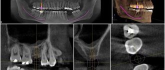
This is followed by diagnostics - facial anthropometry, X-ray studies in the necessary projections (OPTR, teleradiography, targeted images), CT. Based on their results, the volume of bone tissue in the surgical area, the nature of occlusion, and malocclusion are assessed. A treatment plan is drawn up.
If there are concomitant malocclusions that cannot be corrected during mandibuloplasty, the patient may be indicated for orthognathic surgery to correct them.
An important stage of preparation is computer modeling , the purpose of which is to select the optimal surgical strategy, predict its results, and determine the size of the implant.
Before operations, laboratory and instrumental examinations of the patient’s health are also carried out - urine and blood tests (general and biochemical), a coagulogram (to assess blood clotting), bacteriological studies for the presence of viral infections, an electrocardiogram, etc. If required, doctors from other countries are involved in the diagnosis specialties.
15-20 days before surgery, stop taking medications that affect blood clotting. NSAIDs may also be prohibited . It is important that the doctor knows about all medications the patient is taking. If among them there are those that negatively affect hemostasis, the doctor will inform the patient that their use should be stopped for a while.
The length of the preparatory stage is about 7-10 days.