Odontogenic osteomyelitis is a purulent-necrotic lesion of the bone and surrounding tissues as a result of infection of the dental canal and poor-quality dental treatment. It develops 2 times less often on the upper jaw than on the lower jaw. But it is complicated by the spread of infection into the maxillary sinuses. Treatment is only comprehensive - tooth extraction, extraction of infected tissue, medicinal restoration.
Doctor Levin Center specializes in the treatment of odontogenic infections involving the maxillary sinuses. Comprehensive surgical and medical treatment programs are carried out by maxillofacial surgeons with ENT training. The operations are performed in a state of medicated sleep using a gentle ultrasound protocol.
Causes of osteomyelitis
The causes of the development of osteomyelitis of the upper jaw differ in the mechanism of infection
- Odontogenic The result of untimely dental treatment is that infection from the canals penetrates the bone tissue through the root. The inflammatory process spreads to the surrounding soft tissues, and in the upper jaw it involves the maxillary sinuses. Primary sources are advanced caries, pulpitis, periodontitis, granuloma or dental cyst, pericoronitis, alveolitis.
- Hematogenous The infectious process spreads to bone tissue through the bloodstream. Primary sources are tonsillitis, purulent otitis, carbuncles and boils of the maxillofacial area. The process also develops on the basis of a chronic focus of infection in the sinus, polyps, and mucous cysts. Inflammation of the tooth roots is secondary in nature - the tooth is affected by the inflamed bone.
- Traumatic Domestic and sports injuries to the facial area contribute to fractures and infection of the bones of the maxillary sinus. As a result, inflammation and gradual destruction of the sinus septa and bone structures of the entire jaw develop. The entry of bone fragments into the sinus complicates the situation and provokes the growth of cysts and polyps.
Odontogenic osteomyelitis occurs most often - in 85% of cases. But all forms of disease in the upper jaw require treatment by an oral and maxillofacial surgeon with ENT training, since the maxillary sinuses are involved.
Causes and stages of the disease
The causes in most cases, excluding rare forms, are:
- Destruction of teeth and pulp by caries.
- Tooth trauma usually occurs when the bone tissue is already weakened due to poor nutrition or bad habits - alcohol, smoking.
- Diseases of the ENT organs - ears, paranasal sinuses, throat.
Osteomyelitis of the lower jaw is more common and more difficult to treat.
Blood diseases contribute to the rapid development of infection, since red blood cells carry little oxygen or their total quantity is extremely small to meet all the needs of the body.
Immunity is also affected by infectious diseases - tuberculosis, syphilis, AIDS, herpes. They weaken the body, and it gradually ceases to fight pathogenic flora. Therefore, for osteomyelitis of the jaw bones, vitamins and immunomodulators are always prescribed to strengthen the defenses.
Mandibular osteomyelitis develops very quickly. In the morning a person feels nothing, by the evening one side of the face swells, a tooth hurts, the temperature is 39 degrees. In this case, you must immediately contact a dental surgeon. The situation is worse if, due to a non-healing wound on the body, a person exhibits symptoms of osteomyelitis of the jaw.
The acute stage usually lasts from 10 to 14 days, depending on the state of the immune system. Diffuse osteomyelitis lasts longer - up to 3 weeks, then the process enters the subacute stage. In cases of chronic disease, all symptoms appear gradually.
Symptoms
The clinical picture depends on the stage
of Acute Pain, mobility of the causative tooth and those located nearby. Opening the mouth, difficulty swallowing, breathing with a putrid odor. Swelling and redness of the gums. Feverish state, lack of appetite, lethargy, headache. Inflammation of the lymph nodes. Subacute
The inflammatory process subsides, suppuration decreases, the mouth opens freely.
Teeth mobility is maintained. Improvement in general condition, decrease in temperature (slightly above 37°C). Regional lymph nodes are dense but painless. Chronic
Reduced tissue infiltration, subsidence of pain.
The jaw in the area of the inflamed lesion is thickened; upon incision, fistulas with purulent contents are revealed. CT scan reveals roughness and unevenness of dying bone tissue. Exacerbation
The clinical picture of the acute stage is repeated - general signs of intoxication suddenly appear. Local symptoms increase - swelling of the inflamed area, soreness and mobility of teeth, the formation of purulent fistulas.
Secondary chronic
In children, rapid chronic inflammation is noted. After 3 weeks, acute symptoms disappear, but the child does not become healthier.
When it comes to chronic inflammation, there is an active process of destruction, as well as melting of the elements of bone substance. After which areas of necrosis form. In addition, thanks to the intraosseous structures and periosteum, bone tissue is actively restored. The peculiarity of this type of osteomyelitis is the presence of the rudiments of permanent teeth. If they are involved in pathology, they may then die, and their behavior will resemble sequestration.
Symptoms of chronic osteomyelitis
When the inflammatory process becomes chronic, acute symptoms decrease. 10 days after the disease began, the child feels better (appetite and sleep return to normal, fever and symptoms of intoxication disappear). But children still feel weak, they get tired quickly, they sweat profusely, and their skin is pale. Sometimes children say that their jaw hurts slightly on the side that is affected.
During the examination of such a patient, you can notice certain manifestations of the inflammatory process:
- over the place where the focus of osteomyelitis was, there are soft tissue infiltrates;
- pain is felt when the child's jaw is probed;
- There are fistulas in which pus is released. They can be multiple or single;
- lymph nodes in the neck and jaw increase in size and hurt;
- The sockets of the tooth that were removed do not heal well. It may also leak pus;
- teeth are very loose.
When the disease worsens, the symptoms resemble acute osteomyelitis.
Diagnosis of chronic osteomyelitis
An x-ray should be taken to confirm the diagnosis. It will allow you to see the foci of destruction, those rudiments of teeth that have died, sequestration. If the lesion is extensive, then jaw fractures may even be detected.
Treatment of chronic osteomyelitis
When it comes to secondary osteomyelitis of a chronic form, a conservative treatment method is used:
- antibiotic therapy. Which drugs are suitable? All this is determined after the patient undergoes a culture of the pus discharged from the fistulas. Thus, the type of microorganism that provoked the pathology and its sensitivity to different drugs will be determined;
- desensitizing therapy. This includes antihistamines, which will remove the allergic reaction and also increase the body's resistance;
- therapy that can be used to stimulate the immune system and strengthen the body as a whole;
- physiotherapy. For this purpose, UHF therapy and laser irradiation are performed.
To perform an osteotomy or sequestrectomy, you must have the following indications:
- sequesters that are large in size and do not undergo spontaneous lysis for a long time;
- there are rudiments of permanent teeth that have died. They support inflammation;
- there was a risk of amyloidosis of internal organs.
If the disease worsens, the treatment will be identical to that in the case of acute osteomyelitis. But the main method is surgical intervention, during which lesions with pus are opened and drainage is performed.
What is dangerous - complications
With osteomyelitis, bone tissue melts, starting from the alveolar process. An untimely treated disease becomes hidden, but does not lose its aggressiveness, dissolving the bone to the state of cotton wool. Gradually, all bone structures are involved in the process; in the upper jaw, adjacent formations are involved - the septum of the maxillary sinus, the TMJ. As well as the surrounding soft tissues - gums, oral mucosa, the infection spreads to the brain.
As a consequence of the spread of the process, the following are possible:
- deformation, fracture, reduction of jaws;
- soft tissue phlegmon;
- TMJ ankylosis;
- thrombosis of facial vessels;
- thrombosis of the cavernous sinus;
- meningitis, meningoencephalitis, brain abscess.
In the chronic form, the infection spreads throughout the body, disrupting the functions of the body as a whole. Often complicated by amyloidosis of the heart muscle and kidneys. The advanced course of the disease provokes a lung abscess, inflammation of the mediastinal organs and sepsis with a risk of death.
Classification
Osteomyelitis can be classified according to location, route of penetration, and severity of the process.
If we talk about the course of the disease, then there is an acute or chronic type.
If we are talking about how the bone tissue became infected, then it is worth noting three types of osteomyelitis - post-traumatic, hematogenous, odontogenic.
If we take into account the localization of the purulent process, then it is worth distinguishing between the disease of the lower jaw and the upper jaw.
Why you should entrust treatment to the ENT department of dentistry
Treatment of osteomyelitis of the jaw should be carried out comprehensively - surgery to eliminate the source of infection, treatment of the affected bone, drug treatment and restorative therapy.
The usual tactics for treating osteomyelitis with inflammation penetrating the maxillary sinuses is to redirect the patient from one specialist to another. First, specialized specialists eliminate the source of infection - a dental surgeon removes the causative tooth, an otolaryngologist treats inflammation in the sinuses. Then, in the inpatient departments of otolaryngology or maxillofacial surgery, a surgical operation is performed to excise the infected soft and bone tissues of the maxillary sinus and remove sequesters.
ENT dentistry combines several areas of medical services and allows you to combine otolaryngological, surgical and dental treatment options. This is an optimized approach to the rehabilitation of patients with combined pathologies of the maxillary sinuses of a “dental” and external nature. Allows a comprehensive surgical and treatment program to be carried out in one place.
We have been treating combined ENT and dental pathologies for more than 20 years
When the maxillary sinuses are involved in the process, treatment is carried out by experienced maxillofacial surgeons with ENT training, candidates of medical sciences. A full-fledged ENT department is equipped with modern equipment to perform complex operations.
Levin Dmitry Valerievich Chief physician and founder of the Doctor Levin center
Diagnostics
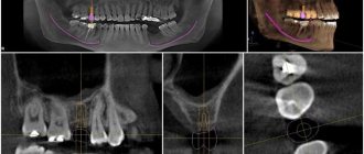
Sight radiography for odontogenic osteomyelitis of the upper jaw shows the presence of inflammation around the tooth root, but does not give a complete picture of bone damage. Therefore, in our Center, computed tomography is performed on a high-precision SIRONA-SIEMENS tomograph, with the GALILEOS diagnostic software package and ENT mode settings .
Unlike conventional radiography, a 3D examination with a computed tomograph gives the most accurate picture even at the initial destruction of bone tissue. Allows you to determine the extent of damage to bone tissue, the percentage of involvement of the maxillary sinus - characteristic foci of sequestration and the coarse fibrous structure of the bone are clearly visible on the 3D image. In the presence of neoplasms and foreign bodies, it allows us to clarify their nature and location.
CT with ENT mode settings gives an accurate picture already in the initial stages of the disease
In parallel, clinical laboratory tests are prescribed:
- blood biochemistry for ESR, C-reactive protein, globulin and albumin levels;
- urine test for red blood cells and traces of protein;
- bacteriological culture of discharge from the inflammation site to identify the pathogen.
Differential diagnosis is carried out to exclude other diseases - jaw tumor, purulent periostitis, festering cyst, acute periodontitis, bone lesions due to tuberculosis, syphilis, actinomycosis.
Stages of treatment
Treatment at our Center is comprehensive; whenever possible, we strive to combine all activities into one visit.
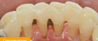
- Oral preparation It is important to establish a sterile environment before surgery to avoid re-infection. Hygienic cleaning and retreatment of teeth are carried out.
- The operation uses ultrasound to remove teeth that have become a source of infection. According to the selected protocol, access to the sinus is formed, infected areas and elements that support inflammation are eliminated.
- Prosthetics If a tooth was removed during the treatment of an odontogenic form of osteomyelitis, on the same day we manufacture and install temporary orthopedic structures to hide the defect.
After surgery, X-ray monitoring is required to assess the quality of the operation.
Recovery after surgery
No hospitalization
We do not use general anesthesia, which often leads to complications and requires a hospital stay of 1-3 days. Sedatives for immersion in medicated sleep have no effect on the body and are completely eliminated after 30-40 minutes.
A low-traumatic ultrasound protocol in combination with microscopic surgery, performed by experienced maxillofacial surgeons, allows you to avoid trauma and perform the operation as carefully and delicately as possible.
When you contact our Center, you will spend the first night after the operation at home.
Accelerated rehabilitation
Recovery after surgery occurs within a week. For those who want to speed up the process, we offer our own accelerated rehabilitation program to eliminate pain, bruises and tissue swelling on the day of the visit:
Home care
After surgery, medication is prescribed to relieve pain and prevent inflammation. A course of antibiotics is prescribed separately. You will receive the entire package of drugs free of charge to prevent the purchase of counterfeit products.
All necessary medications and instructions with rules of behavior for the rehabilitation period are issued immediately after the operation
Please do not skip taking medications, treat the operated area and follow the rules of behavior during the rehabilitation period - the instruction is in the bag with the medications.
1.5-2 weeks after the operation, we will invite you to remove the sutures and take a control CT image to assess the condition of the sinus and bone.
Consequences and rehabilitation
Osteomyelitis can have quite serious consequences in childhood:
- defects associated with bone tissue;
- jaw fractures that cause abnormal joints to form;
- the jaw is deformed;
- absence of permanent teeth, edentia;
- arthrosis, ankylosis, arthritis that relate to the joints of the temples and lower jaw;
- the jaw grows much more slowly, microgeny is noted;
- soft tissues are deformed and look like scars.
Due to the above complications, cosmetic defects may occur, but they do not have any effect on the masticatory apparatus or its functioning.
In order to restore the anatomy of the face and jaw to the maximum and ensure its normal functioning, it is necessary to take certain rehabilitation measures:
- sometimes plastic surgery is used. But this is done only after the facial bone stops growing;
- If adentia develops, then temporary dental prosthetics can be done. And as soon as the skeleton is finally formed, then permanent prosthetics are allowed;
- To improve the temporomandibular joint and its functioning, it is worth applying physiotherapeutic procedures.
All children who have suffered osteomyelitis that affected the jaw area are registered at the dispensary. They are required to visit the dentist at least 2 times a year.