The facial skeleton of each person is individual; its basis is made up of the jaws - upper and lower. The shape of the jaws determines the oval of the face and its aesthetics. But besides this, the jaws perform several important functions in the human body.
If their injury occurs, it is important to diagnose as early as possible and begin appropriate treatment to avoid serious complications that can affect other vital organs, disrupt numerous processes and remain for life, turning the person into a disabled person.
It is equally important to know the peculiarities of the structure of the jaw and the location of the teeth in a child in order to promptly identify deviations from the norm and, if possible, eliminate them painlessly.
Why are jaws needed - main functions
The beauty of a person’s face is largely determined by the anatomy of the lower jaw. If it is wide, protruding, square in shape, the face looks rough. But at the same time, it should not be too narrow and weak, since it is assigned various functions.
- Chewing - on each jaw there are 14-16 teeth (in a child 8-10), directly involved in biting, gnawing and chewing food. The bone must withstand stress even when eating hard foods that require prolonged and thorough grinding by teeth.
- Swallowing. Thanks to the structure of the jaw joints, a person can perform swallowing movements.
- Conversational. The jaws are directly involved in the articulation and pronunciation of sounds. If they are injured, the person's diction is almost always impaired.
- Respiratory. The jaw is indirectly involved in the breathing process, but, nevertheless, if it is damaged, a person begins to experience difficulty breathing, and in severe cases it is completely impossible.
- Fixing. The jaws create a cavity that serves as the location of numerous human sensory organs; structural disturbances inevitably lead to disturbances in vision, hearing, smell, etc.
An interesting fact is that after lengthy research and comparisons, scientists discovered: in terms of their physical capabilities and anatomical structure, human jaws are more similar to the jaws of ruminants than of predators. This indicates that man was originally conceived by nature as a creature that eats softer plant foods, rather than coarse meat.
Prevention of the development of micrognathia and macrognathia
Let's talk about possible measures to prevent the development of maxillofacial anomalies. Since the influence of negative factors on the health of the child’s future dental system is especially active during the mother’s pregnancy and in the first months of the baby’s life, preventive measures must be followed even before the birth of the child and in the first year of life . To promptly prevent the development of dentofacial deformities, you should adhere to the following:
- carefully monitor the course of pregnancy, try, if possible, to avoid the risk of infectious diseases of the expectant mother;
- try to avoid long stays of a pregnant woman, mother and baby in an unfavorable ecological environment;
- eat a nutritious and varied diet and lead a healthy lifestyle during pregnancy;
- give up bad habits in the process of planning pregnancy, during pregnancy and breastfeeding;
- adhere to proper feeding of the baby (use an anatomically correct nipple and discard it in a timely manner; when artificially feeding, make sure that the bottle with the mixture does not put pressure on the alveolar processes);
- if possible, stop bad habits (sucking and biting fingers and cheeks, tongue, various objects) that provoke deformations of the dentition, disturbances in the growth of the jaw bones;
- observe the timing and sequence of teething, monitor the development of speech;
- promptly identify and eliminate congenital pathologies in the form of a shortened frenulum, cleft lip, palate, etc.;
- wean the child from sucking fingers, objects, etc.;
- conduct myogymnastics with the child to prevent disorders of respiratory, swallowing, and speech functions; ensure that the child does not breathe through his mouth;
- timely treatment of baby teeth and periodontal diseases;
- adhere to preventive measures and promptly treat common diseases (rickets, diseases of the ENT organs, endocrine pathologies, etc.)
Thus, in the prevention and timely identification of risk factors that contribute to the occurrence of various dental anomalies, the most important thing is the active and careful participation of parents at all stages of the child’s growth and development.
Upper jaw - structure
The structure of the upper jaw is completely different from the lower jaw. The temporomandibular joint is multifunctional; there is nothing superfluous or ill-conceived here. The upper jaw is connected to almost every bone of the facial part of the skull. She participates in the formation of:
- The walls of the eye sockets;
- Oral and nasal cavities;
- Temporal fossa;
- Pterygopalatine fossa.
The upper jaw is a pair; it is considered the largest cavity in the skull. The organ has a body and four branches:
- Frontal – directed upward.
- Palatal – facing medially.
- Zygomatic – located laterally.
- Alveolar.
If you look at an x-ray, you can see the connection of the frontal process with the frontal bone. On the surface of the upper jaw there is a ridge on which the nasal concha is attached, and under it the palatine groove is visible, which is the wall of the palatine canal.
The upper jaw has three surfaces in addition to the anterior one: nasal, orbital and infratemporal. Unlike the lower, the upper is connected by other cranial bones motionlessly. Its shape is concave, on the lower part there is an alveolar process - it contains recesses for teeth. These recesses contain the roots of the teeth, separated from each other by partitions.
The highest notch is for the fang. In the center of this area, next to the infraorbital foramen, there is a depression, the diameter of which varies from 2 to 6 mm. The muscle responsible for raising the corner of the lips departs from it; the optic nerve and blood artery run through the center.
The nasal notch is the medial boundary between the fused frontal and anterior parts of the jaw. There is a tubercle on the temporal surface. It is separated from the upper part of the jaw by the zygomatic process. On the upper part there are four alveolar openings for large chewing teeth. Nerve fibers pass through them.
Inside the upper part there is an air cavity, which opens into the nasal cavity and is lined with mucous membrane. The upper surface of the jaw smoothly passes into the nasal. It has palatine processes, which form the bottom of the nasal sinus. There is also an orbital surface on the upper jaw. The lacrimal crest runs along the frontal process, the medial edge is formed from the lacrimal notch and the lacrimal ossicle entering into it.
The constituent element of the jaw is bone plates, which can form seals to cope with the loads of the air cavity and perform chewing functions. Based on the size and thickness of these plates, the number of grooves for teeth, anthropologists from human remains can determine a person’s race, population and level of evolution.
Structure of the lower jaw
The lower jaw in humans also has a body and two processes. From an anatomical point of view, these are two arches connected to each other - the major basal and minor.
The structure of the lower jaw with milk teeth in a child is not the same as in an adult. At birth, both arches of the child’s lower jaw are a single unit, they are connected, but later they separate and differ markedly in size, thickness and height. On the lower jaw there are many protrusions, tubercles, depressions and roughnesses that are needed to attach the bone to the muscles and ligaments. This is necessary to ensure its high mobility.
The lower jaw is inferior in strength to the upper jaw. This is how nature intended it so that when clenching teeth, blows, or injuries, the lower jaw takes most of the load, while the upper jaw, which protects the brain, remains intact.
On the front of the lower jaw there is a chin protrusion, an opening and a tubercle. The roots of the lower teeth of a person are located in the mental foramen. In total, the spirit has 16 alveolar openings for teeth. On the inside, in the center, there is the mental axis; it can be either single or bifurcated. Also through the mental foramen there is a canal with blood arteries and nerves.
The temporal part of the lower jaw is an element of the temporal joint. At its edges there are cartilaginous tissues and ligaments. They are the ones who ensure the movement of the lower jaw in various planes - moving forward, backward, lowering, moving to the side.
Why do you need to know the structure of the upper and lower jaw?
Usually a person does not think about the importance of the jaws and their functions. Many people do not even realize how helpless they would be if the jaw were designed differently or if it were suddenly severely injured. Therefore, it is important to pay due attention to the formation of the jaw from the birth of a child.
With developmental disorders, the child often experiences malocclusion, speech defects, and more serious pathologies associated with the influence of the jaw structure on the functions of the organs of vision, hearing, and breathing. Most child jaw defects can be successfully corrected in childhood without surgery or with minimal surgical intervention.
But to restore the correct shape and structure of the jaw in an adult, a long and serious operation will be required to remove or implant excess or insufficient jaw bone plates. At the same time, there is a very high risk of damage to the facial muscles and nerves, the cerebral cortex, and blood arteries, which can lead to the most dire consequences. Therefore, jaw development must be monitored.
Another important point is dental prosthetics, which almost every adult person, and sometimes even a child, has to undergo. Indications for prosthetics and its type are largely determined by the structural features of the upper or lower jaw, the thickness and structure of bone tissue.
Permanent bite
After all primary teeth have been replaced and the second molars (“sevens”) have erupted, the formation of a permanent dentition is considered complete. In an ideal permanent dentition, after changing teeth, the upper incisors should overlap the lower incisors by 2 mm. To assess the correct bite, it is also necessary to pay attention to the relationship and contact of the upper and lower teeth. At this age, your child is most likely already visiting a dentist, who, if necessary, will provide a referral for a consultation with an orthodontist. The last stage of teething will occur at the age of 18-20 years, when wisdom teeth appear. Sometimes there is not enough space for them to grow, or they may be in an incorrect position. In these cases, you may need to consult a dental surgeon.
A person needs a correct bite not only for beauty and beautiful speech, but also for the normal functioning of the maxillofacial apparatus. Poorly chopped food can lead to diseases of the gastrointestinal tract and the entire body as a whole. Over time, malocclusion can cause headaches and poor health. An uncomfortable psychological state also plays an important role for a person with malocclusion. That is why we recommend that you pay attention to the condition of your child’s teeth and promptly listen to the advice of doctors. The sooner you start orthodontic treatment, the better the results will be.
The Dial-Dent clinic has a newly created orthodontic department, headed by orthodontist O.A. Baranova. She specializes in correcting bites in adult patients and solves any, even the most complex cases. If you are concerned about your child’s malocclusion, then it is best to contact pediatric specialist E.V. Rada, the owner of numerous diplomas, certificates and certificates.
Dial-Dent specialists, whom you can contact for advice on the condition of your child’s oral cavity:
- Borisova Yulia Aleksandrovna, pediatric dentist.
- Nazarenko Evgenia Yurievna, pediatric dentist.
- Rada Elizaveta Valerievna, dentist-orthodontist, correction of bite in children.
- Selector Olga Nikolaevna, dentist-orthodontist, bite correction in children.
- Sleptsova Maria Pavlovna, dentist-orthodontist, bite correction in children.
- Tsukor Tatyana Borisovna, speech therapist - child psychologist.
- Smirnova Elena, dental hygienist.
Who to contact
If an adult’s jaw is injured, or signs of impaired development of the facial skeleton are noted in a child, you need to make an appointment with a traumatologist, orthodontic surgeon or dentist. In a child, anamolies, if they are not clearly expressed, begin to appear at the age of 5-6 years.
You should not delay examination and treatment - it is during this period that defects are easiest and fastest to correct. With the help of a small procedure and course of treatment, future problems with bite formation, growth and quality of teeth, hearing and speech in children can be completely avoided.
In some cases, on the contrary, it is recommended to wait several years for the bone tissue to become stronger and the facial skeleton to be fully formed, and then carry out the operation. But this is usually required in the case of the development of serious congenital pathologies and, fortunately, is quite rare.
It is important to know:
Changeable bite
Between the ages of 6 and 12 years, a gradual change of teeth occurs when baby teeth fall out and are replaced by permanent ones. Doctors call this condition a mixed dentition. If your child is already being seen by an orthodontist, then changing the bite is an important stage, which should also take place under the supervision of a specialist. The process of changing the bite looks like this: the first molar erupts behind the last milk tooth. It is important that baby teeth are healthy - only in this case the change in bite occurs normally.
It is not recommended to remove baby teeth prematurely without special medical indications. It is also not recommended to ignore the treatment of baby teeth, believing that “they will fall out anyway.” The first large permanent molar—the “six”—erupts behind the baby teeth when baby teeth have not yet begun to fall out.
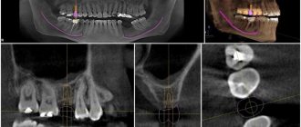
Using a panoramic photograph of the jaws, the doctor monitors the change of teeth, the presence of the rudiments of permanent teeth and the depth of their location.
The Dial-Dent clinic places great importance on dental treatment for young patients. Healthy baby teeth are the key to healthy permanent teeth. In addition, timely and regular observation of the child by a pediatric dentist will allow his dental and orthodontic problems to be resolved in a timely manner. Unfortunately, it is still not uncommon now for a desperate mother to come to the clinic and ask to have her baby’s bad teeth treated under anesthesia. It is difficult for a dentist to understand why the parents did not pay attention to the child’s condition earlier, when the treatment would not have been so difficult and traumatic for the little patient...