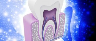
Enamel composition
- Minerals (up to 95%);
- organic matter (1.2%);
- water (3.8%).
Depending on the thickness, the enamel changes color - from white to yellow. In the thinnest places where the dentin is visible, the tooth looks yellowed. The degree of mineralization affects the transparency of the tissue. The more minerals in the composition, the more transparent the surface will be. For this reason, baby teeth, which are less saturated with minerals than molars, look whiter.
- Enamel is a tissue that is not capable of regeneration, since it does not contain cells.
- It constantly metabolizes ions coming from saliva, dentin and pulp.
- The tissue is characterized by mutual permeability, with the least permeability characteristic of the external surfaces adjacent to the oral cavity.
Types of dentin
- Peritubular
- surrounds the dentinal tubules, forms their walls, and is characterized by a high content of minerals. If the level of mineralization decreases due to caries, this leads to rapid destruction of peritubular dentin, which contributes to the expansion of dentinal tubules and increased tissue permeability. - Intertubular
– located between the tubules. It contains mineralized collagen fibers 50-200 nm in diameter. - Predentin
is the inner portion of peripulp dentin adjacent to the odontoblast layer. Under normal conditions, predentin does not mineralize and constantly increases in volume even in an adult, which leads to a narrowing of the pulp chamber. - Secondary dentin
is formed after tooth eruption. It is characterized by a less ordered arrangement of tubules and less mineralization. - Substitute (tertiary)
– is formed as a reaction to irritants locally, in certain areas of the pulp, mainly in the area of the horns. The structure often lacks dentinal tubules, and mineralization is episodic. - Sclerotic, or transparent
, is a consequence of the deposition of peritubular dentin in the tubules, due to which they gradually narrow. The cause of such changes is the age factor or caries. Such dentin is less permeable due to the deposition of lime salts on the walls of the tubules, so it prolongs the vitality of the pulp.
Other structural features of enamel
The enamel also contains other components:
- Gunther-Schreger stripes - light and dark formations 100 microns wide, located perpendicular to the surface of the enamel (the result of longitudinal and transverse opening of enamel prisms);
- lines of Retzius in the form of symmetrical oblique arches, which in cross sections resemble growth rings on a tree trunk (these are the so-called growth lines, signaling the periodicity of mineralization processes);
- neonatal line - a dark stripe that separates the enamel formed before and after the birth of a child (visible in all baby teeth and in the first molar);
- enamel bundles and plates - enamel that contains hypomineralized prisms and the same interprismatic substance, with a high protein content.
Enamel plates are located from the surface inward, mainly in the area of the tooth neck, bundles are located in the inner layers, near the enamel-dentin junction. It is believed that microorganisms penetrate into the thickness of the enamel through these formations, contributing to the development of caries.
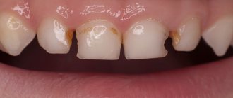
Damage to enamel: what to do
One of the most common phenomena is damage to the enamel. There are several types of lesions. It is worth considering that all types of lesions can be classified into two groups: those that formed before the eruption of baby teeth, and those that arose after. The first category includes the following pathologies:
- Dysplasia – there are a lot of symptoms of the disease: the formation of spots, thinning of the enamel layer. Most often, this pathology is associated with genetics and is often accompanied by problems with bone development. Teeth affected by dysplasia erupt with an initially irregular shape.
- Hypoplasia. Tooth tissues atrophy or are absent altogether. The formation of spots, changes in shade, thinning of the enamel are the main signs of the disease. To treat this problem, medications are prescribed that help restore mineral balance.
- Hyperplasia is excess tissue growth. It can also be caused by a mineral imbalance. Various blood diseases and hormonal imbalances can lead to the development of hyperplasia of the tooth buds. Enamel “drops” appear on the tooth surface. In rare, particularly difficult cases, hypertrophied areas can be filled with dentin or pulp.
- Fluorosis. The disease is characterized by the appearance of spots and furrows. Small pits or streaks may appear. Pathology is caused by excess fluoride content in the body. Often the disease affects baby teeth.
Below we will consider pathologies that are not associated with genetics and congenital anomalies:
- Erosion refers to non-carious lesions of the enamel layer and dentin. Erosion can be triggered by gastrointestinal diseases, excessive consumption of too acidic foods, and taking certain types of medications. Drug treatment is prescribed.
- Hypersensitivity. When a person reacts painfully to too cold or hot, this indicates the development of tooth sensitivity, which is often accompanied by thinning of the enamel. In this case, the task is to strengthen the layer. Minerals and vitamins are prescribed.
- Wedge-shaped defect. The pathology is that the neck of the tooth is gradually exposed, and the base is destroyed. The disease can be triggered by thyroid disease, gastrointestinal problems, and gum disease.
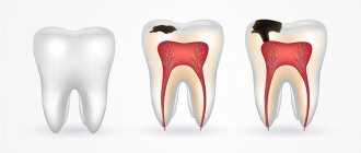
- Caries. If the carious lesion of the enamel layer is not eliminated in a timely manner, the disease will spread to the dentin and soon reach the pulp. The disease is treated by cleaning the cavity and installing a filling there.
- Impacts of a mechanical nature. Chips and cracks often form on the enamel. Modern dentists, using advanced equipment, can restore the damaged area.
Features of the structure of the enamel of baby teeth
The thickness of the enamel of a temporary tooth is approximately half that of a permanent tooth, but this is not the only reason for the need for more frequent treatment of dental caries in children. It contains fewer minerals, and the Retzius lines are much less pronounced. Unlike the apically directed prisms of molars, in milk teeth they are located horizontally.
Due to the weakly defined layer of final enamel, prisms are clearly visible on the surface; pores and microcracks are present in large quantities due to numerous fascicles and plates. This is why children's teeth are more quickly damaged and suffer from caries more often.
Methods of strengthening
To eliminate pathologies associated with tooth enamel, dentists use various techniques, both therapeutic and orthopedic. For severe lesions, crowns or veneers are used. But the best option for every person is to try to preserve their own healthy teeth. What options for strengthening enamel are suitable for an adult:
- Take vitamins and minerals regularly.
- Use preventive and therapeutic agents – gels and pastes containing calcium and fluoride.
- Regularly visit the dental office for preventive examinations and professional cleanings. This will not only protect the enamel, but also prevent the development of caries.
- If the enamel is damaged and visual appeal is lost, you can undergo a whitening procedure, but only after prior consultation with your doctor.
For children, dentists recommend fluoridation, fissure sealing and other procedures aimed at comprehensively strengthening teeth. Parents should pay attention to their child's diet. It should not contain sweet soda or excessive amounts of sweets. Since tooth enamel contains a large number of microelements, it is worth taking vitamins periodically, after consulting with a pediatrician and pediatric dentist.
Tooth enamel, types and methods of restoration
Tooth enamel is the natural mineral shell of the tooth. The ratio of minerals in it depends on the age, nutrition and habits of a person, as well as on the general condition of his body. The main components of tooth enamel are calcium and phosphorus.
Teeth enamel color
The color of tooth enamel is initially almost transparent. There are three types of natural shades of tooth enamel:
- white;
- white with a bluish tint;
- white with a yellowish tint (considered the most durable).
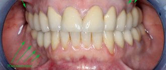
The color of teeth depends on the quality of dentin (the main tissue that makes up the tooth). If the color of tooth enamel changes, darkens, or spots appear, this indicates its destruction or internal diseases of the tooth.
In dentistry, a change in the natural shade of enamel is called discoloration. Its main reasons:
- congenital discoloration, hereditary predisposition;
- poor ecology, drinking water with mineral impurities;
- smoking, drinking alcohol, tea and coffee in large quantities, abuse of soda and energy drinks, as well as foods containing acids;
- insufficient oral hygiene;
- mechanical damage to the enamel;
- diseases of the internal tissues of the tooth.
The tooth acquires a red tint With minor injuries, the enamel may become pinkish .
Another variant of enamel redness is porphyria (a rare disease in which tissue pigmentation is disrupted).
Blue (blue or violet) tooth enamel indicates pulp necrosis or hyperthyroidism (increased thyroid function). A blue tint to teeth appears with prolonged use of tetracycline or water saturated with iron.
Tooth enamel turns green Another reason is prolonged contact with certain metals, for example, among metallurgists.
Brown enamel reveals a smoker and coffee drinker. Brown spots on the teeth may indicate acid necrosis, and caries may occur. In a child, brownish enamel of baby teeth can be a consequence of Rh conflict with the mother.
The yellow color of tooth enamel appears after mechanical injuries, taking antibiotics, sugar abuse, and smoking. With age, if the enamel becomes thinner, yellowish dentin shows through. Yellow teeth can signal problems with the adrenal glands, hematogenous jaundice, and Addison's disease.
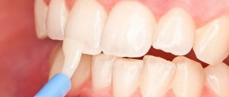
How to restore tooth enamel?
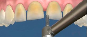
If a change in the color of tooth enamel is associated with its thinning or microtraumas, cracks and chips, modern dental technologies can correct this. An indication for strengthening enamel is also tooth hypersensitivity.
Among patients, the name of the procedure has stuck - coating teeth with enamel. In fact, this process is remineralization - restoration of the mineral composition of tooth enamel.
The most common method is dental fluoridation. There are several options for the procedure.
At first, the teeth are coated with a fluoride-containing varnish using a brush.
The second option is that individual applicators are made for the patient according to the shape of the teeth, which are filled with a fluoride-containing composition. They can be used at home.
Another method is the use of electrophoresis, when fluoride and calcium ions are introduced into the tooth tissue under the influence of a weak current discharge.
Innovative developments have made enamel coating a reality. The technique is called “tooth enamel implantation.” It allows you to change the color of the enamel, reduce tooth sensitivity and correct their forum. An enamel implant is as similar in composition to the natural mineral enamel of teeth. It is implanted at the cellular-molecular level and lasts a very long time.
An experienced dentist will tell you how to restore tooth enamel in a particular case.