Many people often ask the question - how many roots does a molar have? This question is relevant for most doctors. Because the complexity of many medical procedures depends on the number of roots, ranging from treatment, restoration and ending with removal. After birth, after birth, every person begins to grow milk teeth, of which there should be 20 by the age of 3. Then, after 6-7 years, the dairy ones are replaced by radical units, which should already increase by almost 1.5 times - 32. At the same time, dairy units can only have one root, but radical ones grow with several roots.
Features of the structure of teeth, their roots and canals
There are no two identical root dental systems, which is explained by the purely individual structure of a person’s teeth. In addition, the root system of incisors, canines and molars is arranged in accordance with their purpose:
- Ones and twos (incisors) are needed for biting food.
- Fours and fives (premolars) perform the initial chewing function.
- Sixes and sevens completely grind food.
Based on this, it becomes clear that the seventh tooth requires more nutrients than the fifth. It must be strong and hardy, therefore it has a more developed channel system. Despite the fact that the 6th tooth in the lower jaw performs the same functions as the seventh, it usually has fewer canals. This is due to the fact that there is less chewing load on it.
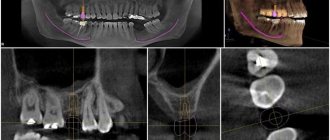
For a detailed study of the structure of the dentofacial apparatus of a particular patient, radiographic examination is used.
Tooth structure
Each dental unit consists of:
- crowns - the area above the gum;
- neck - the area between the crown and the root;
- root - the area under the gum.
Inside the crown is the pulp, which passes into the root canals. At the end of the root there is a small apical opening through which blood vessels and nerve endings pass, starting from the main neurovascular bundle and ending in the pulp.
When a person’s pulp becomes inflamed, not only it, but also all root canals need to be cleaned of infected tissues, since they are “communicating vessels.” If even one canal is left uncleaned, pathogenic microorganisms will continue to develop inside the dental unit, which will lead to its removal. That is why the doctor must know the exact number of canals in the tooth.
How many nerves are there in a human tooth?
Thanks to the nerve, the tooth can respond to external stimuli. After removing the pulp and filling the canals, the dental unit loses sensitivity, as it is deprived of a nerve. But due to the removal of blood vessels, problems begin with its blood supply and mineralization. The crown becomes less durable and more prone to various chips and breaks. The enamel quickly darkens, and it cannot be properly bleached even with strong chemicals.
Before removing the pulp, the patient is sent for an x-ray to find out how many canals are in the operated tooth: a person has only one dental nerve in a tooth, but there may be several canals . This preparation allows for depulpation to be carried out competently and quickly.
Types of Root Canals
There are several options for the structure of dental canals:
- in the root there is one canal passage, which corresponds to one apical foramen;
- in the root there are several canal branches that connect in the area of a single apical foramen;
- two different branched passages have one mouth and two apical openings;
- canal cavities in one root merge and diverge several times;
- three root canal passages emerge from the same orifice, but have 3 different apical openings.
There can be as many channels as there are roots, but often their number differs. Several types of canals may be present in one molar and premolar.
How many canals in a person’s teeth - table
According to statistics, the number of canals depends on the depth of the tooth : the deeper it is located in the jaw, the more canals it has. This is due to the increased load on the molars located at the base of the dentition.
Typically, teeth in the upper jaw have more canals. But this pattern is not observed in all patients.
The table below presents average statistical data on how many canals are in a person’s teeth above and below.
Number of canals in teeth in the lower jaw
The teeth on the lower and upper jaws are significantly different from each other. This is partly due to the uneven load and different functions. Typically, teeth in the lower jaw have fewer canals. But each specific case requires detailed study. Therefore, the dentist first sends the patient for an x-ray, and only then proceeds to open the crown and treat pulpitis.
It is impossible to start treating caries and pulpitis based only on encyclopedic information, because:
- The 6th tooth of the lower jaw can have any number of canals - from 2 to 4;
- in the 5th tooth below there is usually only 1 canal, but in approximately 10% of patients there are quints with 2 canals;
- In the 4th tooth there is usually only 1 canal, but in about a third of cases there are 2.
The eighth tooth on the lower jaw is the most “unpredictable”. Exactly how many canals are in the wisdom tooth located below can only be determined using x-rays. Officially, there are no more than 3 of them, but during the treatment of caries, additional cavities usually open. It is precisely because of its incomprehensible structure and inconvenient location that the figure eight is most often removed.
It is impossible to treat a dental unit without studying the structure of its root and canal system. This can only aggravate the pathology and lead to complications.
Number of canals in teeth in the upper jaw
The root system of the teeth of the upper jaw is more complex and branched. This explains the longer treatment of upper molars and the frequency of repeat visits due to incompletely sanitized dental cavities.
Features in the structure of the canal system of teeth in the upper jaw:
- The 6th tooth of the upper jaw is most often three-channel. But sometimes there are also four-channel first molars.
- The fourth and fifth teeth from above are most often two-canal, but sometimes single-canal and three-canal premolars are found.
- The 4th upper tooth usually has 2 canals, but sometimes there are premolars with 1 or 3 canals.
The “wise” eight on the upper jaw is a four-channel tooth. Third molars with 5 canals are extremely rare. However, in dentistry, even cases of the presence of eight-channel wisdom teeth located at the top have been recorded.
How does a human tooth work?
The elements of a human tooth can be divided into:
- crown,
- neck,
- root.
The crown is located above the gum and has a special coating called enamel. Under the enamel there is a durable layer of dentin, which in its structure resembles bone tissue.
The cavity of the tooth located inside the crown is called the “pulp”. It passes into a narrow canal of the tooth root, at the base of which there is a small hole. Nerve endings and blood vessels pass through it into the tooth cavity. Inflammation of the pulp is called pulpitis. It is an indication for opening the tooth cavity and cleaning the root canals. The most difficult thing to treat is pulpitis in the cavity of three-channel units (for example, in the sixth). In advanced cases, it is necessary to remove the tooth, and if it is also on top and in the last rows (6, 7 or 8), then this is also inconvenient.
Channels in baby teeth
There are as many nerves in baby teeth as there are in molars—one. In addition, temporary units are similar to permanent ones in the structure of the root system. That is, a milk tooth such as the upper six or second molar has a canal system similar to its molar brother, the second premolar.
Nerve endings perform standard functions:
- signal about developing caries;
- responsible for the growth and development of teeth;
- control the flow of water and nutrients to dentin and enamel.
The root canals of baby teeth are also treated and filled, but the tactics of their treatment depend on how long ago they erupted. Under the temporary units, permanent ones are formed, so treatment should be aimed at preserving them. Milk teeth can only be removed if the permanent teeth are ready to emerge.
The roots of permanent incisors, canines and molars do not form immediately, but over the course of about 3 years. Treatment of permanent teeth with unformed roots also differs from the standard one. The canals in the teeth of patients four, five, six years old (depending on the rate of formation of the dentoalveolar apparatus) are filled with a special paste with calcium and fluoride, which helps close the roots.
Read also: Inflammation of the wisdom tooth hood
Number of canals in a tooth of different jaws
Doctors initially start from the fact that the same teeth on both jaws are significantly different. The first three upper incisors usually have one canal each. On the lower jaw the situation with these teeth is somewhat different. It can be represented in the following percentage:
- The first incisor usually has one canal (70% of cases). Only every third patient has 2 of them. The second tooth in an equal percentage may have one or two canals (56% to 44%). In the lower jaw, the third incisor requires special attention. Almost always it has a single channel, and only in 6% of cases there are two.
Premolars are characterized by a larger structure and bear a heavier load. It can be assumed that the number of channels in them also increases exponentially. However, everything is not so simple here either.
How many canals are there in tooth 4? This number usually denotes the first premolar. In the upper jaw, only 9% of teeth have a single canal. In 6% of cases their number can increase to three. The rest are usually found with two branches. The next premolar is the 5th tooth. How many channels does it have? This tooth experiences even more pressure. However, this does not affect the number of channels. Only in 1% their number is three.
On the lower jaw the situation is different. The first and second premolars are not three-channel at all. In 74% of cases, the four and 89% of the five have only one branch.
Molars are considered larger teeth. Therefore, the number of channels they have is rightfully increasing. Sixes on the upper jaw can have either three or all four branches. The probability in this case is approximately the same. It is extremely rare that the picture changes in the lower jaw. Usually there are as many canals in the upper teeth as there are in the lower ones.
Posterior molars are characterized by the following percentage:
- Top seven: 30% to 70% four and three channels respectively. Bottom seven: 77% to 13% three and two branches.
The posterior molars are not very different in structure. Therefore, any dentist can almost 100% correctly say how many canals are in the 7th tooth of a particular person.
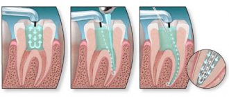
Root canal treatment
The treatment plan for dental canals consists of several stages:
- First, access to the problem area is freed: using a special dental instrument, the filling or the area of the crown damaged by caries is removed.
- Then the contents of the pulp are removed, and the canals are cleaned mechanically using antiseptic drugs.
- After this, the root is prepared for filling. At this stage, the dentist can form the correct conical shape of the canal passage.
- Then the canals are carefully sealed. If baby teeth are treated, the dentist uses a special filling paste, which gradually dissolves as the root dissolves.
- After this, a filling is placed on the crown.
This treatment regimen is standard and does not depend on exactly how many canals there are in the diseased tooth. The main thing is that all dental canals are cleaned, treated with an antiseptic and carefully closed. If treated incorrectly, it may be necessary to remove the tooth and visit an oral surgeon.
Teeth can be single-channel, two-channel, three-channel and even eight-channel. If one of the ducts becomes inflamed, it is necessary to clean and seal not only it, but also all other canals, since the infection could penetrate into them.
Measures to prevent dental diseases
To avoid any pathologies associated with teeth, it is necessary to monitor oral hygiene.
Dentists do not recommend brushing immediately after eating. It's better to wait 20-30 minutes. To avoid the accumulation of pathogenic microbes, you need to use special rinses. If it is not possible to purchase a ready-made product, you can make it at home. For this, regular chamomile tea or a decoction of oak bark is suitable. You should brush your teeth no more than 2 times a day, since the enamel tends to gradually thin out.
Such simple recommendations allow you to avoid dental diseases. However, do not forget that you should visit the dentist twice a year.
How many canals are there in the upper and lower teeth?
Correctly determining the number of canals in a tooth is only possible using an x-ray. Of course, their number depends on where the tooth is located - with a greater chewing load on the teeth in the back of the jaws, the retaining system is stronger; accordingly, they are larger, have more roots and canals. However, this is not a constant indicator, and it does not mean that the upper or lower incisors will have only one canal; it all depends on the individual characteristics of the jaw structure of each person. Therefore, how many canals in a diseased tooth require filling can be determined by the dentist during an autopsy or with the help of an x-ray.
Why does a tooth need a nerve?
The contents of the dental canals are covered by a network of nerve fibers grouped into branches. Each base is endowed with a nerve branch, and often several at once; the branch can be divided at the top.
How many nerves are there in a molar tooth? This directly depends on the number of roots and canals present in it.
Nerve fibers influence the development and growth of teeth and ensure their sensitivity. The presence of nerves allows the masticatory organ to be not just a piece of bone, but a living organ.
Dental mathematics is very exciting. Compared to the cost of dental procedures, each molar is worth its weight in gold.
Interest calculation
Due to the fact that each person is individual and there are no clear norms and rules for determining how many canals are in the teeth, in dentistry data on this issue are given as a percentage. Initially, they are repelled by the fact that the same teeth of the upper and lower jaws are very different from each other. If the first three upper incisors in almost one hundred percent of cases have only one canal, then with the same teeth of the lower jaw everything is much more complicated, and they have approximately the following percentage ratio:
- In the first incisor, most often there is only one canal - this is in 70% of cases from the general statistics, and only in 30% there can be two of them;
- The second tooth can have either one or two canals in almost equal proportions, or more precisely, a ratio of 56% to 44%;
- The third incisor of the lower jaw almost always has only one canal, and only in 6% of cases can there be two.
Premolars have a larger structure, more pressure and load are already placed on them, so it is logical to assume that there are more canals in the tooth, however, not everything is so simple here. For example, in the fourth tooth of the upper jaw, only 9% of teeth actually have one canal , in 6% of cases there may even be three, but the rest most often have two. But at the same time, the next premolar (fifth tooth), which seems to bear an even stronger load, most often has one canal and only in some cases more (of which only 1% is accounted for by three branches).
At the same time, in the lower jaw the situation is completely different - the first and second premolars are not three-canal at all, but most often have only one canal (74% - four and 89% - five) and in only 26% of cases for four and 11% for five - two.
The molars are already larger and the number of canals is increasing. The sixes of the upper jaw can have either three or four branches with equal probability. On the lower jaw, a two-canal tooth can sometimes be found (usually no more than 6% of cases), but most often there are three canals (65%) and sometimes four.
Posterior molars usually have the following relationship:
- Top seven: 70 to 30% three and four channels;
- Bottom seven: 13 to 77% two and three channels.
How many roots does each tooth have?
The root is located under the gum, below the surface of the neck and makes up approximately 70% of the organ. The number of chewing organs and the roots present on them is not identical. A system has been developed according to which they find out how many roots there are, for example, the 6th tooth on top or a wisdom tooth.
How many roots do adult teeth have? Their number in each chewing unit depends not only on its position, but also on hereditary factors, a person’s age, and race. Mongoloids and Negroids have one more root than Caucasians, and they grow together more often.
Dentists numbered each chewing organ. If you visually dissect the jaw vertically so that the section line passes through the middle of the skull, then to the left and right of it there will be central incisors. From this area the organs are numbered towards the ears. If we adhere to this principle of classification, then the root system of the chewing organs of an adult individual is as follows.
- #1 and #2 are called incisors, #3 are canines, and #4 and #5 are called molars. They grow on the upper and lower jaws and are endowed with one cone-shaped root.
- No. 6 – 7 and No. 8, located at the top, are called large molars and wisdom teeth. Each of them has three bases. These same units, but present on the lower jaw, can have two roots, except for organ No. 8. He has three, and in some cases four.
This information relates to the root system of adults. What about children, what is the number of roots in baby teeth, do they even exist? Many people think that baby teeth do not have teeth at all. It is not true. They have bases ranging from one to three, with their help the organs cling to the jaw, however, by the time they fall out, the roots disappear, giving rise to the erroneous opinion that they did not exist at all.
Wisdom tooth
The figure eight or wisdom tooth is quite unique and does not fall under standards and statistics.
The upper one can have a completely different structure with channels from one to five. The lower eight is most often found to be three-channel, however, often upon opening during treatment additional branches may be discovered. Among other things, a wisdom tooth differs from others in that its canals are rarely of the correct shape, often very curved and with a narrow passage, which greatly complicates their treatment and filling.
How many roots does each tooth have?
The root is located under the gum, below the surface of the neck and makes up approximately 70% of the organ. The number of chewing organs and the roots present on them is not identical. A system has been developed according to which they find out how many roots there are, for example, the 6th tooth on top or a wisdom tooth.
How many roots do adult teeth have? Their number in each chewing unit depends not only on its position, but also on hereditary factors, a person’s age, and race. Mongoloids and Negroids have one more root than Caucasians, and they grow together more often.
Dentists numbered each chewing organ. If you visually dissect the jaw vertically so that the section line passes through the middle of the skull, then to the left and right of it there will be central incisors. From this area the organs are numbered towards the ears. If we adhere to this principle of classification, then the root system of the chewing organs of an adult individual is as follows.
- #1 and #2 are called incisors, #3 are canines, and #4 and #5 are called molars. They grow on the upper and lower jaws and are endowed with one cone-shaped root.
- No. 6 – 7 and No. 8, located at the top, are called large molars and wisdom teeth. Each of them has three bases. These same units, but present on the lower jaw, can have two roots, except for organ No. 8. He has three, and in some cases four.
This information relates to the root system of adults. What about children, what is the number of roots in baby teeth, do they even exist? Many people think that baby teeth do not have teeth at all. It is not true. They have bases ranging from one to three, with their help the organs cling to the jaw, however, by the time they fall out, the roots disappear, giving rise to the erroneous opinion that they did not exist at all.
Misconception
Since a tooth consists of roots and a pre-crown part, there is sometimes a misconception that there are as many canals in teeth as there are roots . This is far from true, because the canals quite often branch and bifurcate near the pulp. Moreover, several channels can run parallel to each other in one root. There are also cases of their bifurcation at the apex, which means that one root has two apices and this, of course, complicates the work of doctors when filling such teeth.
Taking into account all the features of the individual structure of teeth, dentists need to be very careful when treating and filling, so as not to miss any branch. After all, sometimes without an x-ray it is very difficult, even during an autopsy, to identify how many canals there are in the teeth.
The development of modern medicine and dentistry in particular, today makes it possible to increasingly preserve those diseased teeth that just yesterday had to be removed due to the impossibility of treatment.
The procedure for treating root canals in teeth is itself quite complex, because they are filled with soft tissue - pulp, which contains a large number of nerve endings, blood vessels and other connective tissues.
Today, this is dealt with by a separate branch of dentistry – endodontics, the development of which makes it possible to improve the condition of a person’s teeth and cure even complex problems in more than 80% of cases, preserving the tooth itself. The goals of this treatment are:
- Removing developing infection inside the root system;
- Preventing pulp decay or removing it;
- Removal of infected dentin;
- Preparing the canal for filling (giving it the desired shape);
- Increasing the effect of medications.
Read also: Wisdom tooth grows into cheek
The difficulty of such treatment of the root system is that it is quite difficult for the dentist to get to the diseased canals and control the progress of the procedure. After all, if you do not remove even a microscopic part of the infection, it can develop again over time.
One of the main indicators for such treatment is the inflammatory process, which leads to damage to the soft tissue of the pulp inside the canals. Most often, various diseases such as caries and pulpitis lead to this, but root canal treatment may also be necessary for periodontitis.
The first symptoms of the need for such treatment are tooth pain or swollen gums. However, it is worth considering that if the disease passes into the chronic stage, pain may not be observed, but the disease develops and will ultimately lead to tooth loss. This is why it is so important to have regular dental checkups.
Number of roots in each tooth
Let's find out how many roots teeth have. If you draw a vertical line in the middle of the jaw, dividing it into the right and left parts, then the first from the line in both directions will be 2 incisors, then the canines, then 2 small molars and 2 large molars, and the very last - “wise” » eights.
There are several types of anesthesia used in dental treatment. Find out how local anesthesia is performed.
Do you know how to teach a one-year-old baby to brush his teeth correctly? The answer to this question can be found here.
The number of roots in the teeth of the upper and lower jaws is different. In addition, this indicator can be influenced by the individual characteristics of the body, genetics and race. For example, representatives of the Caucasian race will have fewer of them than the Mongoloid and Negroid races. Therefore, when asked, for example, how many roots the 7th tooth has from below, the dentist will not be able to answer with 100% certainty. Everything is individual. But for the average Caucasian, this is usually the case:
- the central incisors have 1 root both above and below; lateral incisors and canines – 1 each; first premolars from above – 2 each; first premolars from below - 1 each; second premolars and upper and lower jaws - 1 each; 1st and 2nd molars from above - 3 each; 1st and 2nd lower molars – 2 each.
The roots of wisdom teeth or third molars are an individual phenomenon. Experienced dental surgeons say that people’s “eights,” like appendixes, are unique. “Wise” molars can appear in old age or adolescence. Moreover, the number of their “roots” can vary from 2 and up to 5.
Interesting: some people mistakenly believe that baby teeth do not have “roots”. In fact, just like molars, temporary bone formations can have from 1 to 3 “roots”. It’s just that by the time the baby teeth are replaced by permanent ones, they dissolve.
Formation of roots
The first of the indigenous ones to appear in childhood are the “sixes”. This happens around the age of 5-6 years. You already know how many roots the 6th tooth will most likely have. Did you know that a tooth begins to erupt long before its “root” is fully formed? The timing of the formation of the roots of permanent teeth may vary, but on average this process is completed within 2-3 years after the appearance of bone formation above the gum. The order of eruption of permanent teeth and the timing of maturation of their roots looks something like this:
- “sixes” appear by the age of 6, and their roots are formed by 10; the central incisors erupt by the age of 8, and at 10 the “roots” are already formed; lateral incisors grow by the age of 9, their roots by 10; “fours” appear at 10 years old, and their “roots” will form at 12; the roots of the fangs will be formed by 13 years, while the “threes” themselves will appear by 11; “fives” appear by the age of 12, at which time the stage of formation of the root part is completed; “sevens” will grow up by 13, and their roots by 15 years.
Interesting fact: sometimes tooth roots can grow together. For example, it is known exactly how many roots the 6th lower tooth has - there are 2 of them. But on an x-ray it may seem that the “sixth” has one massive root. There is another anomaly in the development of roots - their curvature.
Quick answer: depends on the tooth.
Teeth have a complex structure. Most of the tooth consists of dentin, covered with enamel and cement on the outside. Inside there is connective tissue, permeated with blood vessels and nerves (each tooth has its own nerve apparatus).
Teeth have a special shape and structure; they occupy a certain position in the dentition. There are both milk and permanent teeth - molars. In the first case there are 20 teeth, while in the second there are up to 32. In addition, in rare cases, supernumerary teeth can be observed.
- From the first to the fifth tooth on both jaws there is one cone-shaped root.
- The 6th and 7th teeth in the upper jaw usually have three roots, the same can be said about the 8th tooth in the upper jaw, but often the “eight” has two or even four roots.
- The 6th and 7th teeth of the lower jaw have two roots, the “eight” has the same number as may be the case with the upper jaw.
The wisdom tooth, which is usually called the “eight”, often causes trouble for a person. Firstly, it often causes severe pain when it begins to erupt. Secondly, it often takes the wrong position, which leads to even more severe pain. Thirdly, it is often affected by caries and becomes a source of development of certain diseases. By the way, access to it is difficult, as a result of which it is not so easy to clean the tooth of plaque. To do this you need to use a special brush.
Process and stages of root canal treatment
The root canal treatment process has a clear sequence of stages:
- Initially, it is necessary to correctly diagnose the disease. In difficult cases, the dentist sends the patient for an x-ray.
- After this, the doctor carries out the necessary preparatory procedures in the oral cavity for treatment.
- Canal treatment is a rather painful procedure, so the tooth must be numbed by injecting anesthesia (usually into the gum near the diseased tooth).
- After this, complete asepsis of all instruments is carried out and the diseased tooth is separated from the rest using a special rubber film (caffedram).
- Next, the diseased tooth is opened using a drill, providing maximum access to the diseased canals, after which they are initially cleaned, the infected pulp is removed, and in parallel the canals are treated with special medications.
- Afterwards I dry them and seal them with special materials.
If the doctor has any doubts (usually this happens when the tooth is inconveniently positioned and instruments have difficulty accessing it), he puts a temporary filling , after which he sends the patient for an x-ray, using a photo of which he checks whether all the infection has been removed and whether all the canals have been removed. cleaned it. The permanent filling is then placed approximately two weeks later.
This whole procedure, of course, is not very pleasant, but it allows you to save the tooth. Its duration depends on the location of the tooth, the number of canals in it, the complexity of the developed infection and usually takes from thirty minutes to one hour. And success depends on the professionalism of the doctor and the high-quality work he has done, since it is necessary to remove all the affected pulp from the canals without leaving a drop of infection, otherwise it can develop again and tightly seal the tooth so that nothing else can get into the cleaned cavity.
Tips from dentists for disease prevention
The development of dental pathologies can be prevented only by following the advice of qualified doctors and observing the rules of oral hygiene.
So, for prevention purposes, dentists recommend:
- do not abuse the rules of hygiene, brush your teeth only in the evening and in the morning. More frequent exposure to tooth enamel contributes to its wear;
- hygiene procedures should be carried out half an hour after eating;
- use rinses to destroy germs remaining in the mouth after brushing;
- Cleaning should be carried out for at least 3 minutes, performing circular movements.
The main rule is that if the first signs of the disease are detected, you should immediately contact your dentist. This will help prevent further development of pathology and preserve teeth.
Prevention after treatment
After the root system treatment procedure, you should avoid putting stress on the treated tooth for some time; moreover, you should not eat food earlier than two hours after the therapy, otherwise the filling, which is not completely hardened, may simply fall out.
However, the same thing can happen if the doctor uses low-quality drugs or carries out incorrect treatment (for example, the canals are over-dried or not dried before filling). Also, after filling, for some time (up to several days) the tooth may be painful when pressed or simply ache, cause discomfort, and have increased sensitivity. This is usually normal, but if the pain is severe, you can take painkillers. If the pain does not go away after a certain time, this may also be an indicator of poor treatment (insufficient cleaning of the infection or infected pulp, leaky filling, use of low-quality drugs or materials).
Sometimes there are cases of allergic reactions , which are also accompanied by incessant pain, sometimes itching and rashes appear on the body. It may be caused by a reaction to the drug or the material that was used for the filling. In this case, it must be replaced with another one that will not cause allergies.
In all these situations, it is imperative to consult a doctor as soon as possible for a re-examination and dental prophylaxis in order to identify the cause of deviations from the norm.
Molars and permanent teeth. What are the differences?
Many people are accustomed to thinking that a baby tooth is temporary, and a molar tooth is permanent, already having a nerve. However, it is not. The correct division of teeth involves milk and permanent teeth, which replace them.
But molars are those teeth that never had predecessors, that is, molars: the 6th, 7th, and 8th tooth (1st molar - tooth six, 2nd molar - tooth seven, and 3 1st molar – tooth number eight).
And the premolars (4th and 5th teeth, or 1st and 2nd premolars) are first deciduous, and then permanent, and also molar, because they replaced the deciduous predecessors.
How many canals are there in the upper and lower teeth?
Correctly determining the number of canals in a tooth is only possible using an x-ray. Of course, their number depends on where the tooth is located - with a greater chewing load on the teeth in the back of the jaws, the retaining system is stronger; accordingly, they are larger, have more roots and canals. However, this is not a constant indicator, and it does not mean that the upper or lower incisors will have only one canal; it all depends on the individual characteristics of the jaw structure of each person. Therefore, how many canals in a diseased tooth require filling can be determined by the dentist during an autopsy or with the help of an x-ray.
Why does a tooth need a nerve?
The contents of the dental canals are covered by a network of nerve fibers grouped into branches. Each base is endowed with a nerve branch, and often several at once; the branch can be divided at the top.
How many nerves are there in a molar tooth? This directly depends on the number of roots and canals present in it.
Nerve fibers influence the development and growth of teeth and ensure their sensitivity. The presence of nerves allows the masticatory organ to be not just a piece of bone, but a living organ.
Dental mathematics is very exciting. Compared to the cost of dental procedures, each molar is worth its weight in gold.
Interest calculation
Due to the fact that each person is individual and there are no clear norms and rules for determining how many canals are in the teeth, in dentistry data on this issue are given as a percentage. Initially, they are repelled by the fact that the same teeth of the upper and lower jaws are very different from each other. If the first three upper incisors in almost one hundred percent of cases have only one canal, then with the same teeth of the lower jaw everything is much more complicated, and they have approximately the following percentage ratio:
- In the first incisor, most often there is only one canal - this is in 70% of cases from the general statistics, and only in 30% there can be two of them;
- The second tooth can have either one or two canals in almost equal proportions, or more precisely, a ratio of 56% to 44%;
- The third incisor of the lower jaw almost always has only one canal, and only in 6% of cases can there be two.
Premolars have a larger structure, more pressure and load are already placed on them, so it is logical to assume that there are more canals in the tooth, however, not everything is so simple here. For example, in the fourth tooth of the upper jaw, only 9% of teeth actually have one canal , in 6% of cases there may even be three, but the rest most often have two. But at the same time, the next premolar (fifth tooth), which seems to bear an even stronger load, most often has one canal and only in some cases more (of which only 1% is accounted for by three branches).
At the same time, in the lower jaw the situation is completely different - the first and second premolars are not three-canal at all, but most often have only one canal (74% - four and 89% - five) and in only 26% of cases for four and 11% for five - two.
Read also: Gums become swollen after tooth extraction
The molars are already larger and the number of canals is increasing. The sixes of the upper jaw can have either three or four branches with equal probability. On the lower jaw, a two-canal tooth can sometimes be found (usually no more than 6% of cases), but most often there are three canals (65%) and sometimes four.
Posterior molars usually have the following relationship:
- Top seven: 70 to 30% three and four channels;
- Bottom seven: 13 to 77% two and three channels.
Introduction
In dentistry, since 1971, there has been the so-called two-digit Viola system. According to it, the units of the upper and lower jaw of a person are divided into four quadrants, each of which has 8 teeth (we recommend reading: how often can radiography of the lower jaw be performed?). Quadrants for adults are numbered as 1, 2, 3 and 4, and for children - numbers from 5 to 8 (see table). Therefore, if you suddenly hear from a dentist that you are undergoing medicinal root canal treatment of 46 or 36 units, do not be alarmed.
READ ALSO: structure and anatomy of the human lower jaw
Each unit has its own individual structure. The number of canals and roots depends on where it is located and what function it performs. From this article you will learn what a dental cavity is and why it is affected by pulpitis. Also read about the concept of working length of the root canal. You will learn about methods for expanding dental cavities and their medicinal treatment, and see photos of three-channel pulpitis.
Wisdom tooth
The figure eight or wisdom tooth is quite unique and does not fall under standards and statistics.
The upper one can have a completely different structure with channels from one to five. The lower eight is most often found to be three-channel, however, often upon opening during treatment additional branches may be discovered. Among other things, a wisdom tooth differs from others in that its canals are rarely of the correct shape, often very curved and with a narrow passage, which greatly complicates their treatment and filling.
Number of roots in human teeth
The tooth root is located in the inner part of the gum. This invisible part makes up about 70% of the entire organ. There is no clear answer to the question: how many roots does this or that organ have, since their number is individual for each individual patient.
Factors influencing the number of roots include:
organ location; degree of load on it, functional features (masticatory, frontal); heredity; patient's age; race.
Additional Information! The root system of representatives of the Negroid and Mongoloid races is somewhat different from the European one; it is more branched, which, in fact, is the reason for the larger number of roots and canals.
Dentists have developed a special system for numbering teeth, thanks to which it is almost impossible even for a non-specialist to get confused in the units of the upper and lower dentition. To understand the principle of numbering, you need to mentally divide the skull in half vertically. First come the incisors - the frontal units of the upper and lower rows on the right and left. There are two of them on each side: central (No. 1) and side (No. 2). Next come the fangs or so-called triplets. The four (No. 4) and five (No. 5) are the first and second premolars. These teeth are also called small molars. All of the above units are united by the fact that they have only one cone-shaped “spine” in both the top and bottom rows.
The situation is somewhat different with the first, second and third molars, we are talking about tooth No. 6, 7 and 8. The upper six and seven (large molars) are endowed with three roots, however, in the wisdom tooth located on top, as a rule, there are also 3 grounds. The sixth tooth and the 7th tooth of the bottom row usually have one less root than their upper counterparts. The exception is the lower eight; this tooth may even have not three, but four roots. This feature should be taken into account during the treatment of a four-canal tooth.
Additional Information! Many people mistakenly believe that their children's primary teeth do not have roots. This is absolutely not true. There are bases, and their number can reach up to three; with their help, the chewing organs of babies are attached to the jaw. By the time the milk units are replaced with permanent ones, the “roots” disappear, as a result of which parents have the opinion that they did not exist at all.
Misconception
Since a tooth consists of roots and a pre-crown part, there is sometimes a misconception that there are as many canals in teeth as there are roots . This is far from true, because the canals quite often branch and bifurcate near the pulp. Moreover, several channels can run parallel to each other in one root. There are also cases of their bifurcation at the apex, which means that one root has two apices and this, of course, complicates the work of doctors when filling such teeth.
Taking into account all the features of the individual structure of teeth, dentists need to be very careful when treating and filling, so as not to miss any branch. After all, sometimes without an x-ray it is very difficult, even during an autopsy, to identify how many canals there are in the teeth.
The development of modern medicine and dentistry in particular, today makes it possible to increasingly preserve those diseased teeth that just yesterday had to be removed due to the impossibility of treatment.
The procedure for treating root canals in teeth is itself quite complex, because they are filled with soft tissue - pulp, which contains a large number of nerve endings, blood vessels and other connective tissues.
Today, this is dealt with by a separate branch of dentistry – endodontics, the development of which makes it possible to improve the condition of a person’s teeth and cure even complex problems in more than 80% of cases, preserving the tooth itself. The goals of this treatment are:
- Removing developing infection inside the root system;
- Preventing pulp decay or removing it;
- Removal of infected dentin;
- Preparing the canal for filling (giving it the desired shape);
- Increasing the effect of medications.
The difficulty of such treatment of the root system is that it is quite difficult for the dentist to get to the diseased canals and control the progress of the procedure. After all, if you do not remove even a microscopic part of the infection, it can develop again over time.
One of the main indicators for such treatment is the inflammatory process, which leads to damage to the soft tissue of the pulp inside the canals. Most often, various diseases such as caries and pulpitis lead to this, but root canal treatment may also be necessary for periodontitis.
The first symptoms of the need for such treatment are tooth pain or swollen gums. However, it is worth considering that if the disease passes into the chronic stage, pain may not be observed, but the disease develops and will ultimately lead to tooth loss. This is why it is so important to have regular dental checkups.
Root canal treatment
The development of modern dentistry increasingly makes it possible to preserve those teeth that just 10 years ago had to be removed due to the impossibility of treatment. Root canal therapy is a fairly complex procedure. The branches are located next to the pulp. It is represented by many blood vessels and nerve bundles. Any wrong decision by the dentist can lead to the death of the tooth. Today, root canal treatment is dealt with in a separate branch of dentistry - endodontics.
The most common form of pathology, in which the patient is forced to seek help from specialists in this field, is the inflammatory process. Lack of timely treatment may result in damage to the soft tissues inside the canal. Most often, various ailments such as caries lead to the pathological process. However, appropriate treatment may also be necessary for periodontitis.
Process and stages of root canal treatment
The root canal treatment process has a clear sequence of stages:
- Initially, it is necessary to correctly diagnose the disease. In difficult cases, the dentist sends the patient for an x-ray.
- After this, the doctor carries out the necessary preparatory procedures in the oral cavity for treatment.
- Canal treatment is a rather painful procedure, so the tooth must be numbed by injecting anesthesia (usually into the gum near the diseased tooth).
- After this, complete asepsis of all instruments is carried out and the diseased tooth is separated from the rest using a special rubber film (caffedram).
- Next, the diseased tooth is opened using a drill, providing maximum access to the diseased canals, after which they are initially cleaned, the infected pulp is removed, and in parallel the canals are treated with special medications.
- Afterwards I dry them and seal them with special materials.
If the doctor has any doubts (usually this happens when the tooth is inconveniently positioned and instruments have difficulty accessing it), he puts a temporary filling , after which he sends the patient for an x-ray, using a photo of which he checks whether all the infection has been removed and whether all the canals have been removed. cleaned it. The permanent filling is then placed approximately two weeks later.
This whole procedure, of course, is not very pleasant, but it allows you to save the tooth. Its duration depends on the location of the tooth, the number of canals in it, the complexity of the developed infection and usually takes from thirty minutes to one hour. And success depends on the professionalism of the doctor and the high-quality work he has done, since it is necessary to remove all the affected pulp from the canals without leaving a drop of infection, otherwise it can develop again and tightly seal the tooth so that nothing else can get into the cleaned cavity.
Baby teeth. Initial development
Deciduous teeth are the first set of teeth. At birth they are absent, but are already embedded in the gums. At the 7th week of embryo formation, in the area of future processes of the alveoli, the epithelium thickens, which in turn begins to grow in the form of an arcuate plate into the mesenchyme.
Teething begins after birth, and always occurs in a certain sequence.
As a rule, the primary incisors, the front teeth, erupt first, between 4 and 6 months of a child’s life. But the deciduous premolars are the last ones, both in terms of their location in the deciduous occlusion and in the order of appearance, and erupt within a period of up to 3 years. By this age, the child has all 20 teeth.
But you shouldn’t focus strictly on age. Teeth eruption, loss and replacement depend on many factors, including genetics. Therefore, the process may take place a little earlier, or vice versa a little later.
Then the formation of a permanent bite begins. And the first erupting tooth, which will be a molar, is a six, a permanent tooth, the 1st molar. There is no predecessor in his place. Next, the 2nd molar, or the seventh tooth, begins to appear.
Next, the replacement of all milk teeth, formed by that time, with permanent ones begins, and, as a rule, the process proceeds in the same order as the eruption of milk teeth, that is, starting from the front incisors and ending with the permanent premolars (4th and 5th m tooth).
The replacement process ends between the ages of 8 and 12 years. This is a long period and, as mentioned above, depends on many factors.
And starting from the 1st molar, or the 6th (tooth six), all the new teeth make parents worry about their health and the correct formation of the child’s bite.