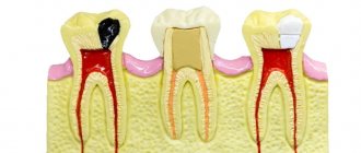
Depophoresis is a procedure for complete cleaning and sterilization of pulpless tooth canals using calcium copper hydroxide. The substance, penetrating into the affected area under the influence of current, decomposes any sources of infection (including necrotic pulp remains). Anatomically complex root processes (curved, twisted, branched) deserve special attention. In this case, the technique allows you to treat even roots recommended for removal, increasing the likelihood of saving the tooth several times.
The Mitino Dental Center has everything necessary for high-quality depophoresis. The experience and knowledge of the staff, modern equipment, certified drugs and the comfortable atmosphere of dental offices will not only help preserve your teeth, but will also do this with minimal stress on the body and with maximum benefit for your budget.
REFERENCE! Conventional endodontic treatment with pulp removal occurs without complications in only 30-60% (depending on the level of the clinic), while depophoresis with calcium copper hydroxide ensures success in 95% of cases.
Depophoresis: indications and contraindications for the procedure
Main indications for depophoresis:
- dental canals of complex shape (curved, deformed, with an extensive network of microtubules);
- the presence of residual pulp tissue in the canals (especially if gangrenous areas are present);
- the need to correct poor-quality fillings;
- the presence of foreign bodies in the dental canals;
- channels that are too wide or too narrow;
- multiple microholes in the tissue structure;
- radicular cysts and granulomas (the method allows you to treat the problem non-surgically, directing a suspension of calcium-copper hydroxide not only from the smallest nooks of the root system, but also into the cavity of the cyst, completely clearing it of pathogens and damaged tissue);
- standard depulpation procedure (for high-quality disinfection).
Several sessions of depophoresis may be needed to achieve results.
REFERENCE! During the process of depulping canals of complex shape, the tip of the pulp extractor may break off. A stuck fragment often cannot be removed without destroying the root tissue. Root canal depophoresis successfully solves the problem by completely sterilizing the fragment itself and the space surrounding it, after which the foreign object can remain inside the tooth without creating a threat of infection.
In addition to the effect of intensive sterilization, calcium-copper hydroxide ions contribute to the restoration of bone tissue. The effect of electric current stimulates the processes of osteocement synthesis, due to which the root canals are quickly sealed, the tissues restore their structure, and the tooth becomes stronger. The list of contraindications to depophoresis is small. These are mainly pregnancy, individual intolerance to the active substance (possible allergy to copper ions) and the acute phase of periodontitis. The reason for failure may be old silver pins in the tooth (risk of severe corrosion with the release of toxic substances).
IMPORTANT! A risk factor during depophoresis is any metal crowns, inlays and brackets, so the dentist must remain extremely careful and avoid contact of a metal element of the dental structure with one of the electrodes of the device.
Research by scientists has shown the effectiveness of depophoresis not only in adults, but also in pediatric dentistry. In the presence of formed roots, the procedure strengthens the weakly mineralized tissues of the dental canals and prevents early tooth loss (in childhood, loss can lead to improper development of the masticatory apparatus and jaw structure as a whole).
Long-term exposure to calcium hydroxide in maxillary anterior teeth (clinical case)
In our dental practice, calcium hydroxide has taken a leading place as medicinal pastes for intracanal therapy only in recent decades, although the use of calcium hydroxide in dentistry has been known since 1838 (Nygren), but this paste has been most widely used in the dental world since 30 years of the last century, with the aim of antimicrobial effects on the microflora of root canals (LRG Fava. WP Saunders. Calcium Hydroxyde pastes: classification and clinical indications. IEJ, 1999). To date, calcium-containing pastes are one of the main drugs in the treatment of infected canals (Ingle's Endodontics 6, Chpt 28, 1009-1010, 2008).
However, in the scientific community there is still no consensus on how long it is possible or necessary to put calcium hydroxide into the root canals. Typically the exposure period is recommended from 1 to 4 weeks. But, works are often published where the exposure period reaches up to 6 months. It seemed like something like that here? Who among us does not have patients who suddenly disappear for six months to a year with calcium hydroxide in the canal, and when we appear we do not find any noticeable exacerbation of the process in them. The inflammatory process either heals during this time, or the temporary restoration we created falls apart and the inflammation persists. In such cases, we blame the failure on failed coronal sealing.
However, among the studies examining prognostic factors affecting the periapical status of the tooth, calcium hydroxide exposure was not even included in the list of prognostic factors. Among such factors, greater importance is still given to the initial periapical status of the tooth and instrumentation with obturation along the entire length of the root canal (NChugal “Endodontics Prognosis”). The additional use of calcium hydroxide adds percentage to the success of treatment (“Reduction of Intracanal Bacteria Using Nickel-Titanium Rotary Instrumentation and Various Medications” G Shuping, D Orstavik 2000 JOE) but is still not able to completely remove the bacterial flora from the root canals (“One-versus Two-visit Endodontic Treatment of Teeth with Apical Periodontitis: A Histobacteriologic Study", Jorge Vera, DDS, Jose F. Siqueira, Jr, DDS, MSc, PhD, Domenico Ricucci, JOE 2014).
In the presented clinical case, a long time investment (more than 6 months) of calcium hydroxide was used in order to obtain sterility of the root canal of tooth No. 11. However, over time, after about six months, the inflammatory process not only did not decrease in size, but repeated exacerbations began to appear. Changes in the root canal treatment protocol, the use of additional irrigants, and an increase in the size of root canal treatment also did not bring any changes to the dynamics of the process. They began to “prepare the patient for resection of the root apex,” additionally “killing” tooth No. 12, since, according to the doctors, curettage of the periapical area could damage its neurovascular bundle.
This patient came to me with approximately the same input data. I decided that perhaps the reason for the failure of the treatment lay in insufficient instrumentation of the root canal of tooth No. 11, so I carried out my chemo-mechanical treatment, increasing the size of the finishing file to ISO 70. Additionally, instrumentation of tooth No. 12 was carried out to size No. 55 according to ISO. However, 10 days after the administration of calcium hydroxide, the tooth worsened the inflammatory process. This became an additional intraoperative factor that reduces the prognosis of treatment (Ng YL, K Gulabivala, T Mann “A prospective study of the factors affecting outcomes of NSRT”, IJE, 2011). Among other factors that negatively affected the prognosis, there was also the factor of the size of the lesion, in particular, according to the study by S Hoskinson (“A retrospective comparison of outcomes of root canal treatment” Oral Patogy Oral Medicine 2002), an increase in the size of the periapical process by 1 mm results in a decrease in success rate by 18%. Therefore, on my part, it was decided to perform apical microsurgery with resection of the apexes of the roots of teeth 11 and 12 after their obturation.
According to the preoperative CBCT analysis, the outer cortical plate of the upper jaw and the lower wall of the nasal cavity were missing, therefore, despite the routineness of the procedure, the curettage process was complicated by perforation into the nasal cavity. This defect was isolated by a biological membrane. The postoperative course was unremarkable. Observations were carried out at 6 and 12 months, after which it was possible to conclude the success of the treatment carried out according to the criteria of the European Society of Society.
Photo report of the work
Video report of the work
Equipment for depophoresis
The active substance is delivered to the affected area using special equipment. At the moment, highly specialized devices for depophoresis are produced solely by the company HUMANCHEMIE, which produces only one model - ORIGINAL II. The device is officially approved by the developer of the method and has a full functional set for high-quality and convenient performance of the procedure in a modern dental clinic:
- sets the duration of the session;
- sets the current strength;
- controls the stage of the procedure (taking into account the current strength and time).
All other examples of devices are complex devices designed to perform several dental procedures.
In addition to technology, to perform the procedure efficiently, you will need a set of specific drugs:
- Disinfectant compositions are represented by various suspensions based on copper and calcium hydroxide - cupral. Various versions come in the form of a powder (diluted in water to form a solution for depophoresis) or a paste (used to temporarily seal canals). Taking into account the main active ingredient, the depophoresis technique is also called cupral-depophoresis.
- Atatsamite is a powder preparation of plastic bactericidal cement for final filling of sterile dental canals.
REFERENCE! In some cases, calcium hydroxide is used to disinfect canals, but a mixture of calcium and copper hydroxides increases efficiency tenfold, which makes the use of cupral relevant as the main drug for depophoresis.
Proper nutrition: calcium for dental health
Lack of calcium in the body leads to multiple health problems, and affects not only the condition of the teeth. A sufficient amount of this macroelement is responsible for the health of the endocrine and cardiovascular systems. Calcium regulates processes occurring in the nervous system and affects blood clotting. If a person does not receive enough of this substance, then the body independently solves this problem by obtaining calcium from the bones.
You can eat many foods high in calcium, but this will not help improve the health of your teeth or slow down tooth decay. However, following the rules of a healthy diet, drawing up a daily diet taking into account all vital vitamins and macroelements is important in order to maintain your body and not aggravate the situation.
How is depophoresis performed in dentistry?
The preparatory stage includes radiography of the tooth. The number of roots, the length and shape of the dental canals, the presence and location of infection are assessed. The more detailed the diagnosis, the higher the effectiveness of treatment.
The depophoresis procedure is performed only on “dead” teeth, so preliminary devitalization (pulp removal) will be required.
The depophoresis procedure itself consists of 2-3 sessions, which are carried out with a break of a week. Sequencing:
- Local anesthesia is performed using application and infiltration anesthesia.
- The tooth is opened to gain access to the canals and the cavity is expanded to the required size.
- The channels are filled with calcium-copper hydroxide.
- Electrodes are installed (a negative charge is inserted into the dental canal to a depth of 8 mm, a positive charge is placed on the inside of the cheek near the problem tooth).
- Electric current is applied, gradually increasing the power until the patient feels warmth in the affected area. On average, the current level reaches 2 mA - this is enough to start the process of electrophoresis of copper and calcium ions into the adjacent tooth tissue.
- The procedure is completed by washing with distilled water (or a 10% hydroxide solution) and temporarily sealing the canal with a paste, again, with calcium hydroxide.
The whole job takes just over 5 minutes. 1 channel is processed per session.
ATTENTION! Simultaneous depophoresis of several channels is undesirable. This will lead to uneven distribution of current and a decrease in the quality of the procedure.
The last session is the final one. It involves re-processing with disinfection of the smallest tubules and sealing the tooth with a plastic cement composition with an antibacterial effect. Finally, the dentist installs a filling, which can be permanent or temporary (with subsequent replacement with an inlay).
For complete safety, a control x-ray of the tooth is taken.
ATTENTION! In case of increased tooth sensitivity (acute pain during a depophoresis session), the breaks between sessions can be increased to 2-3 weeks.
Lack of calcium in the body in pregnant women
Lack of calcium in pregnant women manifests itself in the form of the symptoms described above. There is no scientifically confirmed evidence that during pregnancy calcium is “washed out” and used to form the fetus from the teeth. However, in addition to the fact that the lack of a macronutrient threatens a woman’s health, it also increases the risk of caries in a child in the future. Studies have shown that children of women who received sufficient amounts of this macronutrient during pregnancy were less likely to suffer from caries.
The required daily calcium intake for pregnant women is needed not in order to preserve their teeth, but in order to provide the required supply of macronutrients to the unborn child. The same applies to the period of feeding the baby.
Knowing how important calcium is for teeth, try to adhere to a healthy lifestyle and maintain the recommended balance of vitamins, macro- and microelements in your diet for your age. To maintain your smile, see your dentist at least twice a year.
What is calcium filling?
For filling, a special composition is used - copper-calcium hydroxide. It has the consistency of a paste, which is injected into the recess with a special tool - a channel filler.
Calcium hydroxide paste completely closes the canal, preventing pathogens from getting inside
To ensure that there are no voids left in the cavity, the tooth is lightly exposed to electric current.
This method performs a therapeutic function: it disinfects tooth tissue, killing bacteria.
The patient walks with this calcium filling for some time. Then the dentist takes it out, rinses it, thoroughly dries the root canal and performs the final filling with a permanent filling.