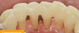
Anatomy of the canines The canine of the upper jaw (Fig. 33) has an irregularly cone-shaped crown.
The cutting edge resembles a triangle in appearance, bounded by three teeth, two outer ones and one middle one, well defined. The tubercle has two slopes, the medial slope is smaller than the lateral one. The vestibular surface is convex, has a longitudinal ridge that divides the labial surface into two facets, of which the lateral one is larger. The lingual surface is convex, also divided into two facets. Longitudinal enamel ridges on both surfaces of the crown pass into the cutting cusp. The lateral edges form two angles with the cutting edge, of which the medial one is more obtuse than the lateral one. The contact surfaces have the shape of a triangle. The root is slightly compressed laterally. Its lateral surface is more convex than its medial surface. All three signs are well expressed.
Canine of the lower jaw (Fig. 34). The structure is similar to the upper one, but somewhat shorter and smaller. The crown, while partially retaining its rhombic shape, is narrower and elongated. The besticular surface is convex, the lingual surface is flat and slightly concave. On the cutting edge, the central cutting main cusp stands out, in the area of which the edges of the crown converge. The medial part is shorter than the lateral one. The medial angle is acute and located further from the neck. From the main cusp towards the premolar there is a small notch that separates the medial cusp. The height of the crown of the vestibular and lateral surfaces is slightly higher than the height of the lingual and medial surfaces. There is one root, shorter than that of the upper canine . There are deep longitudinal grooves on the lateral surfaces. On cross cut oval shape. All three signs are well expressed.
The canine of the upper jaw (Fig. 1) has an irregularly cone-shaped crown. The cutting edge resembles a triangle in appearance, bounded by three teeth - two outer ones and one middle one, well defined. The tubercle has two slopes, the medial slope is smaller than the lateral one. The vestibular surface is convex, has a longitudinal ridge that divides the labial surface into two facets, of which the lateral one is larger.
The lingual surface is convex, also divided into two facets. Longitudinal enamel ridges on both surfaces of the crown pass into the cutting cusp. The lateral edges form two angles with the cutting edge, of which the medial one is more obtuse than the lateral one. The contact surfaces have the shape of a triangle. The root is slightly compressed laterally. Its lateral surface is more convex than its medial surface. All three signs are well expressed.
Canine of the lower jaw (Fig. 2).
The structure is similar to the upper one, but somewhat shorter and smaller. The crown, while partially retaining its rhombic shape, is narrower and elongated. The vestibular surface is convex, the lingual surface is flat and slightly concave. On the cutting edge, the central cutting main cusp stands out, in the area of which the edges of the crown converge. The medial part is shorter than the lateral one. The medial angle is acute and located further from the neck. From the main cusp towards the premolar there is a small notch that separates the medial cusp. The height of the crown of the vestibular and lateral surfaces is slightly higher than the height of the lingual and medial surfaces. There is one root, shorter than that of the upper canine. There are deep longitudinal grooves on the lateral surfaces. On cross cut oval shape. All three signs are well expressed.
Anatomy of the maxillary canine
The incisors, dentes incisivi
, four on each jaw, have a crown shaped like cutting chisels that cut food into inappropriate sizes. The crown of the upper incisors is wide, the lower ones are twice as narrow. The root is single, compressed from the sides of the lower incisors. The root apex is deviated somewhat laterally.
Upper medial incisor
the largest of the group of incisors. The labial surface of its crown is convex in the transverse and longitudinal directions. It has 3 small longitudinal ridges, each of which ends at the chewing edge with a clove. On both sides of the median ridge there is one longitudinal depression. The lingual surface of the crown is concave in the longitudinal and transverse directions.
In the cervical region it has a tubercle, tuberculum dentale
, From which ridges extend, heading along the distal and mesial edge of the lingual surface to the chewing edge of the tooth. Of the 3 signs of a tooth, the sign of crown curvature is the most pronounced. The root is conical and longer than the crown; the lateral grooves on it are weakly expressed. It has 3 surfaces: labial and two approximal.
Upper lateral incisor
smaller than the medial incisor, from which it differs in the following features: on the labial surface of the crown there is often a median longitudinal groove, on both sides of which there is one small tuberculate elevation on the cutting edge of unworn teeth. On the lingual surface, the lateral ridges are usually better defined than those of the medial incisors. Often on this surface there is a depression located occlusally (below) from the dental tubercle. The mesial surface is longer than the distal one and passes into the cutting edge almost at a right angle, while the distal one forms a significant rounding. The sign of the crown angle is well expressed. The root is shorter than that of the medial incisor, compressed in the mesiodistal direction; in most cases it is straight and has lateral grooves. Its distal surface is more convex than the mesial one.
Medial and lateral lower incisors.
The lower incisors are the smallest in both jaws. In this case, the medial incisor is smaller than its distal neighbor. Both teeth have characteristics characteristic of all incisors. Their crown has the most typical chisel shape. On the front surface it is slightly convex in the longitudinal direction and flattened in the transverse direction, on the rear surface it is concave in the longitudinal direction and flattened in the transverse direction. The ridges are weakly expressed and sometimes absent. The root is significantly flattened.
At the medial incisor
there are no signs of curvature of the corner and root. To distinguish the right medial incisor from the left, a better defined lateral longitudinal groove on the root is important.
At the lateral incisor
the crown is wider than that of the medial one, and the root is more massive. At the same time, this tooth has quite clearly expressed signs of angle and root and weakly - curvature.
Description of the dentition
Incisor teeth. The human jaw is symmetrical and contains the same number of teeth of each type. But there are certain anatomical features of the upper and lower jaw. Let's look at them in more detail.
Incisors are the front teeth. A person has eight of them - 4 at the bottom and 4 at the top. Incisors are needed to bite food and separate it into parts. The peculiarity of the structure of the incisors is that they have a flat, chisel-shaped crown with rather sharp edges.
On anatomical sections there are three tubercles, which are erased throughout life. On the top of the jaw there are two central incisors - the largest of all the incisors in their group. The lateral incisors are similar in shape to the central ones, but smaller in size.
What is noteworthy is that the immediate cutting edge of the lateral incisor also has three tubercles, and often takes on a convex shape as a result of the development of the central tubercle. The root of the incisor takes the shape of a cone, and is flat and single. A distinctive feature of the incisor is that on the side of the tooth cavity there are three tips of the pulp, corresponding to the tubercles of the cutting edge.
The anatomy of the upper teeth is slightly different from the structure of the lower dentition, so in the lower jaw everything is exactly the opposite. The middle incisors are smaller, unlike the lateral ones, and have a shorter and thinner root than the lateral incisors. The outer surface of the cutter is slightly convex, while the inner surface is concave.
The crown of the incisor is curved from the side towards the lips and is very narrow. The cutting edge has 2 angles - in the center, more acute, and inside - more obtuse. The roots have longitudinal grooves.
Fangs are used to break down food into smaller pieces. The anatomy of the fangs is such that there is a groove on the inside of the crown; it disproportionately divides the crown into 2 parts. The cutting edge of the fangs has one pronounced and developed tubercle, this makes the cone-shaped crown often similar to the fangs of a predator.
The canine on the lower jaw is narrower in shape, the ends of the crown are concentrated in the medial tubercle. The canine root is flat, inclined inward and the longest, unlike the roots of other teeth. A person has 2 fangs on both jaws. The lateral incisors with the canines form an arch, where the transition from the incisors to the chewing teeth begins in the corner.
Let us consider the structure of the small chewing tooth first, and then the large chewing tooth. Their main task is to thoroughly process food. This function is performed by molars and premolars.
Premolars
The first premolar (No. 4 in the dental formula) differs from the incisors and canines in its prismatic shape, and there are convex surfaces on the crown. The surface has 2 tubercles - lingual and buccal, with grooves between them.
The buccal tubercle is much larger in size than the lingual one. The root of the first premolar has a flat shape, but with a slight bifurcation into the lingual and buccal parts.
The second premolar is similar in structure to the first, but its buccal surface is much larger, and the root has a compressed anteroposterior direction and a conical shape. In the first lower premolar, the chewing surface is inclined towards the tongue.
The second premolar is larger than the first due to the fact that both tubercles are symmetrical and equally developed, and the depressions in the enamel between them have the shape of a horseshoe. The root is the same as that of the first premolar. A person has 8 premolars in the dentition, four on each side (on the lower and upper jaws).
Molars
In the upper jaw, the first molar is the largest. Its crown is similar to a rectangle, and the chewing surface is diamond-shaped with 4 tubercles. This molar has three roots: one straight - the most powerful, and two buccal - flat, deflected in the posterior direction.
During the closing of the jaws, the first molars rest against each other and form a kind of “limiter”; because of this, they undergo significant loads throughout a person’s life.
The second molar is smaller. The roots are the same as those of the first molar. The structure completely coincides with the location of the premolars described above.
On the lower jaw, the first molar for chewing food has five cusps. This molar has two roots - the anterior one with two canals, the posterior one with one. In this case, the anterior root is larger than the posterior one. In the lower jaw, the second molar is similar in structure to the first. The number of molars in humans is the same as the number of premolars.
The third molar is called a “wisdom tooth,” and a person has four of them in the dentition, two on each jaw. On the jaw below, the third molar has many variations in cusp development. As a rule, there are five of them. And, in general, in a person the structure of a “wisdom tooth” is the same as the structure of a second molar, but the root usually resembles a very powerful and short trunk.
Fangs
Fangs, dentes canini,
two on each jaw, have a long single root, compressed from the sides and having lateral grooves. The crown has two cutting edges that meet at an angle; there is a tubercle on its lingual surface near the neck. It is flattened in such a way that the lingual and labial surfaces converge towards the cutting edge. Its vestibular surface is convex in the transverse and longitudinal directions. There is always a well-defined longitudinal ridge on it, especially at the cutting edge, dividing the surface into a smaller one, the mesial segment, and a larger one, the distal one. On the lingual surface, lateral ridges are clearly visible, converging towards the neck at the tubercle of the tooth. The cutting edge of the crown consists of 2 halves: the smaller - mesial and the larger - distal, converging towards the apex of the edge. The distal half of the edge descends towards the corresponding approximal surface more steeply than the mesial half. Canines are characterized by all the signs of teeth (root, angle and crown curvature).
Upper canine.
The crown is massive. Its contact surfaces diverge significantly towards the cutting edge. A powerful middle ridge runs along the lingual surface of the crown, which, starting at the dental tubercle, thickens significantly and expands towards the cutting edge. The contact surfaces are wide at their bases, but relatively short. The root is massive and is the longest of the roots of all teeth. Its approximal surfaces are wide. The labial edge is blunt and wide compared to the lingual.
Lower canine
less than the top. It has less pronounced longitudinal ridges on both the vestibular and lingual surfaces of the crown. The vestibular surface of the crown is slightly convex, the lingual surface is slightly concave; the contact surfaces run almost parallel, and the mesial one does not converge at all towards the neck, while the distal one is somewhat inclined towards it. The incisal edge of the crown is shorter than that of the upper canine, and its mesial segment differs little in length from the distal one. The root is shorter than that of the upper canine, more flattened, and has better defined longitudinal grooves. There may be a bifurcation of the root at the apex, sometimes turning into a double root.
The teeth sitting in front of the fangs underwent changes in one direction - their crown became flat and a cutting edge was formed - the tip of the tooth, cuspis dentalis,
those sitting behind them changed in a different direction: they acquired a well-developed crown, which serves for crushing and grinding food, and the fangs seemed to be in a neutral zone and retained the original conical shape and the ancient function of the tooth - to prick and tear food. Therefore, they sit on the border between the front teeth (incisors) and the back teeth (molars).
Text of the book “Fundamentals of clinical dental morphology: a textbook”
4.1.4. Mandibular lateral incisor
The mandibular lateral incisor occupies the second position in the lower dental arch. It is larger than the lower medial incisor, with a well-defined crown angle feature.
The child erupts at 8-10 months. The change to a permanent lower lateral incisor occurs at 7–8 years.
In vestibular and lingual norms
(Fig. 68) the shape of the crown is close to the shape of an irregular quadrangle. The cutting edge line is relatively straight. The mesial angle of the crown is less than the distal one. The distal corner of the crown is rounded.
Rice. 68. Lateral incisor of the lower jaw, right.
a – vestibular norm; b – language norm.
The approximal contours noticeably converge towards the enamel-cementum junction. The medial contour is longer than the distal one.
The curvature of the enamel-cementum border in the direction of the root is more pronounced on the lingual side.
The transition of the approximal contours of the crown to the corresponding contours of the root is better visible from the distal side.
The root is cone-shaped, deviated distally (a sign of root position). The approximal contours of the root are somewhat convex in the middle third, smooth or concave in the apical and cervical thirds. The “waist” of the tooth is more pronounced along the distal contour. The apex of the root of the formed tooth, as a rule, is located distal to the USV.
On the vestibular surface
On the crown there is a vertical median ridge, which is separated from the lateral vertical ridges by weakly expressed depressions. The median and lateral vertical ridges in the cervical third merge with a clearly visible tooth belt.
The transition of the contact contours of the crown to the corresponding contours of the root is more pronounced on the distal side.
Lingual surface
more prominent than that of the medial incisor of the lower jaw. The marginal scallops, of which the distal one is the most developed, are delimited from the median scallop by pits. The distal fossa is deeper than the medial one. The median and marginal ridges in the cervical third of the crown merge with the lingual tubercle and the belt of the tooth.
The lingual surface of the root is already vestibular. The mesial and distal root surfaces converge lingually, so that in the lingual norm both approximal root surfaces are visible.
In medial and distal norms
(Fig. 69) the crown is shaped like a triangle, the most acute angle of which is located at the cutting edge.
The vestibular contour is convex at the tooth belt and relatively straight to the cutting edge. The lingual contour is more extended than the vestibular one, with a convexity at the level of the lingual tubercle and a concavity further to the incisal edge.
In the distal norm
The lingual contour of the crown is formed by the distal marginal ridge and has a concavity above the lingual cusp.
The line of the enamel-cementum border is convex in the direction of the cutting edge, and with a greater amplitude of curvature in the medial norm than in the distal one.
Rice. 69. Lateral incisor of the lower jaw, right.
a – medial norm; b – distal norm.
The vestibular and lingual contours of the crown, passing into the corresponding contours of the root, form the “waist” of the tooth. The transition of the contours of the crown to the root on the lingual side is smoother than on the vestibular side.
The contours of the root can be either convex or concave and reflect the uneven topography of the surface. The root apex is located near the USV (usually on the vestibular side of it). On the cone-shaped root, a vertical groove is visible on the distal side, which can serve as an additional sign of lateralization of the tooth.
In occlusal norm
(Fig. 70, a) the mesial-distal size of the crown somewhat prevails over the vestibular-lingual one.
Rice. 70. Lateral incisor of the lower jaw, right.
a – occlusal norm and section at the level of the base of the crown; b – tooth cavity.
The vestibular and lingual contours of the crown are convex. The degree of curvature of the lingual contour is greater than that of the vestibular one. The point of greatest convexity of the vestibular contour is slightly shifted to the medial side, and the point of greatest convexity of the lingual contour is distally.
Along the vestibular contour, a medial-distal slope is noticeable (a sign of crown curvature). The approximal contours are approximately equal in length. The cutting edge is located closer to the vestibular contour.
On horizontal sections, the root has the appearance of a laterally compressed oval, on the distal contour of which a concavity is visible.
Tooth cavity
(Fig. 70, b) corresponds to the external shape of the tooth. The cavity of the crown is narrowed in the vestibular-lingual direction and imperceptibly passes into the root canal. In the cavity of the crown there are small depressions that continue to the corners of the crown.
The lumen of the unformed root canal is relatively wide. A tooth with a resorbing root has a narrow canal. The diameter of the hole at the root apex corresponds to the width of the canal.
Anatomical options.
In
the vestibular and lingual norms,
the shape of the crown resembles an irregular quadrangle, usually trapezoidal. There are options for a rectangular or oval crown.
On the cutting edge, tubercles are more common in unformed teeth. The distal angle of the crown is usually larger than the medial one and is often rounded. The ridges and tubercle on the lingual surface vary slightly in size. There are sometimes vertical grooves on the lateral surfaces of the root.
In medial and distal norms
The crown is close to the shape of a triangle. The cutting edge is located along the USV or is slightly shifted to the lingual side.
The height of the tooth can be from 14.4 to 16.5 mm, while the height of the crown is 5.2–7.3 mm, the height of the root is 9.2–10.6 mm. The mesial-distal size of the crown ranges from 4.1 to 5.5 mm, the neck - from 3 to 3.7 mm. The size of the crown in the vestibular-lingual direction is from 4 to 4.8 mm, in the cervical area - from 3.2 to 4.5 mm.
4.2. Group of fangs
Milk fangs –
single-rooted teeth with a crown pointed on all surfaces, which are located in the middle part of each half of the dental arch distal to the incisors (third position). Primary canines erupt at 16–22 months and are replaced by permanent canines at 12–13 years.
The child has 4 primary canines (two in each dental arch):
– canines of the upper jaw
(right and left);
– fangs of the lower jaw
(right and left).
What is common in the anatomy of primary canines, as well as permanent ones, is the presence of a crown pointed on all surfaces and the longest root.
Primary canines differ from permanent canines in their smaller size and more symmetrical location of the main tubercle in relation to the approximal surfaces of the crown.
The upper milk canine is larger than the lower one.
The main signs of lateralization are uninformative.
To determine whether a canine belongs to the right or left half of the dental arch, the combination of structural features of the crown and root is taken into account.
4.2.1.
The canine of the upper jaw The canine of the upper jaw occupies the third position in the upper dental arch, just like the permanent tooth of the same name, it has a crown pointed on all surfaces and the longest root. The primary canine is less variable than the permanent canine.
The upper primary canines erupt at 16–20 months, and are replaced by permanent upper canines at 11–13 years.
In vestibular and lingual norms
(Fig. 71) the crown is close in shape to a pentagon.
Rice. 71. Maxillary canine, right.
a – vestibular norm; b – language norm.
The occlusal contour consists of two segments, which at the junction (the position of the tip of the “tearing cusp”) form an angle close to a right angle. The mesial segment of the occlusal contour is located more vertically and is somewhat longer in length than the distal one. The apex of the main tubercle coincides with the USV. The angle formed by the IVS and the mesial segment (slope) of the occlusal contour is smaller (sharper) than the angle between the IVS and the distal segment of the occlusal contour.
The approximal contours are relatively short, with the distal contour being longer than the mesial contour.
The line of the enamel-cementum border is slightly curved towards the root on both the vestibular and lingual sides.
The transition of the approximal contours of the crown and root is clearly visible and more pronounced on the mesial side. The approximal contours of the cone-shaped root are relatively smooth. The root apex is located near the USV.
On the vestibular surface
The crown has a vertical median ridge located along the length from the “tearing tubercle” to the tooth girdle. On both sides of the median ridge there are less protruding ridges, separated from it by depressions (mesial and distal).
On the lingual surface
The marginal ridges are well defined, which are delimited from the median ridge by triangular pits. The median ridge runs from the top of the main cusp to the base of the crown, where it merges with the marginal ridges, the lingual cusp and the tooth belt. The zygomatic tubercle is located approximately midway between the approximal contours of the crown. The lingual surface of the root is narrower than the vestibular one, therefore there is a convergence of the approximal contours of the root towards the lingual side.
In mesial and distal norms
(Fig. 72) the shape of the crown resembles a triangle, the base of which faces the neck of the tooth.
Rice. 72. Maxillary canine, right.
a – mesial norm; b – distal norm.
The line of the occlusal contour is connected to the vestibular and lingual contours on the vestibular side of the USV. The vestibular contour is convex with the greatest curvature near the neck (in place of the belt). The lingual contour is convex in the cervical third at the location of the lingual tubercle and relatively smooth throughout the rest of the length.
The line of the enamel-cementum border is slightly curved towards the occlusal contour. On the distal side, the enamel-cementum border is less convex than on the mesial side.
The transition of the contours of the crown to the corresponding contours of the root is well defined. The angle formed at the junction of the vestibular contours of the crown and root is greater than the angle formed at the junction of the lingual contours of the crown and root.
The contours of the cone-shaped root are uneven. The vestibular contour is often convex throughout its entire length, the line of the lingual contour is flattened. The root apex is rounded and located near the USV.
In the mesial norm
due to the contours of the vertical mesial ridge of the crown, the contour of the vertical median ridge of the vestibular surface protrudes. A mesial depression is noticeable between these ridges.
In the mesial norm, the mesial slope of the “tearing tubercle” is visible, which runs from the top of the main tubercle to the most protruding point of the mesial surface. The outline of the median ridge of the lingual surface protrudes from the contours of the mesial marginal ridge.
In the distal norm
the vestibular surface of the crown is visible with distal and median vertical ridges and a recess separating them.
From the apex of the main tubercle to the most protruding point of the distal surface, the distal slope of the “tearing tubercle” is visible, which is shorter than the mesial slope. From the distal side, the outline of the distal marginal ridge and the outline of the median ridge on the lingual surface are visible.
In occlusal norm
(Fig. 73, a) the contours of the crown resemble a rhombus in shape. The mesial-distal size of the crown prevails over the vestibular-lingual one.
The vestibular and lingual contours are convex. The degree of curvature of the lingual contour is greater than that of the vestibular one. The points of greatest convexity of the vestibular and lingual contours are located quite symmetrically with respect to the approximal surfaces.
The mesial contour of the crown is wider than the distal one, which can be used as an additional sign of tooth lateralization.
Rice. 73. Maxillary canine, right.
a – occlusal norm and section at the level of the base of the crown; b – tooth cavity.
In the occlusal norm, a convex vestibular surface with vertical ridges and a belt is clearly visible. Between the vertical enamel ridges there are depressions, of which the mesial one is more pronounced. On the lingual surface, the median and marginal ridges are clearly visible, converging towards the tooth girdle and clearly visible in the occlusal norm. The median and marginal scallops are separated by pits. The distal fossa is slightly deeper than the mesial one.
Tooth cavity
(Fig. 73, b) to a certain extent repeats its external shape. In the cavity of the crown, in the area of the tubercles and corners of the crown, recesses for the pulp horns are noticeable. The transition of the crown cavity to the root canal is smooth, without noticeable boundaries. The dimensions of the crown cavity and root canal depend on the degree of tooth formation. A tooth with a developing root has a larger cavity than a tooth with a resorbing root.
Anatomical options.
The primary canine is less variable than the permanent canine.
In the vestibular norm
The size of the angle formed by the slopes of the “tearing tubercle” varies less in the primary canine than in the permanent canine.
An additional tubercle is often found on the distal slope, which is less pronounced than that of the permanent canine.
Relief of the lingual surface
determined by the severity of the lingual tubercle and ridges. The zygomatic tubercle can “split” into two parts, which in the middle or occlusal third merge into one and pass into the median ridge.
In mesial and distal norms
the line of the enamel-cementum border can be straight or convex to varying degrees towards the occlusal contour. The bend of the enamel-cementum border on the mesial surface is slightly greater than on the distal surface.
The height of the tooth ranges from 18.2 to 20.2 mm, while the height of the crown is 6.4–7.6 mm, and the height of the root is 11.8–13.5 mm.
The mesial-distal size of the crown ranges from 6.4 to 7.4 mm, the neck - from 4.2 to 5.3 mm. The size of the crown in the vestibular-lingual direction can be from 5.4 to 7 mm, in the cervical area - from 4 to 5.5 mm. 4.2.2.
Canine of the lower jaw The canine of the lower jaw occupies the third position in the lower dental arch, somewhat inferior in size to the upper canine. The sign of crown curvature is weakly expressed.
Erupts at 16–18 months. The change to a permanent lower canine occurs at 11–13 years of age.
In vestibular and lingual norms
(Fig. 74) the contours of the crown form a pentagon, the most acute angle of which corresponds to the apex of the main tubercle.
Rice. 74. Canine of the lower jaw, right.
a – vestibular norm; b – language norm.
The occlusal contour is formed by the slopes of the “tearing tubercle”, converging to its apex at an acute angle. The mesial slope is longer than the distal one. The position of the apex of the main tubercle approximately coincides with the USV.
The occlusal contour line smoothly transitions into the contact contours of the crown, which converge towards its base.
The enamel-cementum border is slightly curved towards the root on the vestibular side and slightly more on the lingual side.
The apex of the root of the formed tooth practically coincides with the USV. The contours of the crown without sharp boundaries transform into the corresponding approximal, relatively smooth and symmetrical contours of the root.
The surface reliefs of the crowns of the lower and upper canines are similar. On the vestibular surface
From the top of the “tearing tubercle” in the direction of the neck of the tooth, an enamel ridge runs in the shape of a triangle, the base of which is the belt. On both sides of the enamel ridge there are depressions, of which the mesial one is better expressed than the distal one.
Lingual surface
less prominent than that of the antagonist of the same name. The marginal ridges at the base of the crown merge with the lingual tubercle. A weakly defined median ridge runs from the top of the “tearing tubercle” to the lingual tubercle. On both sides of the median ridge there are depressions, of which the distal one is larger.
In mesial and distal norms
(Fig. 75) the contours of the crown resemble a triangle, the base of which is located at the neck of the tooth.
Rice. 75. Lower jaw canine, right.
a – mesial norm; b – distal norm.
The apex of the main tubercle is projected, as a rule, on the vestibular side of the USV.
The vestibular contour of the crown is unevenly convex. Its greatest curvature is noted in the cervical third; throughout the rest of its length, its convexity is insignificant.
The line of the lingual contour is concave from the “tearing tubercle” to the lingual tubercle, at the level of which (the cervical third of the crown) the line of the lingual contour is usually convex.
The enamel-cementum border is convex towards the crown and differs little in both norms.
The contours of the crown in the area of the enamel-cementum border have a noticeable transition into the corresponding contours of the root.
The contour lines of the cone-shaped root are often vaguely curved. Often the vestibular contour of the root is somewhat convex in the cervical third and concave throughout the rest of the length, and the lingual contour is, on the contrary, convex in the middle third and concave in the cervical and apical thirds. The root apex is located along the USV, often rounded.
Mesial surface
The root is convex with a smoothed relief. A vertical groove runs along the distal surface of the root.
In occlusal norm
(Fig. 76, a) the contours of the crown are shaped like an isosceles triangle with rounded corners at the base, which is the mesial contour.
Rice. 76. Canine of the lower jaw, right.
a – occlusal norm and section at the level of the base of the crown; b – tooth cavity.
The vestibular contour has a slope in the mesial-distal direction (a sign of crown curvature). The point of greatest convexity of the lingual contour corresponds to the lingual tubercle, which is displaced mesially from the USV. The vestibular and lingual contours of the crown converge towards the distal contour, which can be used as an additional sign of lateralization of the tooth.
The vestibular-lingual size of the crown is not much inferior to the mesial-distal one. On horizontal sections of the root, the vestibular-lingual size significantly prevails over the mesial-distal one.
Tooth cavity
(Fig. 76, b), corresponds to its external outlines. The cavity has small depressions in the direction of the protruding areas of the crown (corners of the crown and the tip of the “tearing tubercle”) and bends in the canal area, repeating the curvature of the root.
The volume of the cavity relative to the external dimensions of the tooth depends on the stage of its development. In a tooth with an unformed root, the relative volume of the cavity is significant. The root canal is large in diameter with the apical opening corresponding in size. A tooth that is in the stage of root resorption has a relatively small cavity volume. The root canal of such a tooth is narrow and opens at the apex with a pinpoint-sized hole.
Anatomical options.
In
the vestibular norm,
the slopes of the main tubercle are often somewhat rounded and converge at an angle close to a straight line. As a rule, the mesial slope of the “tearing tubercle” is longer than the distal one, although there are options when both slopes are equal in size or the distal one is slightly larger than the mesial one.
By changing the relief of the vestibular surface. As a rule, on the vestibular surface there is a large enamel ridge running from the tooth belt to the top of the main tubercle. Often the median enamel ridge is displaced mesially and divides the crown into two unequal parts. The lateral enamel ridges can be expressed differently and are separated from the median ridge by deltoid depressions. More often, the distal depression is less pronounced than the mesial one. With weak development of the lateral enamel ridges, the entire vestibular surface becomes uniformly convex.
The anatomical formations on the lingual surface are variable in degree of development. The severity of the ridges and lingual tubercle is very diverse and determines the features of the relief of the lingual surface. Sometimes the lingual tubercle is absent. In the mesial and distal norms, the line of the enamel-cementum border can be convex towards the crown or almost straight.
The root is slightly flattened in the mesial-distal direction and in cross sections has an oval or close to triangular shape. Root bends are variable.
The height of the tooth ranges from 16.2 to 18.7 mm, with the height of the crown being 6–8.2 mm and the height of the root being 10.6–11.7 mm. The mesial-distal size of the crown ranges from 5 to 6.1 mm, the neck - from 3.6 to 4.2 mm. The size of the crown in the vestibular-lingual direction can be from 4.8 to 5.7 mm, in the cervical area - from 3.8 to 5 mm.
4.3. Molar group
Primary molars –
teeth with a multi-tubercular chewing surface and several roots. Molars are located in the distal parts of the dental arch and occupy the fourth and fifth positions.
Primary molars erupt from the 14th to the 30th month and are replaced by permanent small molars (premolars) from 8 to 13 years.
The child has eight primary molars:
– first and second molars
upper jaw (right and left);
– first and second molars
lower jaw (right and left).
Primary molars are the largest teeth in the primary dentition. Second primary molars, unlike permanent teeth, are much larger than first molars.
Primary molars of the upper jaw have three roots - two vestibular and lingual, the lower jaw - two roots - mesial and distal.
A characteristic morphological feature of primary molars is the predominance of the mesial-distal diameter over the height of the crown. In maxillary molars, the vestibular-lingual size of the crown is larger than the mesial-distal size. In mandibular molars, the mesial-distal diameter of the crown is larger than the vestibular-lingual diameter.
The deciduous molars have a well-defined belt, and in the first molars it is most developed.
The roots of primary molars are pincer-shaped. The “waist” of the tooth is well defined. Of the main signs of lateralization, all primary molars are characterized by the sign of crown curvature.
Maxillary canine
The canine, located distal to the lateral incisor, forms the angle of the dental arch - the transition from cutting teeth to chewing teeth. In the dentition, the crown of the canine is slightly deflected vestibularly and, accordingly, protrudes from the arch of the dentition. The shape of the crown is cone-shaped, its anteroposterior size is larger at the base, and its transverse size is larger at the middle.
When examining the canine crown from above, its mesial-distal curvature is clearly outlined. The vestibular surface is convex and... has a vaguely defined longitudinal ridge , better visible at the cutting edge; the roller divides the labial surface into two unequal parts: the smaller - medial and the larger - distal.
The oral surface is narrower than the vestibular one, slightly convex and, like the vestibular surface, has a longitudinal ridge running from the neck to the cutting tubercle. The roller divides the surface into two parts - medial and distal. There are often indentations on either side of it.
In the upper third, the ridge passes into a well-developed dental tubercle. The approximal surface of the canine crown, compared with that of the incisors, is convex.
The medial surface - the cutting edge - of the crown of the maxillary canine ends with a tubercle and has two obtuse angles - medial and distal. The medial angle is located closer to the tubercle than the distal one, as a result of which of the two lines that make up the cutting edge, the distal one is longer than the medial one.
The distal part of the cutting edge is often concave. The medial angle is usually lower than the distal one. First premolar of the maxilla The crown of the first premolar is similar to a prism, the sides of which are convex, has a larger diameter in the vestibular-oral direction, and a smaller diameter in the mesial-distal direction.
It is composed of two halves - vestibular and oral, having rounded surfaces . The vestibular half of the crown is larger than the oral half, it has a well-defined chewing tubercle , similar to the tubercle of a canine tooth, and two smaller ones - medial and distal.
a well-defined cusp . The vestibular surface is similar to the surface of the canine, but it is shorter. I Like the canine, the vestibular surface is often divided by a pronounced ridge into two parts - a smaller (medial) and a larger (distal) part.
| Figure 2. Structure of the crown of the central incisor of the upper jaw |
h - vestibular surface; b - oral surface; c — approximal surface; g - cutting edge
| Figure 3. Vestibular surfaces of the crowns of the central (a) and lateral (b) incisors of the upper jaw |
| Figure 4. Structure of the maxillary canine crown |
a — vestibular surface; b - oral surface; c — approximal surface; g - cutting edge
| Figure 5. Structure of the crown of the first premolar of the upper jaw |
a — vestibular surface; b - oral surface; c — approximal surface; g - chewing surface
The approximal surface has a rectangular shape. The largest convexity on the approximal surface of the crown is located in the upper third. The chewing surface of the premolar, when viewed from above, has an oval shape and two main cusps, which are separated by grooves lying longitudinally and transversely in the shape of the letter “H”.
The transverse line runs in the middle of the chewing surface in the medial-distal direction and reaches the approximal enamel ridges, which end the chewing surface. The grooves have a curvature corresponding to the curvature of the medial side of the crown.
The chewing surface and the crown as a whole are somewhat narrowed in the oral direction. The vestibular tubercle is sharper and higher than the oral one. Second premolar of the maxilla The second premolar of the maxilla is similar to the first premolar, but its crown in all diameters is slightly smaller than the crown of the first premolar.
The differences in the structure of the crowns of the first and second premolars are that in the second premolar the masticatory surface are equal in size. The vestibular surface of the second premolar of the upper jaw has less pronounced similarities with the vestibular surface of the canine: the vestibular surface of the premolar has a more rounded shape.
first molar The vestibular surface of the crown of the first molar is distinguished by the fact that the mesial cusp is higher and larger than the distal one. The vestibular surface seems to consist of the vestibular surfaces of two premolars.
The oral surface of the crown sharply narrows towards the neck, the medial oral cusp is much larger than the distal one, therefore the fissure separating them is located significantly more distal from the middle of the crown.
The approximal surface of the molar is more rounded than the medial one. The approximal line of the largest perimeter of the tooth is located higher on the medial side, and lower on the distal side. The medial surface is more sloping than the approximal one.
The chewing surface has a diamond shape in the diagram. The vestibulo-oral size of the chewing surface is larger than the mesial-distal one. The medialovestibular and distal-oral tubercles have a sharp shape, while others have a rounded shape.
Schematically, the location of fissures on the chewing surface of a molar can be represented in the form of an angled letter “H”, the transverse line of which runs along the long diagonal of the rhombus.
All molar fissures have a bevel towards the middle of the chewing surface and divide the chewing surface of the first molar of the mandible into four cusps: two vestibular and two oral. The medial vestibular tubercle is the largest, and the distal oral tubercle is the smallest.
The fissures of the chewing surface of a molar have different depths; they are depressions located in different places at different levels; in other places, a shallow fissure is located on an elevation of the chewing surface .
Atlas on metal ceramics
https://www.youtube.com/watch?v=https:tv.youtube.com
STRUCTURE OF TEETH AND BASICS OF THEIR MODELING
Recreating dental defects with metal-ceramic restorations is impossible without knowledge of the anatomy of the masticatory apparatus.
frontal (front, biting) and lateral (chewing).
The shape of teeth corresponds to their function. The group of biting teeth consists of incisors and canines.
Incisors are teeth whose crowns have a cutting edge; canines differ from them in that the crown has a pointed conical shape.
There are 12 biting teeth, they are located on each jaw: 3 - from the midline.
The group of chewing teeth consists of premolars and molars.
Premolars - 8: 4 - on each jaw, 2 - after the canines; molars - 12: 6 - on each jaw, 3 - after the premolars.
Chewing teeth have a multi-tubercular chewing surface: premolars - 2 tubercles, maxillary molars - 4 tubercles, mandibular molars - 5 and 4 tubercles.
Figure 1. Tooth structure The structure of teeth and the basics of their modeling
A: 1 - crown of the tooth; 2 - neck of the tooth; 3 - tooth root B: 1 - enamel; 2 - cement; 3 - dentin; 4 - tooth root canal; 5 - periodontium
1. the crown protrudes above the gingival margin, is covered with enamel, its main mass is dentin;
2. the root is immersed in the alveolus of the jaw, consists of dentin covered with cement;
3. The neck of the tooth is located between the crown and the root; the enamel cover ends at its upper border.
1. chewing (occlusal), which come into contact with antagonist teeth; in incisors they are called cutting edges, in canines they are called tearing cusps;
https://www.youtube.com/watch?v=ytabout
2. lateral (approximal), with which the tooth contacts neighboring teeth; the surface facing the tooth in front is called medial, and the surface facing behind is called distal;
3. vestibular, which face the lips or cheeks;
4. oral, which are facing the oral cavity.
On all surfaces of the tooth, the most convex part can be determined. The line connecting the most convex parts of the tooth on all its surfaces is called the equator.
The equator divides the tooth into occlusal and gingival parts. Each tooth has a certain size. It is customary to distinguish between height, width and thickness of the crown of a tooth.
The height of the tooth crown is the distance from the cutting edge or cusp of the chewing surface to the level of the neck of the tooth. The width of the tooth crown is the distance between the approximal surfaces. The cutting surface of the teeth is wider than their cervical part.
We suggest you familiarize yourself with which antibiotics are best to take for pneumonia in adults: list of drugs, rules of use, possible complications
The thickness of the tooth crown is the vestibulo-oral size of the tooth. The size of the height of the teeth decreases from the frontal to the chewing group, with the exception of the canines.
— a sign of crown curvature is manifested in the fact that the greatest convexity of the vestibular (dental, cheek) surface is located medially;
- the sign of the crown angle is expressed in the fact that the medial surface and the cutting edge of the incisors and canines form a more acute angle than the angle formed by the cutting edge and the distal surface;
- a sign of the root is that the roots of the incisors and canines deviate in the posterolateral direction, and those of the premolars and molars - in the posterior direction from the longitudinal axis of the root.
Upper jaw teeth
Maxillary central incisor
The crown of the tooth is spade-shaped.
The lateral surfaces gradually converge towards the neck. The vestibular surface is convex and often has the shape of a rectangle. In young people it is wavy, the waves go longitudinally and seem to divide the vestibular surface into three parts, forming three bends along the cutting edge.
With age, the waviness of the vestibular surface of the crown and cutting edge disappears (erases), and it becomes smooth. The crown is wider at the cutting edge and narrower at the neck of the tooth, the medial angle of the cutting edge is straight, the distal one is slightly rounded.
The outer line of the incisor is rounded on the mesial side, and somewhat concave on the distal side. The oral surface is concave and has the shape of a triangle with the apex directed towards the neck of the tooth. In the upper third there is a tubercle.
https://www.youtube.com/watch?v=ytcopyright
In young people, the palatine tubercle is divided into several small tubercles. The approximal surface has the shape of a triangle with the apex facing the cutting edge. The line of the neck of the tooth (enamel-cementum border) is curved.
The labial surface is convex only in the upper half (closer to the neck), its half, going to the cutting edge, is flattened. Lateral incisor of the upper jaw The lateral incisors are smaller in size than the central incisors, their shapes vary significantly.
The crown of the tooth is spade-shaped. The lateral surfaces of the crown are almost parallel. The crown of the lateral incisor is smaller than the central one in all dimensions (shorter and narrower by about 1 mm). The medial angle of the lateral incisor is more rounded than that of the central one.
The vestibular surface is convex (and the narrower the crown of the tooth, the more pronounced it is) and has the shape of a triangle with the apex facing the neck of the tooth. With a relatively wide crown, its shape is the same as that of the central incisor, that is, flattened in the lower part of the crown. In the dentition, the neck of the lateral incisor is located somewhat distally compared to the cutting edge.
Anatomical structure
Going to the mirror, opening your mouth, you can examine your teeth in detail. A person sees only the vernal shell - these are hard enamel crowns. The protective layer reliably hides vulnerable internal tissues.
Incisors, canines, and molars have different shapes and numbers of roots, but they are united by a common structure.
Let's take a closer look at the histological structure of the tooth in section.
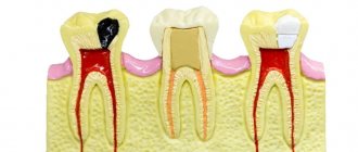
- Enamel is a hard protective layer of a whitish-cream color, consisting of 96% inorganic substances. The fabric has increased strength, but is also fragile, prone to abrasion, and susceptible to adverse environmental influences. If the enamel is injured by the attachment of pathogenic microorganisms, caries develops. On the surface of the chewing teeth there are fissures, depressions and grooves. They most often accumulate food particles that are difficult to remove. This causes the development of the disease. When the pathology occurs, it gradually affects the healthy protective layer, leaving internal tissues defenseless.
- Dentin - located directly under the enamel, consists of 70% inorganic substances. Dentin is a hard tissue, but it is much more vulnerable than the surface protective layer. If the carious process reaches the dentin, the pathological process occurs rapidly, and in the absence of timely assistance from the dentist, inflammation of the neurovascular bundle occurs. When dentin is damaged, medium or deep caries develops. Signs of the disease are clearly visible - violation of the integrity of enamel and dentin, the appearance of pain sensitivity upon contact with negative environmental factors;
- The pulp chamber and root canals contain nerve endings and blood vessels and have a soft, loose structure. Thanks to the pulp, the tooth receives the necessary nutrients. It protects periodontal tissues from penetration of pathogenic microorganisms and promotes dentin regeneration. When the neurovascular bundle is damaged, an inflammatory process occurs. The person feels acute paroxysmal pain. Taking analgesics only briefly relieves the pain symptom. If pulpitis develops, you must immediately visit the dentist. If help is not provided in a timely manner, the dead nerve fiber will become a source of infection. Pathogenic microorganisms will penetrate into the surrounding dental tissues, and periodontitis will develop;
- Cementum is a hard tissue lining the outer surface of the root. Periodontal ligamentous fibers are attached to the cement, which securely fix the tooth in the alveolar socket.
The detailed structure of the tooth and methods of their treatment are described in the video: The crown part of the incisors, canines and molars is located above the surface of the gum, the root is hidden deep in the internal tissues of the jaw.
Types of teeth. Classification
There are certain features in the structure of human jaws and teeth. The incisors are located in the very center, followed by the canines, small molars and large molars.
Each unit performs its primary functions. The incisors help to grasp and bite food, the canines hold and separate it, and the molars and premolars chew and grind it.
External structure of teeth and their types:
- The incisors are the weakest teeth. They consist of a flattened crown and have 1 root. The front surface of the incisor is convex, and the back is slightly curved. At the base of the coronal part there are small serrations (cutting cusps).
- Fangs - located behind the incisors, have 1 powerful root, and are equipped with a pointed cusp at the top of the crown.
- Premolars - capable of withstanding powerful loads, are involved in chewing and grinding food. Dentists call them small molars. The units have a prismatic shape and may contain from 2 to 5 tubercles. The lower premolars have 1 root, and the small molars located immediately behind the canines are equipped with 2 roots. Children have no small molars; they are temporarily replaced by baby molars.
- Molars are chewing units, equipped with a massive crown and designed for chewing food. Molars have 4 - 6 cusps, between which there are deep grooves and fissures. The teeth of the upper jaw have 3 roots, the lower 2. The exception is the 8th molar, in which 3 or even 4 roots can be found.
The value of each tooth is very significant. Important organs, together with the tongue, the inside of the cheeks and lips and the salivary glands, contribute to the formation of the food bolus. If even one tooth is missing, the digestion process becomes difficult. This leads to the formation of gastrointestinal diseases. After removal of an incisor, canine or molar, without subsequent prosthetics, the jaw row shifts, the bite is disrupted, and the likelihood of developing caries increases. With pathology or absence of frontal teeth, a person is embarrassed by his smile, becomes withdrawn and uncommunicative.
General concepts about teeth and their classification
Teeth are special bone formations that carry out the primary mechanical processing of food. People have long been accustomed to eating rather tough foods - meat, grains, plant fruits. This food requires considerable effort to process, and therefore healthy teeth have always been considered an indicator that a person eats a varied and good diet.
To begin with, what you need to know about teeth is the only organs in the human body that cannot be restored. Both their apparent reliability and fundamental nature are quickly violated by bad habits and poor care.
And if milk, primary teeth are fragile precisely because of their temporary purpose, then the molars are given to a person for the rest of his life. In general, the entire dentition in a person is divided into the following types:
- fangs;
- incisors (lateral and central, also called lateral and medial);
- molars or large molars (this also includes the upper and lower wisdom teeth that grow in a person in adulthood or young age);
- premolars or small molars.
As a rule, the location of the dentition on the upper and lower jaw is recorded using the so-called dental formula. For molars and milk teeth, this formula differs only in that the molars are most often designated using Arabic numerals, and the milk teeth - with Latin numerals.
For an average adult, the dental formula looks something like this: 87654321|12345678. The numbers indicate teeth - any healthy person must have one canine, 2 incisors, 3 molars on each side, 2 premolars on the upper and lower jaw. As a result, the total quantity is 32 pieces.
We invite you to read: The lower jaw is smaller than the upper jaw in a newborn
For babies who have not yet had their primary teeth replaced, this formula looks different, since there may be about 20 teeth in total. As a rule, temporary teeth erupt by 2–3 years of age, and by 9–12 years they are completely replaced by permanent teeth. However, not all people can boast of having all 32 sprouted teeth.
Since wisdom teeth or third molars can appear in adulthood, or they can remain completely in their infancy all their lives, in which case a person will have 28 teeth in the oral cavity. Moreover, the structure of the lower and upper jaws has certain differences.
Teeth of the upper and lower jaw. Structural features
On each of the human jaws there are incisors, canines, premolars and molars. The teeth of the lower jaw are smaller in size, have a narrow crown and a flattened root. The incisors and canines of both jaws, as well as the lower premolars, have 1 root. The small molar of the upper jaw, located near the canine, is attached to the socket by 2 roots.
To facilitate diagnosis and treatment, dentists invented a special teeth numbering system. The distinctive features of incisors, canines and molars, as well as their functions, are clearly visible in the photo:
Function and role of fangs
The main function of the fangs is to tear and hold food. In addition, canines are the teeth that are least susceptible to caries. These are the most powerful and strong teeth, which bear quite a large load. If a person suddenly lost his fangs, then:
- perhaps he would have problems with diction;
- the entire load would fall on the weaker neighboring teeth, which in the near future would either become deformed or become loose;
- the dentition would lose its aesthetic appeal;
- the process of infection of teeth with caries has accelerated, since it is the fangs that are the barrier to the “creeping” of this problem from the molars that are most susceptible to it to the incisors;
- Tooth enamel is the hardest human tissue the body produces, but even it can wear down and wear down over time. Fangs are an excellent shock absorber for the contact of the upper and lower jaws; in their absence, tooth enamel would wear down several times faster.
Features of baby teeth
The rudiments of baby teeth appear during the period of intrauterine formation of the fetus. The baby's first teeth begin to erupt at the age of 5-8 months. The appearance of teeth is a long-awaited event in the family, which brings young parents and children not only joy, but also anxiety. During teething, changes in the toddler's behavior (lethargy, moodiness, tearfulness), increased salivation, fever, and disturbances in appetite and digestion may be observed.
Distinctive features of baby teeth:
- small sizes;
- roundness of the crown shape;
- milky color;
- the presence of an enamel ridge near the gum;
- vertical arrangement. Permanent teeth are inclined in the lip and cheek areas.
The incisors, canines and molars of babies have a similar structure to permanent teeth. The significant difference is the thin enamel coating and the voluminous neurovascular chamber. Due to these features, children experience rapid development of caries and the occurrence of pulpitis.