Most patients go to the dentist because of toothache or any other dental problems, but they are not the only ones who can be treated in dentistry. The fact is that the maxillofacial area can present many unpleasant surprises associated with diseases of the neck, mucous membranes and soft tissues of the oral cavity. You may encounter an inflammatory process that will be difficult to associate with your teeth, but they may be the likely cause of the disease. Thus, knowing the signs of inflammatory processes in advance, you will be able to react to the situation in time and not bring the disease to a chronic form by contacting a specialist for treatment.
Causes
The most likely cause of a jaw abscess is mechanical damage , trauma , or periodontal pockets (cracks between the tooth and gum that can become infected).
An abscess can be caused by any infection that enters the damaged area, either from the outside or through the body’s bloodstream. If a patient has chronic tonsillitis, the cause of inflammation may be streptococci and staphylococci, which constantly multiply in the hypertrophied palatine tonsils. In this case, the patient is recommended not only to treat the abscess itself and damaged soft tissues of the oral cavity, but also to remove the tonsils, if their treatment is not possible. Otherwise, infection may recur many times.
Complications from an abscess
The main complications of purulent inflammation are the spread of pus throughout the jaw or throughout the body, as well as the development of sepsis, which can be fatal to humans. With this pathology, pneumonia or even a brain abscess can also develop. The simplest consequence of this condition (compared to other complications) is the loss of a living tooth.
If you seek help from a specialist in a timely manner, it is usually possible to save the tooth. However, a severe form of abscess inevitably leads to the development of serious complications, among which the most dangerous is sepsis or the spread of purulent infection throughout the body, which can even lead to death.
Symptoms and signs
To determine the presence of an inflammatory process, it is enough to know a number of general signs inherent in this disease:
- constant severe headaches, general malaise, chills;
- in some cases, an increase in body temperature, in particular hyperemia of the inflamed area;
- leukocytosis;
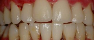
- the presence of fluctuation (accumulation of pus) under the mucosa in the form of a small reddened swelling.
If the above symptoms are present, the patient is advised to immediately consult a doctor for prompt treatment, otherwise the inflammation may intensify, spread to neighboring areas, develop into more serious diseases, or cause respiratory complications.
Kinds
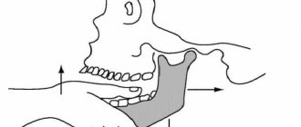
Based on the presence of the upper and lower parts of the jaw in a person, these inflammatory processes can be divided into two types: abscess of the lower jaw (the same type can also include a submandibular abscess, since their sources of origin are the same) and the upper jaw.
Abscess of the upper jaw
The most common source of infection is the upper wisdom teeth. Causes difficulty opening the mouth and swallowing.
Abscess of the lower jaw
Most often, the infection spreads from the lower molars (molars and premolars). The patient's complaints are mostly related to pain when chewing and swallowing.
An abscess of the submandibular region is characterized by visually noticeable and painful swelling in the submandibular triangle, and the shape of the face may be distorted.
Treatment and prevention
Treatment of a jaw abscess consists of opening the abscess and draining the fluid , after which the damaged area is disinfected. In case of high temperature, the patient is prescribed antibiotics; in case of a general weakening of the immune status, immunomodulatory drugs are prescribed; the doctor also gives recommendations for taking analgesics. In rare cases, for better healing of the postoperative incision, physiotherapeutic procedures and ultraviolet radiation are prescribed.
To prevent inflammation of this kind, it is advisable to visit the dentist once every six months, heal periodontal pockets in a timely manner, adhere to a gentle diet enriched with vitamins, and also use appropriate medicated toothpastes.
Some adherents of alternative medicine believe that the above inflammations of the maxillofacial area can be easily cured without resorting to surgery. Of course, there is a possibility that the abscess will open on its own, but if it is not cleaned and the remnants of dead particles and pathogenic bacteria are not removed from the wound, there is a high probability of an acute condition becoming chronic or phlegmon, as well as intoxication of the body with decay products remaining in the untreated abscess .
Secondary chronic purulent osteomyelitis of the mandible
Although recent cases of osteomyelitis of the maxillofacial skeleton are uncommon, clinicians must be able to effectively manage them. We believe that the most suitable treatment method for chronic purulent osteomyelitis (CPO) is antibiotic therapy in combination with surgery.
This article describes a case of successful surgical treatment of CHO of the lower jaw in an 18-year-old patient. Therapy included a preoperative course of antibiotics followed by sequestrectomy, resection of the coronoid process, and removal of the pathologically fractured condylar process on the left side of the mandible. During the postoperative clinical examination, already after one week, healing of the extraoral functioning fistula was noted with improvement in mouth opening.
Osteomyelitis is an inflammation of the bone and bone marrow that affects the jaw after chronic odontogenic infection or due to a number of other reasons. Chronic osteomyelitis can be characterized by a purulent course with the formation of an abscess or fistula, as well as sequestration. Although advances in anesthesia, antibiotic therapy, preventive and therapeutic dentistry, and the availability of combined medical and dental care have reduced the incidence of this disease, it has not yet been completely eradicated.
Based on several reports, we concluded that a successful treatment for chronic suppurative osteomyelitis is antibiotic therapy in combination with surgery, sequesterectomy, or decortication of the affected bone. The purpose of the operation is to remove all infected or necrotic bone tissue. Incomplete surgical treatment can cause persistence of the disease.
Clinical case
To the Department of Oral and Maxillofacial Surgery of the M.R. Dental College and Hospital. Ambedkar was referred to an 18-year-old patient with purulent discharge from a cutaneous fistula on the left lower edge of the body of the mandible. From the dental history it became known that a year ago the patient had a molar tooth of the lower jaw removed. A painful tumor with purulent discharge from the fistula formed in the submandibular region two months after tooth extraction. This problem was solved by the surgeon by opening and draining. Six months later, the discharge of pus resumed. A re-opening and drainage was performed, but did not give a positive result. The patient had no allergic reactions. According to the man, he did not smoke or abuse alcohol. During the examination, a suppurating extraoral fistula was identified, located 2 cm below the lower edge of the body of the lower jaw, with a painful mandibular lymph node on the left. There were two extraoral scars. One on the skin at a distance of 1 cm above the lower edge of the body of the lower jaw on the left. The second large scar was located on the left side of the neck, below and behind the left corner of the lower jaw, which indicated the poor quality of the operation performed by the surgeon. There was difficulty opening the mouth without paresthesia of the lower lip and chin area. Examination of the oral cavity showed a satisfactory condition of the dentition.
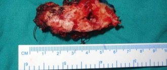
The panoramic radiograph showed a localized mottled area of radiolucency in the area of the sigmoid notch. The site was characterized by an oval shape; its maximum diameter was 20 mm. Based on radiographs, we assumed the presence of sequestration. A band of clearing was observed in the subcondylar region, which could indicate a pathological fracture of the condyle. Based on X-ray data, the patient was diagnosed with chronic purulent osteomyelitis of the condylar region. The treatment plan included a week of preoperative antibiotic therapy, sequestrectomy, and removal of the pathologically fractured condyle.
Under anesthesia, an intraoral incision was made starting posterior to the third molar and extending upward along the anterior edge of the ramus. After lifting the mucoperiosteal flap, doctors were surprised to find a discolored coronoid process with signs of impaired blood supply and infection. On the operating table we planned coronoidectomy and sequestrectomy. To expose the temporomandibular joint, a classic preauricular incision was made, carefully separating the different layers. The pathologically broken condyle was removed. The patient was prescribed antibiotics for one week after surgery.
Histopathological examination revealed chronic inflammatory cell infiltration with areas of bone trabecular resorption. These results, in combination with the clinical picture and radiographic signs, confirmed the diagnosis of chronic suppurative osteomyelitis.
A week after the operation, healing of the functioning fistula was observed with improvement in mouth opening. The preauricular area healed without scar formation. Repeat radiographs were taken three and six months after surgery. There were no clinical or radiographic signs of infection.
Discussion
Osteomyelitis is an inflammation of the bone that affects the medullary cavity and extends into the adjacent cortex, periosteum and soft tissue. The disorder is more common in the mandible due to the presence of dense cortical plates with few blood vessels and the only blood supply from the inferior alveolar neurovascular bundle . The main cause of chronic osteomyelitis is usually microbiological in nature and consists of odontogenic infection, complications after tooth extraction, incomplete removal of necrotic bone, incorrect choice of antibiotics or early cessation of antibiotic therapy, misdiagnosis, trauma, improper treatment of a fracture or irradiation of the mandible. Most often, clinicians receive a bacteriological result such as mixed oral microflora or mixed anaerobic microflora. The localization of osteomyelitis predominates in the lower jaw, most often in the area of the angle or body of the bone. In chronic secondary osteomyelitis, the clinical picture is usually limited to fistulas, hardening of soft tissues, as well as hardening or stiffening of the affected area with pain on palpation. In case of relapse, symptoms often occur directly in the area of decortication. Techniques used to diagnose chronic osteomyelitis include culture, bone biopsy, conventional radiography, radioisotope bone scincigraphy, laser Doppler flowmetry, computed tomography, and magnetic resonance imaging.
Treatment involves a course of antibiotic therapy in combination with surgery. Some authors talk about possible resistance to therapy in cases of chronic purulent osteomyelitis of the mandible. Topazian recommends continuing postoperative treatment for two to four months after symptoms resolve. Bartkowski et al provide ten to twenty-four days of infusion therapy. This is consistent with the published protocols of Van Merkesteyn et al. Some investigators have suggested that antibiotic therapy in combination with surgery is effective for the treatment of chronic suppurative osteomyelitis. Some reports recommend the use of hyperbaric oxygen, especially after irradiation of the mandible. In the present case, the patient was prescribed a course of antibiotics, which, in combination with surgery, gave a successful outcome.
Conclusion
The most important thing for curing the disease is correct diagnosis and treatment planning. In this case, incorrect diagnosis and treatment planning led to recurrence of the lesion with the formation of an unesthetic scar in a young man. Taking into account the clinical symptoms and course of the disease with an unsuccessful outcome of previous therapy, we focused on identifying the source of the infection, for the treatment of which we performed surgery and a course of antibiotic therapy.
Authors:
G.S. Rajkumar, M. Hemalatha, R. Shashikala, D. Veerindra Kumar