Recurrent ameloblastoma: a clinical case
Ameloblastoma is a common, localized, aggressive, benign odontogenic tumor of the oral cavity of epithelial origin. This pathology was first described by Cusack in 1827, and its name was given to Ivy and Churchill in 1930.
According to the 2005 WHO classification, there are 4 subtypes of benign ameloblastoma: (1) solid/multycyst, (2) desmoplastic, (3) unicyst, and (4) extraosseous/peripheral. Histologically, 6 tumor subtypes can be differentiated, including follicular, plexiform, acanthomatous, basal, unicyst and desmoplastic. Treatment is carried out either conservatively or radically, depending on the type, location and size of the lesion, as well as taking into account the age of the patient. A systematic review by Almaida and colleagues found that 50% of tumor recurrences are observed in the follicular subtype, and the incidence of such development is reduced when a radical treatment algorithm is implemented. This article describes two cases of recurrent ameloblastoma in patients who underwent segmental resection of the jaw.
Clinical case 1
A 46-year-old patient sought help at a private dental clinic with complaints of swelling in the area of preliminary surgery, covering the area of the lower jaw on the right. According to the medical documentation, it was established that about 15 years ago the patient underwent a segmental resection of the jaw with further augmentation of the formed defect with an augmentation from the region of the ribs. No data on the histopathological characteristics of the lesion were noted in the patient's medical record. After a CT scan and orthopantomography, a multilocular radiolucent lesion was discovered in the area of the previous surgical intervention (photo 1).
Figure 1: 3-dimensional reconstruction of a CT scan demonstrating recurrent tumor in the area of the augmentation involving the articular process.
Clinically, a diagnosis of recurrent multicystic ameloblastoma with involvement of the bone augmentation was made. An incision in the tumor area was made at a distance of 1 mm from the edge of soft tissues unaffected by pathology through the scar contour already existing from the preliminary operation (photo 2-3). The specimen collected for histology allowed a diagnosis of follicular ameloblastoma with acanthomatous changes and free margins. 1 year after surgery there were no signs of relapse. Therefore, alloplastic reconstruction of the mandible on the right side was planned, given the benign nature of the tumor.
Photo 2: View of the exposed tumor during surgery.
Photo 3: Surgical specimen.
Clinical case 2
A 45-year-old patient sought help from the Department of Oral and Maxillofacial Surgery with complaints of swelling in the right temporal region. The swelling did not provoke pain and developed over the course of a month, increasing in size. There were no signs of infection in the affected area. In 2012, the patient already approached a private dental clinic with a similar problem. An orthopantomogram and CT scans revealed a multicystic lesion. After a biopsy, it became clear that the problematic tumor was an ameloblastoma. The patient underwent a hemimandibulectomy procedure. Directly during surgery, radiography of the surgical tumor specimen was taken to analyze the condition of the tissues at a distance of 1.5 cm from the clinically visualized tumor edge. Histological analysis confirmed the diagnosis of ameloblastoma with atypical features and multicellular structure. In 2021, the patient again experienced swelling in the right temporal region. MRI identified heterogeneous signal enhancement in this area measuring 4.4 cm × 3.7 cm × 4.6 cm with the presence of tiny cystic inclusions (Figure 4). Through access along the zygomatic bone, the tumor was removed with a certain amount of surrounding tissue (photo 5-6). The formed defect was restored using the temporal muscle. After histoanalysis of the surgical tissue sample, the diagnosis of ameloblastoma with non-invasive margins was confirmed. At this time, the patient is at the monitoring stage.
Photo 4: Preoperative CT scan showing recurrent ameloblastoma in the infratemporal fossa.
Photo 5: Visualization of the bone defect after tumor removal, which was then reconstructed using the temporalis muscle.
Photo 6: Excised tissue sample.
Discussion
Ameloblastoma is a locally aggressive tumor of the oral cavity that rarely undergoes malignant transformation. After odontoma, it is the second most common odontogenic tumor. According to epidemiological data, 70% of cases of ameloblastoma were reported in developed countries. The problem of low identification of ameloblastoma in developing countries is obviously associated with the insufficient level of their official registration. The WHO classified ameloblastoma into four subtypes in 2005: multicystic/solid, unicyst, desmoplastic, and extraosseous. Solid/multycystic ameloblastoma is the most common type, which is also the most aggressive and often recurs after conservative treatment such as curettage or enucleation. Ameloblastomas of the lower jaw recur much more often than their counterparts in the upper jaw. Also, the follicular type of tumor is characterized by a higher rate of relapse compared to its plexiform subtype. The pathogenesis of ameloblastoma development remains incompletely understood. The genetic theory of development involves the participation in the process of pathogenesis of the BRAF protein in the structure of the mitogen-activated protein kinase pathway (MAPK), which, as a rule, is already identified as mutated.
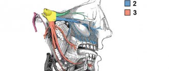
Ahlem et al found that the activity of the biomarkers Ki67 and CD10 was significantly higher in recurrent tumor types compared to primary tumor types. As a rule, ameloblastoma affects the posterior areas of the lower jaw. Infiltration of the lesion occurs into the cancellous part of the bone with less involvement of the cortical component. The CT scanning method for diagnosing a tumor is the most informative when the cortical structure of the bone is damaged and the damage has spread to the surrounding soft tissue. Treatment of the tumor can be conservative or radical. A conservative approach involves enucleation of the tumor with further chemical cauterization or peripheral osteoectomy with expansion of the excision boundaries by 1-1.5 cm from the visualized edge of the tumor. A conservative treatment approach is more effective in cases of monocystic type of formation. According to Pogrel and Montes, unicystic ameloblastomas respond best to conservative treatment using Carnoy or cryotherapy principles. A radical approach is indicated in cases of large ameloblastomas when the lesion has spread to the area of the inferior alveolar nerve, or in cases of the aggressive nature of the tumor. The radical treatment method involves performing marginal or segmental resection with removal of adjacent bone tissue 1.5-2 cm from the visualized edge of the tumor. To confirm that the tumor has spread to adjacent tissues, it is recommended to carry out control radiography directly during surgery. In cases of ameloblastic carcinoma, excision of bone tissue is recommended at a distance of 2-3 cm from the visualized edge of the tumor. Recurrence of ameloblastoma is most often associated with the histological type of lesion and its location. The solid variant of the neoplasm is the most aggressive. Most often, ameloblastoma occurs in the distal areas of the lower jaw and affects the cancellous component of the bone through the mechanism of invasion, reaching the cortical plate and provoking its perforation. Once the tumor penetrates the periosteum, the risk of its further spread to the surrounding soft tissue increases. Inadequate lesion resection increases the risk of possible recurrence during follow-up. Cases of recurrent ameloblastoma have already been described by Grafft, Carvalho, Dolan, Marinelli, Stea, Zacharides, Vasan, Bianchi, Martins and Favaro, Su, Choi, Basat.
In most of the described relapses, restoration of the defect site after surgery was carried out using an iliac graft. Cases of recurrent ameloblastoma after radical treatment were described in studies by Sehdev, Shatkin and Hoffmeister, Mehlisch, Muller and Slootweg, Olaitan, Ueno, Eckhardt, Nakamura. It is known that the relapse rate with conservative treatment is significantly higher (60%) than with radical treatment (13%). The risk of relapse during surgical excision of ameloblastoma may be associated with retention of epithelial conglomerates of the neoplasm in the structure of the surrounding soft tissue. In the first clinical case described, the augmentation tissue succumbed to tumorigenesis, apparently due to the remaining tumor cells in the area of the osteoectomy. Therefore, before starting reconstruction of solid ameloblastomas, it is recommended to monitor the cleanliness of the edges of the excised tissues using control radiography. It is even better to wait for the results of a histological analysis of a tissue sample taken during the operation. Tumors in the infratemporal fossa are usually asymptomatic, and the main complaint of patients remains only signs of swelling and facial deformation. The infratemporal fossa is a pyramid-shaped structure that is located on the lateral surface of the base of the skull, deeper than the temporal arch, masseter muscle and ramus of the mandible. The medial part of the ramus of the mandible and the floor of the skull form the upper surface of the pyramid-shaped structure. The anteromedial part of the infratemporal fossa corresponds to the posterior surface of the upper jaw, and the posteroinferior part is formed by the pterygoid fascia. In fact, the infratemporal fossa is anatomically inaccessible, so it is almost impossible to monitor neoplastic growth in its structure.
Diagnosis and treatment planning for ameloblastomas localized in the infratemporal fossa should include the use of CT and MRI methods, which make it possible to establish an objective level of tumor spread. Conventional radiography in such cases is not sufficiently informative. Access to the infratemporal fossa can be formed transorally, transnasally, transpalatally, through the zygomatic bone, transcervically, and even through an extended maxillary approach. The approach through the zygomatic bone area is one of the most suitable for both resection of neoplasia and the formation of a temporal flap. The choice of surgical approach depends on the tumor type, clinical features, histological subtype, extent of spread and location.
In the second clinical case, relapse may be associated with preservation of the periosteum during a previously performed operation. It is in the periosteum that conglomerates of neoplastic cells could and will remain. A more extensive resection allows you to remove all tissue that has succumbed to neoplastic invasion, and thus minimize the risk of relapse. Carlson and Marx described the need to remove all anatomical structures adjacent to the tumor area for prophylactic purposes. This approach is reasonable when tumor invasion into surrounding soft and hard tissues is confirmed, since cases of recurrent ameloblastomas have been reported even 30 years after initial removal of the lesion. Laborde and Becelli described that in the cases of 7 patients, there was no evidence of tumor recurrence after treatment, and doctors used an approach with extensive excision of surrounding tissue.
Similar reports were also provided by Vayvada, Chaine and Basat. Vaishampayan suggested that recurrence of ameloblastoma after excision from the infratemporal fossa is associated with retention of the weakened process of the mandible after segmental resection procedures. Peacock et al concluded that the completeness of tumor excision can be verified not only by histological examination, but also by plain radiography. Although several publications indicate the resistance of ameloblastomas to brachytherapy, irradiation of recurrent types of lesions with high doses of 110 Gy was characterized by a fairly successful result.
During treatment for ameloblastoma, we recommend adhering to the following recommendations:
- In order to control the degree of invasion of large ameloblastomas into the surrounding soft tissue, it is necessary to use the MRI method.
- To control the completeness of excision of neoplastic tissue during surgery, it is necessary to use control radiography.
- Tumor resection margins should be widened to minimize the risk of future tumor recurrence.
- After confirming the cleanliness of the working field, it is necessary to carry out surgical reconstruction of the bone defect formed after tumor removal.
- Monitoring the area of preliminary localization of ameloblastomas after their resection is mandatory and should include the use of CT and MRI control.
conclusions
Based on the analysis of two clinical cases, we can summarize that the solid type of ameloblastoma is extremely aggressive and is characterized by a high rate of relapse. During treatment, it is necessary to take into account the risk of tumor invasion into the surrounding soft tissue, as well as the fact that the tumor, as it progresses, can provoke erosion of the cortical ball of the mandible. To gain a deeper understanding of the nature of the development of ameloblastomas, it is necessary to conduct a number of additional clinical and histological studies.
Authors: Chithra Aramanadka, Abhay Taranath Kamath, Adarsh Kudva (India)
Microsurgical operation. Ameloblastoma of the jaw.
Key words: jaw defect, dental implantation, restoration of chewing function in patients with jaw defects, computer gnathography.
Ameloblastoma or adamantinoma is a benign tumor belonging to tumors of the maxillofacial localization. Typically, ameloblastoma spreads to the lower jaw. Such a tumor has a slow growth, occurs without any special symptoms, but gradually leads to deformation of a person’s lower jaw. Very often, patients complain of impaired chewing function, loosening and displacement of the dentition. On examination, patients experience facial asymmetry, swelling of the lower jaw, its thickening or protrusion.
The staff of the scientific and clinical department of maxillofacial surgery of the Federal State Budgetary Institution Scientific Center of Otorhinolaryngology of the Federal Medical and Biological Agency of Russia has accumulated a wealth of experience in the diagnosis and treatment of patients with defects of the lower jaw. All surgeons of the department have scientific titles, have completed internships in the world's leading maxillofacial surgery clinics, and are proficient not only in the most modern domestic and foreign techniques, but also actively use their own developments.
In our Center, an important link in the accurate diagnosis of patients with ameloblastoma is the use of biometric examination methods (CT, computed tomography, electromyography). Treatment of patients with defects of the lower jaw is aimed not only at restoring chewing and swallowing functions and correct bite, but also at restoring facial aesthetics.
ameloblastoma of the lower jaw, specialists from the scientific and clinical department of maxillofacial surgery use the technique of using flaps with axial circulation and the technique of preoperative 3-D computer modeling. Based on the modeling results, special templates are made that accurately determine the size of the bone component of the flaps. Our experts believe that such a modern approach is necessary and is due to both functional and socio-aesthetic rehabilitation of patients who have lost their jaw. Successful operations after bone grafting with autografts demonstrate the correctness of an integrated approach to the treatment of patients with defects of the lower jaw, clearly demonstrate the ability of the attached muscles to contract and maintain muscle tone, which creates good conditions for further prosthetics with implants and creating optimal occlusion.
Examples of clinical observation
A patient diagnosed with large ameloblastoma of the lower jaw. Ameloblastoma of the lower jaw was removed with simultaneous surgical reconstruction
BEFORE TREATMENT | ||
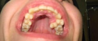
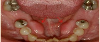
INTRAORAL PHOTOS |
MSCT - 3D SCANS | ||
3D modeling | ||
STL templates |
INTEROPERATIVE PHOTOS |
MSCT after surgery |
Photo 20 days after surgery |
PATIENT WITH DIAGNOSIS: defect of the lower jaw on the left, facial asymmetry, impaired chewing function, condition after subtotal resection for sarcoma
BEFORE TREATMENT |
CT scans of the patient before surgery |
CT scans after surgery: there is a tendency towards an open bite, despite the precise position of the graft in the temporal fossa. |
Patient’s bite at the stage of orthodontic treatment (Bite correction) |
Patient's bite after removing braces |
AFTER dental implantation, formers are installed and temporary prosthetics supported on implants are performed |
CT scans of the patient with muscle visualization: attachment of the masticatory muscles to the bone component of the previously transplanted graft on the left is noted |
Formation of the coronoid process |
Electromyography at rest before relaxation. Significant hypertonicity of the anterior fibers of the temporal muscles. |
EMG after electrical neurostimulation. The tone of all muscle groups is balanced and the indicators are significantly lower. When teeth are closed, the tone of the temporal and masticatory muscles increases. |
The compression test revealed good involvement of muscle fibers of the temporalis muscles in function and decreased involvement of muscle fibers of the masseter muscles, especially on the left. |
EMG compression after electrical neurostimulation. Increased involvement of muscle fibers in function by almost 2 times. |
Computer gnathography. Slight limitation of mouth opening. Deviation of the lower jaw to the left when opening the mouth. |
PATIENT WITH DIAGNOSIS: ameloblastoma of the mandible of the mental region
BEFORE TREATMENT |
| CT scan | Simulation of resection | Position of the fibular autograft |
Photographs in the postoperative period show the patient’s unchanged postoperative appearance, fixed prosthetics, and a CT scan 2 months after surgery. |
At OPTG, dental implants used for fixed prosthetics are determined |
Attachment of the digastric and mylohyoid muscles to the fibular graft. |
Diagnostics on the K7 device |
Patient's myography results |
PATIENT K., 26 years old, was admitted to the clinic with a diagnosis of ameloblastoma of the lower jaw on the right.
The appearance of the patient upon admission, OPG, CT images, 3D modeling of the operation, stereolithographic templates. |
Appearance of the patient after surgery. |
OPTG and CT gram demonstrate the anatomy of the reconstructed alveolar process. |
The OPTG shows installed dental implants 6*16 mm in the projection of 4.4, 4.5, 4.6, 4.7 teeth. |
CT scans show attachment of the masticatory muscles to the iliac autograft. |