25.11.2019
The appearance of various wounds, cysts, growths and tumors on the oral mucosa is a fairly common phenomenon in dental practice, which most often occurs due to mechanical damage to the gum tissue.
When extracting a tooth and not carefully following the recommendations of the attending physician during the rehabilitation period, various complications associated with damage to bone and soft tissue may occur. One of them is the development of jaw exostosis.
GENERAL VIEW
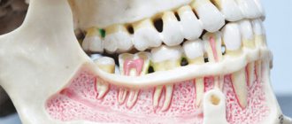
Exostosis of the oral cavity is the proliferation of bone and cartilage tissue in a certain area of the jaw row. Visually, it looks like a lump or nodule that tends to grow rapidly.
The sharp edges of the hardened cartilage tissue that forms exostosis can damage the mucous membrane covering it, leading to injury and severe pain, making it difficult to open the mouth and eat.
On the upper jaw, exostotic formations are most often localized in the area of masticatory elements on the outer or inner side of the row. When the lower jaw is affected, growths form at the base of the incisors, canines and premolars.
As a rule, cartilaginous growths have a benign structure, but there are known cases of their transformation into malignant neoplasms.
A lump appeared on the gum: what could be the reason?
A lump on the gum, by and large, is a compaction that is caused by damage to periodontal tissue in a certain area. The cause of the lesion is another point on which the treatment plan and possible complications in the absence of timely medical care depend. First of all, the doctor determines the nature of the appearance of the lump on the gum, which comes in two types.
- Infectious nature.
Infection caused by pathogenic bacteria is the most common cause of lumps and lumps in the gum area. - Non-infectious nature.
This includes injuries, mechanical, chemical damage, as well as other factors not related to the activity of microorganisms.
A lump on the gum above a tooth or under a tooth (in the case of the lower jaw) appears much more often as a result of infections, however, the treatment regimen is individual for each case, depends on the specific symptoms and is drawn up after collecting an anamnesis. Next, it makes sense to describe in more detail the diseases and pathologies that provoke the appearance of seals on the gums.
REASONS FOR THE APPEARANCE
Most often, the cause of exostosis is heredity. In this case, areas of slight growth of bone or cartilage are observed already in childhood.
If there is no growth and no discomfort, the specialist recommends regular monitoring without surgical intervention.
In addition to the genetic factor, exostosis can be caused by the following reasons:
- injury to the jaw rows, their fractures, causing chipping of bone tissue fragments with subsequent improper fusion;
- infectious inflammatory processes, as a result of which bone tissue atrophies and is destroyed;
- pathological structure of the jaw rows, resulting in the formation of curvature of bone tissue and the formation of growths;
- hormonal disorders, due to which the qualitative composition of the bone changes, causing its destruction and susceptibility to the formation of growths.
Dentists also note that very often the development of exostosis is a consequence of complex tooth extraction.
The reason for the growth of bone tissue in this situation is the lack of natural smoothing of the edges of the socket, improper fusion of bone tissue, or excessive trauma to the periosteum during the procedure.
Varieties of epulis
Conventionally, there are 3 types of epulis:
- Fibrous. The neoplasm is dense, the basis is coarse fibrous tissue. Characterized by slow growth. There is no pain or bleeding.
- Angiomatous. The tumor contains a large number of blood vessels and is localized mainly in the lateral parts of the jaw. With this form of the disease, bleeding is observed even with slight pressure, for example, with a toothbrush or food. When pressed, the tumor is painless and has a soft consistency.
- Giant cell. The tumor can reach large sizes, displacing teeth that are located in the epulis growth zone. The mucous membrane of the epulis is bluish-red in color. On palpation, the neoplasm is painless, but may bleed moderately if injured.
Depending on the nature of the process, epulis can be benign or malignant. In the first case, the neoplasm is characterized by slow development and, as mentioned above, in most cases does not cause pain. The tumor size in this case rarely exceeds 10–20 mm. When the process becomes malignant, rapid tissue growth is observed. In this case, the process is often accompanied by pain, bleeding gums and affects the root canals of neighboring teeth.
SYMPTOMS
The initial stage of development of the disease is characterized by the absence of pronounced symptoms, therefore, at this stage, detection of exostosis is possible only during a dental examination.
A specialist will be able to determine the presence of an area of overgrown tissue and suggest the cause of its occurrence.
As the disease progresses, symptoms become more noticeable:
- on the gum there is a tubercle covered with a mucous membrane, which may look unchanged or be covered with small spines;
- the growth increases in size and can turn from a pea into a large lump, which causes discomfort and interferes with the full position of the tongue;
- painful sensations of varying intensity occur;
- the mucous membrane gradually acquires a rich pink tint;
- there is a violation of the patency of blood vessels located in the oral cavity;
- the degree of mobility of the lower jaw is impaired.
Solitary form of peripheral osteoma on the lower jaw
Osteoma is a rare, slow-growing, benign osteogenic tumor that most often occurs in the maxillofacial region. These types of neoplasms can be found both on the surface of the bone (formed from the periosteum - peripheral, periosteal or exophytic form), and in the structure of the bone marrow space (formed from the endosteum - endosteal or central form). The case of verification of osteoma in the structure of muscle tissue is rare.
The pathogenesis of peripheral osteoma remains a completely unresolved clinical and scientific issue: there are opinions that the neoplasm can be attributed to true tumors or to reactive lesions provoked by trauma, constant traction of the muscles attached to the periosteum, as well as the action of infectious factors. Endocrine changes, in turn, can be a direct etiological factor initiating the occurrence of a neoplasm. Depending on the action of endocrine factors on the tumor, they can be classified as central and peripheral, or extraosseous.
Osteoma is the most common benign tumor of the nose and paranasal sinuses and the most common neoplasm in the frontal sinus. Single peripheral osteomas that occur in the lower jaw are quite rare lesions and are diagnosed in people of different age categories (from 4.8 months to 50 years), equally often in both men and women. Typically, clinically the tumor grows asymptomatically, and radiographically it manifests itself as a distinct contrasting area of a round or ovoid lesion. Depending on the histological picture, compact, spongy and mixed forms of osteoma are distinguished. The presence of many small osteomas in the structure of the lower jaw is diagnosed as Gardner's syndrome, which is also characterized by the presence of perirectal polyposis and associated skeletal abnormalities. These symptoms can be used to differentiate between Gardner's syndrome and multiple dental impaction.
Treatment of osteomas is carried out through surgical excision of the tumor, but only in those patients who complain of accompanying uncomfortable symptoms of the tumor. Until now, only 26 clinical cases of verification of single peripheral osteomas of the mandible not associated with Gardner's syndrome have been reported in the literature.
Based on the extreme rarity of the disease, the purpose of this article is to illustrate an unusual clinical case of a single peripheral osteoma found in the structure of the mandible, as well as to conduct a retrospective analysis of the literature data regarding the pathogenesis, differential diagnosis and choice of treatment for this type of benign tumor.
Clinical case
A 30-year-old patient applied to the Department of Dentistry with the main complaint of swelling on the inside of the posterior part of the lower jaw, which she had noticed since the age of 8. 8 years ago, the formation that had been present since childhood increased to 5 mm in diameter, after which it began to grow progressively. Although the swelling was asymptomatic, due to its large size it provoked a feeling of discomfort when eating. The latter motivated the patient to seek dental care. The patient did not have any hereditary diseases in her own or family history, as well as a recent injury to the problem area of the jaw. Based on the fact that no other symptoms other than a tumor in the mouth were found, Gardner's syndrome was excluded from the list of possible diagnoses. Clinically, a single pedunculated oval node was detected on the lingual side of the jaw in the area of the 36th and 37th teeth. The tumor was more than 1 cm in diameter (Figure 1), and the overlying mucosa showed no signs of change or inflammation. Upon palpation, the tumor was painless, and it was also possible to manually establish its lobular shape and hard consistency.
Photo 1: View of a tumor in the oral cavity.
On a cross-sectional radiograph of the lower jaw, a clear radiopaque area of irregular shape is visualized, connected to the cortical plate of the jaw in the area of the 37th tooth through a pedicle (photo 2). The radiographic density of the formation is similar to the density of the body of the lower jaw. In the process of differential diagnosis, possible variants of bone exostosis, peripheral osteogenic fibroma, osteoid osteoma, osteoblastoma and osteosarcoma were analyzed. After carefully explaining to the patient the essence of the future surgical procedure and the possible risks of manipulation, complete excision of the tumor was performed using rotary instruments and a bone chisel. During surgery, an excisional biopsy of the tumor was also performed under local anesthesia.
Figure 2: Cross-sectional radiograph of the mandible.
Histopathological analysis of the lesion helped to establish that the tumor consists of dense bone tissue, in which concentric layers of bone plates are observed, as well as osteocyte cells in the lacunae (photo 3). The results of histopathological examination along with clinical and radiological findings helped confirm the diagnosis of compact osteoma.
Photo 3: Hematoxylin-eosin staining (10x magnification).
Discussion
According to the World Health Organization classification, osteoma belongs to benign neoplasms, consisting of differentiated mature bone tissue cells with a predominance of laminar structure, and with a very slow growth pattern.
It is not yet clear whether osteomas are true benign neoplasms or hamartomas. Although, on the other hand, the latest WHO classification of diseases defines osteoma as a truly benign tumor of bone tissue, clearly differentiating it from exostoses and other types of hamartomas.
The true etiology of the lesion has not been thoroughly elucidated, although Fetissof and Varboncoeur argue that the tumor develops either from embryonic cartilaginous remnants or from persistent embryonic cells contained in the periosteal structure. According to the embryological theory, osteomas originate from the area of sutures of bone structures filled with tissues of different embryonic origin (membranous or enchondral). Other possible etiological factors are inflammation, trauma or endocrine pathologies. Kaplan et al suggested that the combination of trauma and persistent muscle traction in the periosteal region plays a key role in the development of the peripheral type of neoplasm. But the above-mentioned theories of osteoma formation do not explain the mechanism of its development in the described clinical case. Regezi and Sciubba, as a result of a comprehensive analysis, came to the conclusion that none of the proposed etiological theories fully explains the cause and mechanism of development of the neoplasm.
Histologically, three types of osteomas can be distinguished: hard or compact (consisting of a dense, ivory-like compact substance with a small amount of spongy bone, this type of pathology is quite difficult to resect), cancellous (trabecular), or mature (consisting of soft spongy bone and bone marrow) and mixed. Single peripheral osteomas (PO) of the jaws are quite rare lesions and are most often diagnosed in young people with the same frequency in both men and women. The patient described in the clinical case mentioned above was 30 years old. Typically, osteomas are asymptomatic tumors and can often go undetected until they cause visible facial asymmetry or functional malocclusion. In this case, the pathology must be confirmed by x-ray examination. The most common topography of osteomas is the area of the body of the mandible behind the premolar on the lingual surface and the area of the condylar process. Less often, osteomas occur in the area of the angle, coronoid process and ramus of the mandible, or in the area of the external auditory canal, orbit, temporal bone, maxilla, zygomatic arch and pterygoid processes. Often osteomas can be verified as part of Gardner's syndrome, characterized by the presence of several tumors in the jaw structure, pararectal polyps with a high degree of malignancy, cutaneous fibromas, congenital retinal hyperpigmentation, tooth retention, enostosis, epidermal cysts and supernumerary teeth. Gardner syndrome is an autosomal dominant disease caused by a mutation in the structure of the APC suppressor gene, which is responsible for the occurrence of adenomatous polyposis of the colon. It is extremely rare, but detection of osteomas is also possible in Haberland syndrome (encephalocraniocutaneous lipomatosis).
Radiologically, a peripheral osteoma may have a mushroom-shaped or oval-shaped lesion that is in close contact with the adjacent cortical plate of the mandible. Moreover, the bone density in the jaw area and in the area of the neoplasm is almost identical. Traditional radiography methods, such as occlusal radiographs, are usually sufficient to diagnose osteoma, as in our case. When the tumor is located deeper in the tissues of the maxillofacial region, other highly effective imaging methods can be used to identify it: panoramic radiography, occipitomental projection radiography of the sinuses, computed tomography (CT), magnetic resonance imaging (MRI) or cone -radiation computed tomography (CBCT). Spiral computed tomography provides opportunities for three-dimensional reconstruction of osteoma: using radionuclide imaging, it is possible to distinguish between actively growing neoplasms (“hot”) and stable lesions (“cold”). In the structure of spongy osteoma, the vascular component predominates, while in the compact type of tumor there are almost no vascular elements observed. Differential diagnosis of different types of formation can be carried out using intravenous angiography.
Differential diagnosis of osteoma is carried out with bone exostoses, osteochondroma, osteoid osteoma, periosteal osteoblastoma, peripheral osteosarcoma, peripheral osteogenic fibroma, Paget's disease, fibrous dysplasia and odontoma. Exostoses are hamartomas that are usually located on the lower jaw or in the palate. Typically, exostoses stop growing after puberty, while osteomas continue to slowly increase in size, occupying a limited volume and forming a lobulated radiopaque structure. Osteochondromas consist of areas of endochondral ossification, calcified cartilage, and adipose or bone marrow tissue within the lumen of the trabecular space. Osteoid osteomas, on the contrary, are characterized by rapid growth, are often painful and contain a large number of vessels and an osteoid component. Periosteal osteoblastoma is also a rapidly growing, painful, round or oval tumor attached to a specific area of the cortex. Parosteal osteosarcoma is often diagnosed in the posterior region of the mandible, representing a homogeneous or heterogeneous formation with a poorly verified lobular sclerotic structure. Telangiectasias are often observed at the periphery of the tumor, and Codman's triangle can be seen on the radiograph. Peripheral osteogenic fibroma can be found only in the gum area; it is a reactive lesion of dense consistency, often found in the anterior part of the upper jaw. Symptoms of Paget's disease are more typical for the anatomical areas of the femur, skull and vertebrae, while in the jaw area, bone damage is observed much less frequently and more often in the older age group. In this case, bone damage, as a rule, is always bilateral, and more often affects the upper than the lower jaw. Bone tissue in Paget's disease becomes deformed and increases in size. Fibrous dysplasia is usually seen in the posterior maxilla and radiographically appears as a diffuse, contrast-enhancing mass that may resemble ground glass, orange peel, or cotton wool. Odontomas often complicate the physiological eruption of permanent teeth, while their radiological density is almost identical to that of enamel and dentin, and the affected area is always surrounded by a radiopaque capsule.
conclusions
During the clinical examination of patients with osteomas, significant attention should be paid to the family history to exclude a possible diagnosis of Gardner's syndrome. Asymptomatic osteomas should be under constant observation, and pathologies that cause uncomfortable symptoms should be surgically removed, having previously organized a complex of clinical and radiological studies to accurately make the final diagnosis.
By Rohit Agrawal Shipra Agrawal Shitij Bhargava Mahesh Motlani Rahul Agrawal
INDICATIONS AND CONTRAINDICATIONS FOR OPERATION
Dentists note that mandatory surgical intervention to eliminate exostosis is required in the following situations:
- with accelerated growth of the tumor;
- if the patient experiences severe pain and discomfort;
- during planning of further orthopedic treatment, which becomes impossible in the presence of bone outgrowths;
- with significant cosmetic defects of the facial area associated with an increase in growth.
When planning an operation, it is also worth paying attention to the functioning of the human body, in which removal of exostosis is prohibited.
Dentists name the following contraindications to the procedure:
- some blood diseases, including blood clotting disorders and increased sugar levels;
- diseases of the thyroid gland and adrenal glands;
- hormonal imbalance;
- periods of exacerbation of chronic diseases.
PREPARATION
Surgical intervention is the only option for removing a growth of bone or cartilage tissue from the surface of the jaw row, and therefore requires careful preparation to ensure a favorable outcome.
During the patient’s initial appointment, the dentist clarifies his complaints and conducts a visual examination of the oral cavity in order to identify visible pathologies. At this stage, a number of concomitant diseases are often identified: caries, gum or periodontal inflammation, which require treatment.
To determine the exact location of the growth, its shape, size and nature, the specialist sends the patient for an x-ray examination. The completed image is the basis for making an accurate diagnosis and drawing up a treatment plan.
At the stage of preparation for surgery, the dentist directs the patient to undergo tests, which can be used to identify additional diseases that impede the procedure. Thus, mandatory steps include checking blood for clotting and sugar levels, assessing the balance of hormones and the functioning of the adrenal glands.
At the preparatory stage, the specialist also decides on the option of administering an anesthetic drug.
Surgery to remove exostosis is often performed using local anesthesia, but in some situations general anesthesia may be required.
Before administering it, the specialist checks with the patient for the presence of an allergic reaction and previous experience with pain relief.
What to do if exostosis occurs after tooth extraction
Osteocartilaginous formations in the mouth can only be treated surgically, so if you suspect a pathology, you should consult a dentist. The doctor makes an accurate diagnosis after examining the patient: visual examination and radiography. Differential diagnosis of the nature of the lumps may be required.
Exostosis itself does not disappear, so the doctor decides whether the growth needs to be removed. If the protrusions are small, then the operation can be postponed indefinitely. In this case, you need to undergo regular examination at a dental clinic to monitor changes in tissues.
SURGICAL INTERVENTION
The procedure for removing exostosis begins with anesthetizing the area of the oral cavity that is to be operated on.
Before administering general anesthesia, the specialist determines whether the patient has an individual intolerance to the drug using a special test.
After administering the anesthetic, the specialist performs the following sequence of actions to remove exostosis:
- Disinfection of the oral cavity using special disinfectants, which eliminates the risk of infection of soft and bone structures during surgery.
- Making an incision in the gum tissue to gain access to the cartilage growth. To perform this operation, the dentist uses a scalpel of the appropriate size.
- Removing the build-up. Depending on the chosen method of the procedure, a dental chisel or a laser can be used at this stage, with the help of which the tumor is carefully cut off from the surface of the bone tissue.
- Sanding the treated area. To create a smooth surface and remove small particles of cartilage structure, which prevents recurrence of the disease in the future, the bone tissue is carefully ground using a dental drill. To avoid overheating of the bone, the specialist ensures timely cooling of the treated area.
- Apply sutures to the excised area of gingival tissue and secure with a sterile bandage.
The duration of the operation to remove exostosis often does not exceed 2 hours. When using a laser, the time is reduced to 40-50 minutes.
REHABILITATION
Removal of exostosis is not a simple operation, so the recovery period most often does not exceed a week.
This time is enough for the gum tissue to recover after the cut, and for the bone to take on its natural shape.
To ensure an easy recovery period and to avoid postoperative complications, the patient should follow these rules:
- regularly take antibiotics and other medications prescribed by a specialist;
- avoid eating foods at too high or low temperatures;
- limit physical activity;
- stop smoking and drinking alcohol;
- exercise caution during daily hygiene procedures.
POSSIBLE COMPLICATIONS
Postoperative complications associated with the removal of exostosis are quite rare in medical practice, and according to the experience of dentists, they are associated with the patient’s failure to comply with the rules of the rehabilitation period.
The consequences of unscrupulous adherence to doctor’s recommendations include the following pathological conditions:
- Complete or partial divergence of the suture material. The cause of this phenomenon may be poor diet, smoking, or mechanical damage to the gums during brushing. To fix the problem, you should immediately consult a doctor, who will treat the damaged surface and re-stitch the stitches.
- Inflammation of the gums in the suture area. Due to poor oral hygiene, pathogenic microorganisms can accumulate in the wound area, the result of which leads to suppuration and swelling of the soft tissue. You can only get rid of this complication with the help of a specialist.