23.11.2019
The term “osteoblastoclastoma” was proposed by A.V. Rusakov in 1959. The International Histological Classification of Primary Tumors and Tumor-Like Bone Diseases uses the term “giant cell tumor.”
Until now, there are different names for giant cell tumors: giant cell tumor of the epulid type, brown tumor, gigantoma, giant cell sarcoma, osteoclastoma, local fibrous osteodystrophy.
In the etiology of this tumor, according to some authors, bone trauma plays a significant role. Giant cell tumors are classified as true tumors - blastomas. Currently, both surgeons and pathologists recognize these tumors as benign, although prone to recurrence. This led to the replacement of the terms “giant cell sarcoma” and “giant cell tumor” with the term “giantoma”. Giant cell tumors can have a sarcomatous structure.
Osteoblastoclastoma is a benign osteogenic tumor. Occurs in the jaw bones. The tumor develops mainly in young people, most often in women 11–30 years old. According to Yu. I. Bernadsky, osteoblastoclastomas account for 20.7% of the number of tumors of the facial bones.
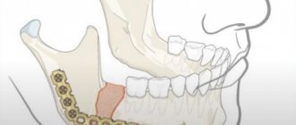
Osteoblastoclastoma can be located along the periphery of the bone and in the thickness of the bone tissue. When localized on the alveolar process of the jaw, it is considered a giant cell epulid. In the lower jaw, the tumor is most often detected in the area of premolars and molars, in the upper jaw - in the area of premolars. Rare localizations include damage to the zygomatic bone.
Osteoblastoclastoma—education clinic
Unnoticed by the patient, a uniform thickening appears in one of the areas of the lower or upper jaw, often painless. Tumor growth continues for 3–10 years, but sometimes it is more intense.
When palpating the formation, its dense areas alternate with softened ones. Its shape is convex, dome-shaped, and sometimes thinning of the jaw walls occurs, which manifests itself as Dupuytren’s symptom (crepitus). The teeth in the formation zone are mobile, their electrical excitability is reduced, and resorption of tooth roots by 1/3 of the length is noted in the lesion. The mucous membrane above the tumor is either unchanged or slightly anemic; the venous network of the mucous membrane in the area of the tumor is expanded.
When an odontogenic inflammatory process occurs, signs of acute inflammation prevail. The diagnosis can be established through a comprehensive clinical and radiological examination.
Based on clinical and radiological data and the morphological picture, three main forms of osteoblastoclastoma are distinguished: cellular, cystic, lytic.
The cellular form is more often observed in adulthood and old age, and is characterized by very slow development. When examining the patient, a dense swelling with a lumpy surface is determined; it is not possible to clinically distinguish the tumor from healthy areas of the bone; the jaw is often spindle-shaped. Teeth located in the tumor area rarely change their position. The electrical excitability of the pulp of intact teeth is not impaired. The mucous membrane covering the tumor is somewhat anemic.
With the cystic form of the tumor, the first symptoms of the disease in most cases are complaints of toothache. During palpation examination, individual areas of the tumor are pliable when pressed (symptom of “parchment crunch”); the thinned bone above the tumor has a smooth-convex, dome-shaped shape.
The lytic form is rare, more often in childhood and adolescence, and accounts for 10% of all osteoblastoclastomas of the jaws. The development of this form of osteoblastoclastoma occurs quite quickly. In some cases, the first sign of a developing tumor is pain. When the cortical layer thins, along with independent pain at rest, pain appears on palpation. The venous network of vessels of the mucous membrane covering the tumor is dilated. Teeth often shift and become mobile, and the electrical excitability of the pulp decreases. Pathological fractures of the jaw may occur in the affected area; when localized on the upper jaw, it is possible to grow into the maxillary sinus, nasal cavity and other bones of the facial skeleton. When the formation is punctured, a brown or yellowish liquid is detected, which is associated with the breakdown of red blood cells and the formation of hemosiderin, sometimes with blood. The punctate does not include cholesterol crystals, which are usually found with cysts.
Causes of development of osteoblastoclastoma of the jaw
The basis of any tumor degeneration of cells is a change in the structure of cellular DNA. And today it is not completely clear what exactly causes such changes. A definitive answer has not been found to the question of why the immune system sometimes loses sight of tumor cells. At the same time, certain patterns of the occurrence of osteoblastoclastoma have been identified and it has been established what factors can provoke the development of this tumor. These include:
- Mechanical injuries - for example, osteoblastoclastoma can be caused by a bruise of the jaw bone or permanent injury to the mucous membrane of the oral cavity with a poorly processed dental filling or a poorly fitted denture.
- Infection in the socket of an extracted or fallen tooth.
- Chronic inflammatory processes in the periodontium, periosteum or jaw bone. Sinusitis or actinomycosis can also contribute to the development of a tumor.
- The presence in the tissues and cavities of the jaw of fragments of fillings, remnants of tooth roots and other foreign bodies.
Osteoblastoclastoma - X-ray picture
Radiologically, with a cellular form of a tumor, a shadow from many small and larger cavities or cellular formations, separated from each other by bone partitions of varying thickness, is noted at the site of the lesion. No reaction from the periosteum is observed. The picture is in many ways similar to the X-ray picture of ameloblastoma.
The cystic form on the radiograph resembles an odontogenic jaw cyst and ameloblastoma. The difference between the cystic form of ameloblastoma is that its border with the bone often has fine scalloped outlines in the form of extremely small bays.
In the lytic form of osteoblastoclastoma, the tumor produces a structureless focus of clearing.
Material and methods
From 2008 to 2014 in the reconstruction of the upper jaw, we used a fibular autograft for the reconstruction of total defects (5 cases) and a radial autograft for the reconstruction of subtotal defects (10 cases). When using a radial skin-bone autograft in the subsequent placement of dental implants, there is a need to recreate the second cortical-spongy layer of the alveolar process on the lingual side, which can be achieved using parietal or mandibular free autoblocks.
With VRGN, relatively small defects of the alveolar process of the frontal part of the upper jaw are noted; more often the problem is the elimination of the defect of the palatal plate. Bone reconstruction requires the alveolar process, represented by genetically modified bone tissue in the projection of the bone cleft. A palatal bone defect does not require repair, since functional loss does not occur in the absence of reconstruction of the bone component. It should be noted that with complete clefts the main problem is communication with the nasal cavity. To eliminate defects of the upper jaw, we have developed a technique for using a free split chin graft and transplanting a fasciocutaneous graft [Application: 2010118686/14, 05/12/2010. Patent No. 2435537 ](4 cases).
In cases where the patient's health condition did not allow microsurgical transplantation, we installed Zygomatic system implants and fixed prosthetics (6 cases).
After a clinical assessment, instrumental diagnostic methods were used to study the pathology of the integrity of the jaw/s: OPTG, TRG, CT (with angiocontrast and soft modes).
If a tumor was present, the extent of resection was assessed and the optimal graft was selected according to the developed algorithm. If the X-ray picture of the tumor was detected, a biopsy was performed and, when the diagnosis was verified, resection of part or all of the jaw was performed. For jaw defects, after clinical and instrumental analyses, treatment planning began.
Preoperative planning was carried out using 3D visualization programs that made it possible to simulate the sizes, shapes, and position of the revascularized autografts relative to the bone structures, taking into account the positioning of the condylar processes of the mandible in the temporal fossae (anterior-superior position in the articular cavities) according to CT studies and the formation of anatomical buttresses for the upper jaw. A special feature of the programs is the absence of distortions in the individual dimensions of the patient’s skull.
We used fibular and radial autografts, since only they allow us to perform 3D modeling of the bone component congruent with the defect in the maxillary defect. Subsequently, when using a radial graft, the technique of transplanting free cortical-cancellous grafts was used to thicken the alveolar process for further dental implantation. Thus, a comprehensive and step-by-step reconstruction of the jaw defect was carried out to create conditions for restoring chewing function with fixed and conditionally removable orthopedic structures, which is important for any jaw defects.
After engraftment and activation of the dental implants, the subcutaneous fat pad of the fibular and radial flaps and the removal of excess skin pad were removed.
Orthopedic rehabilitation consisted of choosing the design of the prosthesis and precise adherence to the accuracy of taking impressions to transfer the complex relief of soft tissues and the position of the implants onto precise plaster models. The usual occlusion and central relationship of the jaws were recorded for the subsequent selection of prosthetic tactics for patients with reconstructed upper jaws. Plaster models were installed into the articulator using a facebow. The existing occlusion and centric relation were assessed. Depending on the type of defect, we selected the desired position of the lower jaw to create optimal chewing function and aesthetic results.
Particular attention was paid to the creation of a prosthetic bed for a future orthopedic design supported by dental implants. In these clinical situations, there is no attached keratinized mucous membrane around the implants in the oral cavity, which makes it difficult to accurately transfer the relief of the soft tissues of the prosthetic bed to create adapted non-removable orthopedic structures.
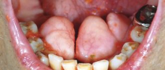
After the manufacture and fixation of temporary crowns on the implants, the presence of tumor-like growths of the mucous membrane around the installed orthopedic superstructures, which were histologically described as polyps, was noted. Patients complained of bleeding and discomfort around the installed crowns. Attempts to change the eruption profile of structures from implant shafts did not produce a positive result.
We used a method of simultaneous surgical correction of the subcutaneous fat area of autografts with the installation of provisional structures supported by dental implants. In a number of cases, we used a removable compression acrylic plate on the gum formers, which held the surgically created profile until permanent prosthetics or installation of provisional fixed and conditionally removable structures.
After 3 months, we made permanent orthopedic fixed or conditionally removable structures.
For the manufacture of orthopedic structures, we used cobalt-chrome alloy, titanium and zirconium dioxide.
As a rule, after the integration of implants and placement of gum formers at the prosthetic stage, we were faced with the need to modernize the prosthetic elements of the implantation system for specific clinical cases.
The gum formers were needed to be longer and predominantly conical in shape. The cylindrical shape of the gum former created parallel walls of the crater of the mucous membrane, which caused rapid collapse of the walls during the placement of the impression coping and caused severe discomfort to patients during manipulations. We attribute this to the presence of excess connective tissue layer, which is located on the autograft bone.
It is important to note that immediately after corrective operations on the skin-fat part of the flaps, orthopedic compression plates were fixed to prevent further growth of the soft tissue component.
Patients were monitored every seven days and appropriate compression plate adjustments were made.
Depending on the chosen permanent structure, a set of further orthopedic manipulations was carried out to make prostheses.
The results were recorded using a Canon D 60 camera, 100 mm lens, and MR-100 ring flash.
Description of clinical observations
Patient U. , 18 years old, was admitted to the clinic on April 1, 2008 with a diagnosis of osteoblastoma of the upper jaw, a condition after subtotal resection of the upper jaw on the left. From the anamnesis: a neoplasm was discovered and removed in early childhood at the Federal State Institution Central Research Institute of Infectious Diseases and Maxillofacial Surgery. The patient was sent to the clinic of the Russian Scientific Center for Surgery named after. acad. B.V. Petrovsky.
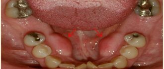
Status localis: the configuration of the face in front and profile is changed, the upper lip is retracted on the left. On palpation, a defect in the alveolar process of the upper jaw on the left is noted.
Mouth opening is not limited, there is a through defect in the alveolar process of the upper jaw with teeth 21-28, the patient wears an obturating denture, the denture teeth are in the bite.
Bite according to the second class according to Angyu.
On OPTG and CT: there is a defect of the zygomatic tubercle, alveolar process on the left, absence of the spine of the upper jaw, the base of the pyriform foramen on the left, defect of the tubercle of the upper jaw on the left. An impacted 28th tooth is noted in the remains of the pterygomaxillary articulation.
Treatment tactics: since the defect of the upper jaw was subtotal and through, a radial skin-bone graft on a vascular pedicle was used. When transplanting a radial cortico-periosteal-skin graft, a zygomatic-maxillary buttress was formed using a free split graft from the branch of the mandible on the right. The second stage involved the formation of the alveolar process using parietal grafts.
Rice. 1. Appearance of the patient, existing defect of the upper jaw, bite, obturating prosthesis, CT scanThe patient was noted to have no nasal lining, and to anatomically restore aeration, before transplanting a microsurgical graft, the nasal lining was formed with local tissues.
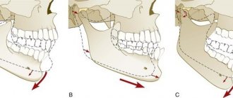
Rice. 2. Stages of planning the bone and soft tissue components of the graft Rice. 3. Stages of surgery Rice. 4. Computed tomography of the patient after surgery and condition on the third day
Rice. 5. Scintigram after surgery
In the postoperative period, there was a divergence of the sutures and a reduction in the volume of the flap, and therefore a protective mouth guard was made to protect the soft tissue component of the flap from food getting into the graft. Kappa also pressed the flap into the defect area to allow healing by secondary intention.
Rice. 6. Postoperative mouth guard2 months later, the patient, being a cycling athlete, fell from the “saddle” and received a fracture of the upper limb at the site of collection of the radial skin-cortical-periosteal graft on the vascular pedicle. Therefore, the patient underwent osteosynthesis with the Ilizarov apparatus.
Rice. 7. Condition after osteosynthesis of the upper limb on the left, condition in the oral cavity Rice. 8. 3D reconstruction of the alveolar process using parietal grafts, CT scans after the second operationAfter 5 months, we installed three dental implants in the area of the reconstructed alveolar process using parietal bone autografts. Thanks to this technique for reconstructing the alveolar process, it was possible to obtain adequate bone thickness and height for placing dental implants.
Rice. 9. Implantation in the area of 3D reconstruction with parietal blocksAfter 5 months, we opened the implants and applied a method of one-stage surgical correction of the subcutaneous fat pad of the autograft with the installation of elongated gum formers in dental implants, which simplified the formation of soft tissues during surgery.
After completion of the surgical stage, the alginate impression was removed from the upper jaw and a compression plate was made using cold polymerization at a pressure of 3 atmospheres from acrylic plastic.
The patient was advised to wear the plate constantly, removing it only for hygiene procedures. Inspection and correction of the plate fit were carried out every seven days for a month.
Thus, we were able to form a stable mucosal contour around the gingival formers.
The next stage was the production of a conditionally removable prosthesis supported on a beam structure on implants.
Rice. 10. Correction of the subcutaneous fat area of the autograftConsidering the fact that microsurgical elimination of the jaw defect and reconstruction of the alveolar process was carried out by transplantation of free parietal cortical autografts, the further stage of orthopedic rehabilitation is practically no different from classical prosthetics for patients with an extended terminal defect of the dentition.
In order to be able to produce a conditionally removable beam-type prosthesis supported by implants, it was necessary to produce an accurate plaster model that displayed the entire relief of the prosthetic bed and accurately reproduced the position of the implants. To do this, we took primary impressions of the upper jaw using Clip-transfers for implants. The primary plaster model gave us the opportunity to make a custom impression tray with prepared transfer checks for precise transfer of the position of the implants.
Having made an accurate working model of the upper jaw and a model of the antagonists, we installed them in the articulator according to the average parameters. Registration of the central ratio was carried out using an occlusal hard wax plate with refinement on ALUWAX (soft wax with the addition of aluminum filings for long-term heat retention and elasticity).
Rice. 11. Impressions and plaster modelsAfter analyzing the relationship of the plaster models in the articulator, we manufactured a beam frame supported by three implants. The beam frame was made by vacuum casting from cobalt-chrome alloy. To achieve a passive fit of the frame, we used adapters for external connection with the implants.
We fixed titanium ball locks into the cast part of the frame. Then a model of the upper jaw was made from a fire-resistant mass for modeling and casting the mating part of the prosthesis itself.
Rice. 12. Beam frame and mating part of the prosthesisTaking into account the possibility of changing the relief of the soft tissue component of the autograft, the frame of the response part was modeled in such a way that it was possible to change the acrylic base part, changing the fit of the prosthesis to the bed.
After fitting the beam and frame in the oral cavity, we manufactured the base part of the conditionally removable denture with the installation of acrylic artificial teeth, using the method of cold polymerization of plastic under a pressure of 3 atmospheres and a temperature of 50 degrees Celsius. We took into account all possible characteristics of the patient at rest and when smiling, creating the most natural appearance of the entire orthopedic restoration.
The final fixation of the prosthesis was carried out in a certain sequence:
- Cleaning and disinfection of all components of the prosthetic restoration.
- Removing the gum formers, irrigating the internal shafts of the implants with a solution of 3% hydrogen peroxide and 0.05% chlorhexidine solution, installing adapters on the external connection.
- Installation of the beam structure on three implants, tightening the fixing screws with a force of 30 N/cm2.
- Fixing the actual removable denture on the beam frame, checking the occlusion, teaching the patient about hygienic measures.
Osteoblastoclastoma - microscopic picture
Microscopically, in osteoblastoclastoma, a large number of small, slightly elongated cells with a rounded nucleus (such as osteoblasts) are distinguished, among which massive accumulations of giant multinucleated cells (osteoclasts) are identified. In mononuclear cells, mitoses are observed; in multinucleated cells, they are absent. The formed elements of the tumor are also represented by fibroblasts and xanthoma cells. Areas of hemorrhage undergo a macrophage reaction with the corresponding phagocytic cells. In some areas of the tumor, islands of osteoid tissue are found. The tumor is saturated with decayed red blood cells and is imbibed by the blood pigment - hemosiderin, which gives it a brown color. The presence of the latter and multiple hemorrhages, often in the form of blood cysts, is due to the peculiarity of blood flow in the tumor.
Blood in osteoblastoclastoma circulates outside the vascular bed through intertissue gaps. The absence of endothelium allows it to penetrate the tumor tissue and accumulate there. In this case, the formed elements of the blood are destroyed, forming an accumulation of hemosiderin. Sometimes an abundance of fibrous tissue may be found in the tumor.
Osteoblastoclastoma - differential diagnosis
Osteoblastoclastoma (cystic and cellular forms) must be differentiated from ameloblastoma and radicular cyst, the lytic form - from osteogenic sarcoma (in contrast to osteolytic sarcoma, thinning and swelling of the cortical layer of the jaw are noted in osteoblastoclastoma) and fibrous osteodysplasia (characterized by slow growth and preservation of the continuity of the cortical layer on the radiograph). layer of the jaw), fibromyxoma, central fibromas of the jaws.
Clinical and radiological examination without histological comparison is sometimes insufficient to establish an accurate diagnosis of osteoblastoclastoma. The frequency of discrepancy between the clinical diagnosis and pathohistological diagnosis is 30%; the most common discrepancies arise when differentiating osteoblastoclastoma from ameloblastoma - 16%
Diagnosis
In the diagnosis of osteoblastoclastoma, great importance is attached to x-ray examination. X-ray, O.'s picture is typical in most cases. The tumor is solitary and, as a rule, initially has an eccentric location. According to the characteristics of the growth and localization of O., its structure on the radiograph can be different. There are cellular and osteolytic variants of the structure.
Rice. 2. Radiographs of the bones of the forearm (a, b) and lower leg (c) with osteoblastoclastoma: a - direct radiograph of the proximal forearm with the cellular version of osteoblastoclastoma; the epimetaphysis of the radius is sharply swollen, with a thinned cortical layer and cellular restructuring of the spongy substance; b — direct radiograph of the distal forearm with an osteolytic variant of osteoblastoclastoma; the epimetaphysis of the radius is sharply swollen, the cortical layer is thinned or completely absent, the cellular structure of the bone substance is not determined; c — direct radiograph of the knee joint and the proximal part of the leg bones with a marginal location of osteoblastoclastoma; eccentric swelling (indicated by arrows) of the epimetaphysis of the tibia, the cortical layer is thinned, but preserved throughout.
With the cellular (trabecular) variant, the most characteristic sign is swelling of the epimetaphysis with thinning, but preservation of the cortex (Fig. 2, a). The affected bone takes on a club-shaped form. At the same time, as a result of the osteoblastic function of the tumor, a coarse cellular restructuring of the spongy substance of the epimetaphysis is observed - the appearance of large and few septa, as well as a more delicate and dense mesh. In this case, it is sometimes possible to distinguish O. from an aneurysmal bone cyst only histologically. In the structure of the tumor, as a rule, there are no deposits of calcium salts, the contours of the swollen area of the bone are quite smooth, unlike enchondroma, the edges are often characterized by the presence of speckled shadows of calcification and the wavy contour of the affected area of the bone. The affected area is clearly delimited from the adjacent unchanged parts of the bone by a thin sclerotic border, which, together with the thinned cortex, forms a tumor capsule. When the tumor spreads towards the diaphysis, the so-called telescopic transition: the diaphysis is inserted, as it were, into the swollen epimetaphysis hanging over it. Often strips of ossifying periosteum are also identified here. The fragility of the cortical substance covering the tumor, in some cases, can cause the occurrence of pathological breaks with reparative, often fringed periostosis.
The osteolytic variant of the tumor structure is observed either primarily, from the very beginning of tumor growth, or secondarily, as a result of the transition from the cellular variant. The X-ray picture is somewhat reminiscent of osteolytic sarcoma. However, in contrast to it, thinning and swelling of the cortical substance covering the changed area of the bone is observed (Fig. 2, b). The cellular structure is completely or partially absent. The swollen and thinned cortical substance can undergo almost complete resorption, but at the base of the tumor, at the site of its transition into the unchanged bone, remnants of the expanded and thinned cortical substance are usually visible, which is usually absent in osteolytic sarcoma. So-called telescopic transition of the diaphysis into the tumor in this case rentgenol. The variant is observed even more often than with the cellular one.
Radiologically, centrally located and marginal tumors are distinguished. Regional tumors (Fig. 2, c) usually do not involve the central parts of the epimetaphysis. Diagnosing them is difficult, but it is quite possible on the basis of taking into account rentgenol, signs - swelling, cellularity, absence of a periosteal visor, etc.
Osteoblastoclastoma of short and flat bones can present significant difficulties for differential rentgenol. diagnostics, since an identical picture can be given by benign Chondroblastoma (see), enchondroma (see Chondroma), reticulosarcoma of bone in the initial phases of development (see Primary reticulosarcoma of bone), in the sacrum - chordoma (see) and some -ry metastatic tumors, primarily metastases of clear cell kidney cancer and thyroid cancer, local fibrous osteodystrophy (see) and bone dysplasia (see). O. in the initial phases of malignancy cannot be radiologically distinguished from its osteolytic variant. The possibility of O.'s malignancy is indicated by the rapid growth of the tumor and the disappearance of the cellular structure. Based on the complete destruction of the tumor capsule bordering the diaphysis, the appearance of a typical periosteal visor and metastases, it can be assumed that the tumor has transformed into sarcoma.
Osteoblastoclastoma - treatment
Treatment for osteoblastoclast of the jaws is surgical. In cystic and cellular forms, careful curettage of the lesion is usually sufficient. If the tumor occupies large areas of the jawbone, it is possible to perform resection of the lower jaw, if necessary, with simultaneous bone grafting. In the lytic form, partial resection of the jaw without breaking the continuity of the mandibular bone (continuum resection) or resection of a fragment of the jaw with simultaneous bone grafting is more often used. Radiation therapy does not provide sufficient therapeutic effect. However, this method of treatment is prescribed to patients for whom any surgical intervention is contraindicated due to their general somatic status. Usually short-focus X-ray therapy, remote gamma therapy, bremsstrahlung and high-energy electron radiation are used. The average radiation dose ranges from 30–50 Gy/kg. The prognosis for treatment is favorable, but the possibility of malignancy and metastasis of the tumor due to the benign nature of the tumor cannot be excluded. Incorrect treatment tactics usually lead to relapses of the tumor process.
Published in Articles
Forecast
The prognosis for the benign form of Osteoblastoclastoma is favorable in most cases - after treatment, complete recovery usually occurs. However, taking into account the structural features of O., its ability to recur and give metastases even with a typical (benign) histological structure, caution should be exercised in each specific case when determining the prognosis.
Bibliography:
Baran L. A. Complex treatment of malignant bone tumors, Kyiv, 1971; Vinogradova T. P. Bone tumors, p. 76, M., 1973; Odesskaya - Melnikova L. A. Giant cell tumor (osteoblastoclastoma), Vestn, rentgenol, i radiol., No. 3, p. 50, 1957; Pereslegin I. A. and Sarkisyan Yu. X. Clinical radiology, p. 298, M., 1973; Reinberg S. A. X-ray diagnosis of diseases of bones and joints, book. 2, p. 322, M., 1964; Guide to the anatomical diagnosis of human tumors, ed. N. A. Kraevsky and A. V. Smolyannikov, p. 393, M., 1976; Rusakov A.V. Pathological anatomy of the skeletal system, p. 308 and others, M., 1959; Atlas radiologique des tumors osseuses, t. 1-2, P., 1974-1976; Bone tumors, ed. by DC Dahlin, p. 8 ao, Springfield, 1967; Glanzmann G.u. Horst W. Stellung der Strahlentherapie in der Behandlung von Riesenzelltumoren des Knochens (Osteoklastome), Strahlentherapie, Bd 154, S. 81, 1978; Jacobs P. The diagnosis of osteo-clastoma (giant-cell tumor), Brit. J. Radiol., v. 45, p. 121, 1972; Jaffe HL Osteoid-osteoma, A benign osteoblastic tumor composed of osteoid and atipical bone, Arch. Surg., v. 31, p. 709, 1935; McNerney DP a. Maddlemiss JH Giant-cell tumor of bone, Skeleton. Radiol., v. 2, p. 195, 1978; Murray RO a. Jacobson HG The radiology of skeletal disorders, v. 1, p. 564, Edinburgh, 1977; Schajowicz F. Giant-cell tumors of bone (osteoclastoma), J. Bone Jt Surg., v. 43-A, p. 1, 1961.
Yu. N. Soloviev; Yu. S. Mordynsky (rad.), I. L. Tager (rent.).