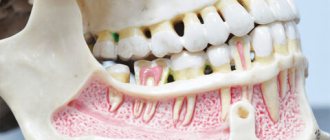
Odontoma is a benign tumor consisting of fragments of dental tissue. It appears as a concomitant phenomenon during the growth of permanent teeth.
The development of such a neoplasm is typical for childhood or adolescence. Odontoma is localized near the sinuses of the upper jaw or in the angular part of the lower jaw. There are known cases of their bilateral lesions.
Large formations grow into the dental bone, thereby leading to its thorough destruction, the formation of ulcers and purulent fistulas. It often develops without pain, is characterized by gradual growth and the absence of complaints from the patient.
General information
Odontoma is an odontogenic formation that is the result of an abnormal development of dental tissue.
Odontoma is translated from Greek as “tumor consisting of teeth.” Already from this definition it becomes clear that this tumor is organ-specific and develops only in the jaw bones. Most often, odontoma affects the bone in the area of premolars and molars, both in the upper and lower jaw. Maximum tumor growth is observed either at the age of 6 to 11 years, which is associated with the period of eruption of permanent teeth, or later - at the stage of eruption of third molars. Most often, the tumor is detected at a young age (up to 20 years) with equal probability in both sexes. Its growth stops with the end of the formation of the tooth germ, so in adults the tumor is discovered accidentally during an X-ray examination or during the development of an inflammatory process in the tissues of the neoplasm. Odontoma accounts for about 22% of all odontogenic tumors and is one of the most common diseases in this group. Early diagnosis and treatment of odontoma helps prevent the development of complications. Odontoma
Methods of treating the disease
Odontoma is a benign neoplasm that develops from the tissues of the dental pulp, and the causes of the pathology are not precisely understood. In many cases, the pathology is diagnosed in early childhood, since the neoplasm often develops precisely during the period of teething. Experts strongly recommend not delaying the start of treatment, since the progression of the disease is associated with an increasing risk of malignancy of the tumor and its degeneration into sarcoma. Therapeutic methods are selected taking into account the nature, size and localization of neoplasia, and a number of other factors determined during the examination of the patient.
Surgery
Relatively recently, surgical removal of odontoma has been the main method of treatment. Today, surgery is indicated in cases where the tumor reaches a significant size or is localized in a place inaccessible to non-invasive treatment. As a rule, the tumor is excised without affecting the surrounding tissue, but if malignancy is suspected, it is removed in combination with a fragment without pathological changes. Thus, prevention of relapse is ensured.
With a certain localization of odontoma, the tumor is eliminated using maxillectomy - removal of a fragment of the jaw. If a small defect is formed, autotransplantation is permissible, during which the cavity is filled with the patient’s bone tissue. In case of significant defects, an obturator is implanted - a special plate that replaces the removed area, individually manufactured for the patient using computer modeling. Such implants are most suitable for the patient and allow you to restore chewing and speech functions in a short time.
Laser ablation
A minimally invasive treatment method based on the destruction of pathological tissues using laser radiation. Under the influence of the laser, the local temperature rises sharply, which causes the death and literal evaporation of cells with pathological changes. In this case, there is no damage to surrounding tissues or the formation of cosmetic defects. Since such treatment can be carried out on an outpatient basis, the patient can return home after a short period of time.
HIFU therapy
Indicated for the removal of small odontomas, HIFU therapy (high-intensity focused ultrasound ablation) is also based on heating and destruction of pathological tissue under the influence of intense ultrasound waves. Before the procedure, the required wavelength and exposure mode are selected.
Cryoablation
A minimally invasive technique, also recommended for eliminating small tumors. The death and necrotization of tumor tissues is caused by freezing under the influence of ultra-low temperatures, achieved by introducing liquid nitrogen into the tumor. If necessary, it is possible to conduct several cryotherapy sessions without the threat of side effects. The procedure is performed on an outpatient basis, without hospitalization of the patient.
Radiotherapy
When identifying the reasons that make surgical intervention impossible, radiotherapy is one of the main treatment methods. Radiotherapy sessions are also prescribed after surgical removal of the tumor in case of a high risk of malignancy. Irradiation of the pathological area helps to reduce the tumor, up to complete disappearance, relieves painful symptoms and normalizes the patient’s condition. In Israeli clinics, radiotherapy sessions are carried out on modern TrueBeam and Novalis installations, using gentle techniques that provide precise irradiation of the tumor with the ability to modulate the intensity of irradiation in different areas, subject to minimal negative effects on healthy tissue.
Chemotherapy
It is prescribed in case of relapse of the pathology, as well as in case of an extremely high probability of malignant degeneration. Clinics in Israel use the latest types of chemotherapy drugs, which are characterized by low toxicity, a gentle effect on the patient’s body and a small number of mild side effects.
Causes of odontoma
There are two theories of the occurrence of odontomas. Some dentists believe that odontoma is a malformation of one or more tooth germs, which consist of epithelial or mesenchymal tissues, representing a mixed odontogenic tumor. Others believe that this type of tumor is exclusively a tissue malformation because epithelial and mesenchymal cells can also be found in healthy tissue. A major role in the development of odontomas is assigned to general factors - genetic predisposition, as well as local reasons - the presence of infection in the tissues of the oral cavity, injuries.
The morphological basis of the tumor, according to different authors, varies; mainly isolated from Malasse cells, tissue of the dental plate, oral mucosa and all dental tissues (normal and supernumerary teeth). During histological examination of the tumor, you can often see parts of the tooth tissue: pulp, dentin, enamel, cement, located in a chaotic order. Sometimes there are layers of connective tissue between the tooth tissues. Also, odontoma can be represented by homogeneous dental tissue. Thus, an important factor in the etiology of odontomas is high odontoblastic activity, which is responsible for the development of tooth buds.
Reasons for tumor development
Most often, a tumor is discovered accidentally - during an x-ray or during a tooth extraction procedure.
Sometimes, instead of bone of the usual degree of density, a compacted formation is found that is difficult to process with conventional dental instruments. There are several reasons for the development of tumor formation. It may appear due to an abnormal development of the dental embryo or adjacent tissues, genetic predisposition, trauma or infection of the oral cavity.
When studying the composition of the tumor, remains of epithelial islands of Malasse, cells of the oral mucosa, particles of enamel, pulp, and dentin are found in it. In addition, the odontoma may contain homogeneous dental tissue.
According to traditional medical beliefs, odontomas are classified as benign tumors. However, in 4% of cases there is a chance of their degeneration into malignant ones. Soft odontoma is especially susceptible to this process.
What is the disease?
Benign tumors of dental tissue are classified as dental diseases only because of their location.
All types of cementoma consist of rough, fibrous connective tissue similar to the cementum of tooth roots. It replaces bone tissue and expands the periodontium. Areas of increased mineralization appear in the tumor, and it gradually ossifies.
According to statistics, cementoma appears much more often in women than in men. And in children - more often than after twenty years. In adults, they are usually located on the sides of the lower jaw, covering the roots of the molars and premolars.
This is what it looks like in the photo:
In children, cementomas are not associated with dental roots, tend to grow endlessly, and more often than in adults, they affect the center of the upper jaw.
Cementoma develops very slowly, without the appearance of any symptoms.
Only when it reaches a significant volume can the following appear:
- Pain while chewing food.
- Sensitivity to palpation.
- Changing the shade of tooth enamel.
If the tumor grows strongly, ruptures of the gum epithelium at the site of the tumor and changes in the shape of the face are possible. When the jaw bone becomes thin, there is a high chance of fracture.
Modern classification
Depending on the causes of odontoma, there are:
- simple;
- complex;
- cystic.
A simple form of tumor formation is formed as a result of a violation of the development of one dental embryo; it can be complete (formed from all tooth tissues) or incomplete (containing one type of hard dental substance).
Complex odontomas are formed by chaotic connections of teeth or tooth-like tissues. The development of this type of tumor is associated with disturbances in the formation of several dental germs. The x-ray shows a tumor with pronounced protrusions of round or irregular shape.
Complex odontomas are divided into compound and mixed. A compound complex odontoma contains several correctly formed teeth, which at the same time have a deformed shape. Mixed teeth consist of randomly located hard dental tissues.
The cystic form of formation develops in the form of a cyst, the shell of which contains a tumor. The presence of a transparent halo is noted around the formation.
Depending on the structure of the tumor, they are divided into soft and hard.
Soft odontoma is less common than hard odontoma. It is often diagnosed among young patients. The stage of formation of permanent teeth is most “favorable” for tumor development.
The tumor can develop in both the lower and upper jaw. It looks like a formation consisting of delicate fibrous connective tissue. Soft formations are considered precursors to the appearance of hard ones and are prone to relapse and transformation into malignant tumors.
The hard tissue contains dental and periodontal tissues, which are arranged randomly and have varying degrees of maturity. Hard odontoma is diagnosed several times more often than soft odontoma.
Record composite odontoma for 232 teeth:
Symptoms
The clinical picture of odontoma depends on the type of tumor. For example, the symptoms of soft odontoma are similar to those of ameloblastoma; the main difference between these two types of tumors is that the first is more common at an early age. The tumor develops slowly, but this period is limited, and its cessation is associated with the end of tooth formation. Continuous growth is characteristic only of hard complex odontoma. Due to the increase in tumor size, the bone swells and enlarges, after which destruction of the cortical plate is observed, due to which the tumor penetrates the periosteum and then into the soft tissues. With pronounced thinning of bone tissue, a symptom such as parchment crunch is sometimes detected. In this case, the bulging fabric has a dark color and a soft-elastic structure. During palpation examination of such a neoplasm, bleeding may occur. As these tumors develop, they are prone to ulceration.
This is interesting: Removing tartar - how does ultrasonic tartar cleaning work?
Clinical signs of hard odontoma depend on its location. The tumor is characterized by expansive growth, but even when it reaches a large size it remains painless. In the initial stages, a tumor is detected with an uneven, bumpy surface. As the formation develops, damage to bone tissue, periosteum and mucous membrane is noted. Sometimes a decubital ulcer is found with a bottom formed from tooth tissue. Tumors are prone to infection, which leads to the formation of a focus of chronic inflammation in the soft tissues that surround them. The infectious process in soft tissues occurs with periods of remission and exacerbation, and therefore fistulas with pus are often formed. Regional lymphadenitis is accompanied by inflammatory phenomena in the area of odontoma. Sometimes hard odontomas can move and damage formed teeth and their rudiments.
Symptoms
In most cases, odontomas develop painlessly, unnoticed by the patient. Having reached a certain size, they stop growing. The absence of severe symptoms results in later, often accidental, detection. And if a neoplasm is detected, it occurs in childhood or adolescence, in the mixed dentition.
The manifestation may be retention or missing teeth. It is precisely such cases that often force the parents of a young patient to consult a dentist.
Not always, but quite often, the result of an examination for missing or impacted teeth is a diagnosis of odontoma.
Although much less common, there are cases when odontomas manifest themselves with symptoms that clearly indicate the presence of a problem and force the patient to see a doctor. Such cases include the following:
- Pain or discomfort in the gum area where the tumor is located. Typically, painful sensations are neurological in nature, and are generated by the pressure of a growing tumor on the nerves.
- Jaw deformation. The cause is also a significant amount of tumor, which thins and deforms the jaw bone.
- The appearance of a dense, lumpy swelling on the gum. Usually it is painless when pressed and does not cause discomfort, but its very presence indicates trouble and forces you to see a doctor.
- Formation of fistulas in the area of odontoma. The cause of their appearance may be other pathologies of the dentofacial apparatus. But, the main thing is that they force the patient to see a doctor, who will take the necessary measures to diagnose and determine the root cause of the formation of fistulas.
- Some manifestations characteristic of other diseases of the oral cavity - periostitis, osteomyelitis, periodontal inflammation.
And yet, most often, hard odontoma does not manifest itself with pronounced symptoms, and is discovered in adulthood by chance, during an examination for another reason.
If a bluish tumor appears on the gum, which from time to time becomes inflamed and bleeds when injured by food or teeth, there is a possibility that this is a soft odontoma, the growth pattern and development of which is completely consistent with a true tumor.
This neoplasm is elastic to the touch, spindle-shaped, has a bumpy surface, is easily injured and bleeds.
During the process of growth, the soft form gradually destroys the bone and penetrates into the surrounding soft tissue, so the attitude towards it should be more attentive and strict than towards hard formations.
General information
Cementoma (periradicular osteofibrosis) is an odontogenic malformation of connective tissue. In adulthood, cementomas are more common in women. The tumor in most cases is localized in the area of the body and angle of the lower jaw. In this case, the roots of large and small molars are involved in the pathological process. In children, cementomas are not associated with roots and are prone to unlimited growth. In childhood, the maxillary bone is most often affected. Cases of benign neoplasms growing into the cavity of the maxillary sinus and the base of the skull have been recorded.
If the cementoma reaches a large size, it can lead to a jaw fracture. One of the types of cementoma, periapical cement dysplasia, is detected mainly in women of the Negroid race between the ages of 30 and 50 years. Teeth associated with lesions are invariably vital and rarely have restorations. The prevalence ranges from 1 to 5.9%.
Cementoma
Causes of cementoma
Gigantoform cementoma is a genetically determined disease in which multiple lesions are detected in several family members. Reliable causes of other types of cementum have not been identified, however, a number of factors and events that may precede the appearance of formation have been studied in clinical dentistry. The history of patients with cementoma often includes:
- oral injuries
: damage to the mucous membrane from the edges of destroyed teeth, chipped fillings, poorly fitted orthopedic structures, jaw bruises; - the presence of foreign bodies
: filling material, instruments, pins in the root canals; - chronic inflammatory diseases of the oral cavity
: periodontitis, osteomyelitis of the jaws, actinomycosis of the maxillofacial area, etc.
Pathogenesis
The pathogenesis of cementoma remains unknown. Scientists admit that a reactive or dysplastic process is taking place. At the pathological level, cementoma involves progressive resorption of normal alveolar bone and its replacement by fibrous tissue consisting of fibroblasts, collagen, and varying amounts of bone or cement-like calcifications.
The initial stage of cementoma development is characterized by the proliferation of fibroblasts and collagen fibers in the apical region of the periodontal ligament, resulting in resorption of the medulla surrounding the apex. At this stage, the radiograph shows zones of rarefaction of bone tissue, which are very similar to periapical foci of infectious transdental origin. Subsequently, during the second stage of cementoma development, differentiation of cementoblasts occurs. This, in turn, contributes to the appearance of areas of increased mineralization. At the third stage, ossification of fibrous tissue is observed. The lesion appears as calcification of limited density, surrounded by a narrow radiolucent rim.
Localization
Most often, the pathology is located in the area of the canines and incisors, that is, in the central parts of the jaws. Moreover, in most cases, the lower jaw is subject to such changes.
Quite rarely, detection of such a formation is possible in the upper section. This is a more complex version of the disease, since the tumor can affect the sinuses and affect the functioning of the respiratory system.
Classification
In dentistry, there are 4 main types of cement:
- True cementoma
. The tumor develops without obvious symptoms. More often, the neoplasm is diagnosed accidentally during an X-ray of the tooth associated with the cementoma or neighboring teeth. Histological examination of cementoma reveals coarse fibrous connective tissue with varying degrees of calcification. The tumor is surrounded by a membrane that separates pathologically altered tissue from healthy bone. Microscopically, the neoplasm is similar to osteoid osteoma and Paget's disease. - Cementing fibroma
. The disease occurs with mild symptoms. As the cementoma grows, the cortical layer of the bone becomes thinner, and mild pain occurs from pressure on the periosteum. Histologically, cementing fibroma is a benign neoplasm consisting of intertwined cells and connective tissue fibers with pronounced zones of mineralization. Cementing fibroma, like true cementoma, has an external capsule. - Periapical cement dysplasia
. Most often localized in the frontal part of the body of the lower jaw. It is characterized by the appearance of multiple pathological foci that do not exceed 1 cm in diameter and do not extend to the cortical plate. In its development, the tumor goes through 3 stages: osteolytic, calcifying and mature. On an x-ray, along with the destruction of bone tissue, areas of increased mineralization are revealed. - Gigantoform cementoma
. It is characterized by intensive transformation of connective tissue into cement tissue. An x-ray reveals a round or oval shaped area tightly fused to the root of the tooth. The degree of density of gigantoform cementoma is very similar to the structure of the hard tissues of the tooth. The periodontal gap is not visible, cellular elements are practically absent.
This is interesting: Sedation is a safe alternative to general anesthesia
Medical Internet conferences
Relevance. Currently, there is a high incidence of such a tumor disease of the maxillofacial region as odontoma. Its incidence in children is lower than in adults [1]. The reason for this is the slow asymptomatic growth of odontoma. Unlike adults, removal of this tumor in children should be carried out immediately after its diagnosis, since further development of the tumor can lead to consequences such as disruption of the formation and eruption of permanent teeth, displacement of tooth germs, changes in the position of teeth in the dentition, thinning of the cortical plate jaw bones. That is why great attention should be paid to the timely detection of this disease and its monitoring at any age in order to prevent complications.
Purpose: to consider a clinical case of lower jaw odontoma in a 3-year-old child.
Tasks:
1) Analyze the symptoms, clinical picture, methods of examination and treatment of patients with odontoma.
2) To describe a clinical case of lower jaw odontoma in a 3-year-old child.
Materials and methods. A standard examination protocol was used in a pediatric dentistry clinic. Modern sources on this topic were studied, and an analysis of the design of case histories was carried out.
Results and discussions. A benign tumor, built from dental tissue, was first described and named P. Broca odontoma in 1867 [1].
Odontomas make up about 22% of the total number of odontogenic tumors, being one of the most common tumors in this group of neoplasms. There are several ideas about the origin of odontomas. According to some scientists, odontoma is a malformation of one or more tooth germs, consists of epithelium, mesenchyme and their derivatives, and is a mixed odontogenic tumor. According to other data, odontomas are not true neoplastic formations, but rather belong to hamartomas, since mesenchymal and epithelial cells can be found in small quantities in the structure of healthy tissues. The reasons for the formation of odontomas can be the remains of cells of the dental plate, trauma, or a local infectious process. A significant role in the etiology belongs to odontoblastic hyperactivity and changes in the genetic component responsible for the process of dental development [2]. Odontomas can form during the formation of temporary and permanent teeth. As the primary morphological basis from which odontoma is formed, some authors name: 1) epithelial cells of Malasse;
2) tissue of the dental plate, folded into a ball; 3) oral mucosa (this hypothesis is explained by the frequent absence of one or more teeth in the area where odontoma is located); 4) all tissues of normal or supernumerary tooth buds.
There are no differences in occurrence between males and females.
Most often, odontoma occurs among young people, and rarely in old age. According to our observations, odontoma was more common in children during the period of mixed dentition from 6 to 12 years [3, 4]. The reason for visiting a dentist was most often the retention of permanent teeth, less often this neoplasm was an accidental x-ray finding.
Odontoma usually grows slowly; faster tumor growth may be associated with the eruption of the first permanent teeth at the age of 6-11 years or the eruption of third molars. According to our data, odontoma is most often localized in the upper jaw in the area of incisors and canines, less often in the lower jaw, mainly in the area of molars.
Odontoma results from the growth of fully differentiated epithelial and mesenchymal cells and is characterized by slow development and a benign course [5]. It consists of all the components of hard and soft tissues of the tooth: enamel, dentin, cement inclusions, pulp. Neoplasm tissues may have a disorganized structure with a predominance of one tissue over others[6].
The World Health Organization (WHO) classified odontomas in 2005 based on histopathological features into complex and compound. Complex odontomas consist of several teeth, the tissues of which are well differentiated, but arranged randomly. Composite odontoma is represented by small teeth and tooth-like formations that have a certain arrangement [7].
Different types of odontomas are characterized by a number of features. Complex odontoma can grow throughout life; more often detected in adolescence; localized in the area of the molars of the lower jaw; there is no pain or swelling of the jaw; The radiograph reveals a high-intensity, inhomogeneous shadow, which is limited by a thin band of rarefied bone tissue. During the growth process, the tumor can displace tooth germs and formed teeth [8].
A compound odontoma is represented by a combination of several rudimentary tooth germs; interferes with teething; The x-ray shows several small tooth-like formations surrounded by a strip of sparse bone tissue; after excision there are no relapses. Compound odontoma is more common than complex odontoma. After reviewing 104 clinical cases, 67 were diagnosed with compound odontoma and 32 with complex odontoma. It is very rare to find both types of odontomas in one person. Of all those examined, there were only 5 such cases. In the anterior part of the jaws, the majority of odontomas are compound combined (61%), while in the distal part of the jaws complex combined odontomas are more common (34%). Moreover, both types of odontomas are most often located on the right side of the jaw (composite – 62%, complex – 68%) [9].
Odontoma can be hard or soft [10]. Hard odontoma is represented by solid elements of the tooth, pulp and periodontium in various quantitative ratios and combinations. Soft odontoma is the initial stage of the formation of hard odontoma.
A.I. Evdokimov classifies hard odontomas into simple, cystic, complex and complex-mixed forms. In turn, simple odontomas are divided into: 1) complete (tooth-like and rounded in shape) and 2) incomplete (crown, root, periodontomas).
Simple odontomas may involve the joining of tissues from a single tooth germ or consist of a single tooth-like formation. Complete simple odontomas consist of a whole tooth germ; incomplete simple odontomas are represented by a part of a tooth.
Complex mixed odontomas have the appearance of a continuous conglomerate consisting of chaotically mixed hard and soft tissues of the tooth, with separately occurring teeth at different stages of formation [10].
The main method for making the diagnosis of odontoma is x-ray. On an x-ray, a complex odontoma appears as a voluminous, heterogeneous, dense shadow with jagged edges or as small structures resembling teeth. Along the periphery, this formation has a zone of clearing, which passes into limitedly sclerotic bone tissue. Simple complete odontomas produce a round or tooth-like intense shadow on the radiograph. Simple incomplete odontomas appear as an intense shadow of the crown or root part of the tooth, altered in shape and size.
The presence of an inhomogeneous shadow of an odontoma on an x-ray image is explained by the heterogeneity of the histological structure of odontomas (alternation of soft and hard tissues of the tooth).
Cystic odontoma is localized in a follicular cyst, with a characteristic clinical and radiological picture. The only difference from a follicular cyst is that with a cystic odontoma, the cavity of the cyst contains an odontoma, not a tooth [11].
The macroscopic specimen of odontoma can be represented by a formation resembling a deformed or underdeveloped tooth, or complexes consisting of several tooth-like formations. They can easily separate or firmly merge with each other [12].
The most characteristic clinical symptoms of odontoma are the absence of one or more permanent teeth, less often - the absence of temporary teeth, or bone enlargement. The bulge in the jawbone area is usually painless, dense, with an uneven surface [13].
Treatment for odontomas involves removing the tumor along with the connective tissue capsule. After tumor removal, it is advisable to fill the bone cavity with some osteotropic substance [14, 15].
The prognosis is favorable.
Clinical case.
Patient G., 3 years old, came to the Department of Pediatric Dentistry and Orthodontics with complaints, according to her mother, of swelling of the gums in the lower jaw area on the left, which her mother noticed 1 week before going to the doctor. Previous and concomitant diseases: the child is registered with a neurologist (tone disorder syndrome), an ophthalmologist. There was a history of an allergic reaction to Actovegin (urticaria).
The general condition of the child at the time of examination was satisfactory.
The configuration of the face is not changed, regional lymph nodes are without signs of inflammation. The dental formula is age appropriate. The dentition lacks teeth 5.5 and 7.5. In the retromolar region behind tooth 7.4 there is a deformation of the alveolar process, extending to the vestibular surface, with a diameter of 1.5 cm, dense elastic consistency, covered with unchanged mucous membrane, painless on palpation.
After conducting a visual examination, it was not possible to diagnose the disease due to the overlapping of many shadows on each other.
A tomographic examination (CBCT - cone beam computed tomography) was performed (Fig. 1).
CT scan revealed a heterogeneous radiopaque mass surrounded by a thin radiolucent line. The formation is closely related to the germ of tooth 7.5, which is at the stage of root formation. Perforation of the lingual cortical plate was detected (Fig. 1). Interestingly, the absence of tooth germs 1.5, 2.5, 3.5, 4.5 is determined by X-ray, which confirms the relationship of the development of odontoma with a violation of the formation of tooth germs and can indirectly serve as confirmation of the presence of a common etiological factor in their development [16].
A preliminary diagnosis was made: Complex odontoma of the lower jaw on the left. Anomaly in the number of teeth.
A plan of treatment and preventive measures has been drawn up:
1. Removal of odontoma under general anesthesia in a hospital setting.
2. Clinical observation with repeated X-ray control after 1 year.
3. Treatment by an orthodontist.
Treatment was carried out in a hospital setting.
An intraoral approach was created under general anesthesia. The formation was completely enucleated along with the second primary molar. After careful curettage, the wound was packed with iodoform turunda.
Based on the examination of the material - odontoma obtained during the operation (Fig. 2), the diagnosis was clarified: Complex mixed odontoma of the lower jaw.
The turunda was changed once a week for one month. Intraoral healing proceeded without complications. After the intervention, no anesthesia or paresthesia was observed in the areas innervated by the inferior alveolar nerve.
The child was registered at the dispensary. The bone tissue defect was completely restored after 6 months. A repeat computed tomography scan was performed 1 year later. The photographs show complete restoration of the bone structure in the surgical area (Fig. 3).
Further treatment of this child will be carried out by an orthodontist.
Characteristics of varieties
Each of the histological types of this group of formations should be described in more detail.
Benign cementoblastoma
Under the name benign cementoblastoma or true cementoma lies a neoplasm consisting of coarse fibrous tissue of varying degrees of mineralization. The tumor is located in a capsule that separates it from healthy cells.
Cementoblastoma is defined by slow but unlimited growth. At the initial stage of the disease, there are no symptoms.
Over time, resorption of the cortical plate occurs, which entails discomfort and pain during palpation and eating.
At an advanced stage of the disease, deformation of the jaw may be observed, as well as the eruption of a tumor, which leads to its infection and the development of inflammation.
Most often, cementoblastoma is localized on the lower jaw row in the premolar area.
Periapical fibrous dysplasia
Photo: mandibular cementoma on CT
In this case, there is a defect in the formation of cement
. In addition to the roots, the process of destruction also affects the bone of the jaw itself. However, despite this, the prognosis is usually favorable.
Detection, as in other cases, usually occurs by accident. Most often this is associated with the treatment of a tooth when its root is fractured, since the neoplasm can make it more fragile.
The main distinguishing characteristic of this species is the absence of a periodontal fissure, although changes occur near the apex (or apexes) of the root. In the affected areas of the bone there may not be clear boundaries of newly formed dense tissue.
The degree of mineralization of these areas can be different, so the consistency also changes - from soft to very dense.
The best way to diagnose is an x-ray examination of the desired area with four or five times magnification.
Cement-forming fibroma
As in other cases, this formation is not classified as malignant. Diagnosis is carried out in the same way. However, the picture during X-ray examination and histology is somewhat different.
First of all, the growth of this tumor is determined by the processes occurring in the bone. More precisely, it is the process of mineralization.
In the image you will be able to see an area where the bone tissue is somewhat sparse. This is the source of the lesion. It has smooth outlines.
The sparseness is explained by the fact that this type of formation has rather weak mineralization. Histology shows a fibroplastic structure.
A cement-like structure can only become in the last stages of the disease, if it has not been identified and treated earlier.
Gigantoform cementoma
It has been reliably revealed that this variety is a hereditary pathology
. In this regard, most often it can be observed in several family members.
The image will show that large areas of spongy bone are dense and dark. In most of the described cases, pathological changes occur symmetrically on the jaw.
Diagnosis and treatment
Sometimes odontomas manifest themselves in a clear way, allowing one to make an assumption about their presence already at the initial stage of diagnosis - during questioning and examination. This could be a bulging gum, distortion of the jaw shape, or patient complaints of discomfort or pain.
However, in any case, a final diagnosis requires an x-ray, which, as a rule, resolves all doubts about the presence or absence of pathology.
On an x-ray, the hard type manifests itself as a heterogeneous (due to the presence of soft and calcified tissues) shadow with clear, uneven edges, surrounded by a white stripe (capsule). The shading density is close to the dentin of an intact tooth.
What are denticles in the dental pulp?
This publication contains all the most important information about acid necrosis of hard dental tissues.
Read here about the dangers of hereditary dentinogenesis imperfecta.
Differential diagnosis
The differential diagnosis is based on the presence of a capsule (manifests itself on an x-ray as a white stripe about 1 mm thick around the shading), and the nature of the shading.
Odontomas are differentiated from:
- impacted teeth (the true nature of the pathology can be established by filming from different angles);
- salivary stones (diagnosis is carried out by localization, salivary stones are usually located in the sublingual area);
- osteotome - unlike odontomas, they do not have a capsule (there is no white strip along the shading contour), and the shading itself is more uniform.
Soft neoplasms are differentiated from periostitis, chronic periodontitis, and osteomyelitis.
Treatment
There are differences in the treatment of soft and hard odontomas. The former require mandatory surgical removal, since they have the property of destroying bone and growing into soft tissue. Damaged bone tissue is removed along with the tumor.
A selective approach is used for hard odontomas. The main group of patients for whom treatment is considered mandatory are children. And not so much because of the danger of the neoplasm itself, but because of its effect on the development and growth of the dentofacial apparatus.
Even the non-growing type can interfere with teething and disrupt the bite. Formations that manifest themselves by bulging on the gums, curvature of the jaws, discomfort and pain leave no other choice but to remove them.
Hard odontomas are removed surgically, always together with the capsule, the remains of which can provoke a recurrence of the tumor.
In adults, the decision for surgical removal is made based on the clinical picture. Asymptomatic formations that have stopped growing and do not cause any discomfort are left untouched, limited to observation. Others that manifest themselves with one or another negative symptoms are removed.
Soft odontoma is subject to mandatory surgical removal due to its tendency to infiltrating growth, infection, inflammation and fistula formation.
When removing a soft tumor, resection of the surrounding inflamed, necrotic and granulomatous bone tissue is required.
The depression formed in the bone at the site of the removed tumor is filled with osteo-replacement material. After several months, new high-quality bone is formed in place of the pathological tissue.
Etiology of occurrence
Dental experts are still working on the causes of cementoma.
To date, it has been possible to identify several factors that contribute to the formation of tumors according to medical indicators. These include:
- long-term chronic inflammatory processes of the oral cavity - fibrous periodontitis, osteomyelitis, actinomycosis, sinusitis, etc.;
- injuries of a chronic nature - damage to the mucous membrane of teeth destroyed by caries, incorrectly installed dentures, sharp areas of fillings;
- immediate injuries - bruises, blows, mechanical damage to the jaws and gum tissue.
Experts do not rule out a genetic predisposition to the formation of cementoma. A version of the long-term harmful effects of certain unfavorable factors is also being considered. For example, smoking or radioiodine therapy.
This is interesting: Is it possible to take dental x-rays during pregnancy - expert opinions
Symptoms
The etiology of the disease has no obvious signs for a long time, which complicates its early diagnosis. Often, patients in dental clinics do not even suspect that there is a group of interconnected tumors in their oral cavity.
Therefore, cementoma is discovered by x-ray by chance, when they come to a specialist with a completely different problem.
However, at the peak of the development of the disease, those affected by the pathology note some symptoms:
- the presence of a feeling of discomfort and pain while chewing food or when talking;
- segmental change in the shade of enamel in the affected area;
- increased sensitivity to palpation.
Cementoma is not always localized in the depths of the tissues of the oral cavity. Sometimes, neoplasms are located closer to the mucous membrane and over time come to the surface, damaging the gums.
Reviews
Most odontomas found in adults are stunted, do not cause discomfort and do not require surgical removal of the formation.
Therefore, most often adults deal with odontomas of their children, which are clearly indicated for removal.
If you personally find yourself in this situation, please tell us what type of neoplasm was discovered in your child, and how did the treatment end? The comment form is located at the bottom of this page.
If you find an error, please select a piece of text and press Ctrl+Enter.
Tags jaw
Did you like the article? stay tuned
No comments yet
Diagnostics
As has already been said more than once, it is not possible to determine the presence of cementoma in the oral cavity based on any specific symptoms.
Pathology can be detected visually only in certain cases, when neoplasms erupt onto the surface of the mucosa. On an x-ray, which is often taken for another reason, specialists identify dark and dense oval or round areas. Accordingly, the contour of the pathology, in most cases, is smooth, the tumors differ significantly from the structure of healthy tissues. In the photograph, the cementoma appears to have a common wall with the root of the tooth.
In addition to radiographic diagnostics, the patient is subsequently assigned a microscopic examination. As a result of this analysis, the nature and structure of tissue similar to bone is determined.
The pathological formation has a yellow tint, a low level of mineralization or a significant amount of calcifications in the composition. When viewing tissue through the lenses of a microscope, laboratory technicians note the heterogeneity of the structure; it has many different inclusions.
Due to the fact that benign neoplasms are very similar in nature to many other pathologies, the final point in making a diagnosis is a histological examination, which is prescribed without fail.
How is the disease diagnosed?
It should be remembered that since odontoma is characterized by asymptomatic initial stages, in order to detect the pathology in a timely manner, you should regularly visit the dentist. A comprehensive diagnostic examination, necessary to develop an effective treatment program, takes no more than three days in Israeli clinics.
- The first day
- Second day
- Day three
At the initial consultation, the attending physician gets acquainted with the patient’s medical history, clarifies the symptoms, examines the teeth and oral cavity, and then prescribes the necessary examinations.
Completing prescribed examinations, including:
- general and biochemical blood tests;
- visual research methods (radiography, CT, MRI, PET-CT) - are carried out to identify a tumor, accurately determine its size, location and structure, assess the condition of distant organs if a malignant nature of the tumor is suspected;
- biopsy followed by histological analysis of pathological tissues.
A medical council consisting of the attending physician and a number of highly specialized specialists reviews the research results, makes a final diagnosis and develops a treatment regimen.
Treatment
Treatment of cementoma takes a fairly long period of time.
Specialists remove tumors step by step, tracking the dynamics. This measure is necessary to avoid destruction of enamel and deformation of the maxillofacial bone. It is worth noting that only a highly qualified specialist should carry out treatment. Before proceeding with surgery, the dentist excludes the presence of diseases and inflammatory processes in the oral cavity and carries out professional cleaning.
In general, the procedure for removing cementoma goes without complications, however, identifying root fractures or the presence of infection in an unsealed canal can significantly aggravate the rehabilitation period after surgery.
Complications
As a rule, complications are encountered by patients in whom the area of cementoma localization was found in the central part of the upper jaw.
During surgery, an inexperienced specialist can cause harm to nearby sinuses. The growth of dense neoplasms in this organ is fraught with difficulty breathing or various diseases of the nasopharynx.
The emergence of tumors on the surface of the gum tissue leads to the formation of holes that stretch from the mucous membrane to the jaw bone.
Failure to provide timely assistance can result in infection of oral tissues due to the penetration of harmful microflora through the formed “tunnels”.
Prevention
Cementoma, like any other pathology, is easier to prevent than to treat later. In order not to encounter such an unpleasant problem, it is necessary to carefully care for the oral cavity - brush your teeth twice a day, use dental floss and other hygiene products.
You should monitor the slightest changes in the condition of the teeth and mucous membranes, undergo preventive examinations at the dentist (at least once every six months), which will make it possible to identify pathology at an early stage of its development.
Remember that sometimes the disease can result in irreversible deformation of the jaw, which will result in difficulty chewing food, disturbances in the articulation apparatus, and an aesthetic defect in the appearance of the face.
Forecast and prevention of the disease
Detection of tumors at the initial stage and timely contact with medical professionals contribute to a favorable prognosis for the outcome of the disease.
You should not postpone a visit to the dentist if the initial signs of the disease appear. This will greatly worsen the situation and lead to uncontrolled growth of the tumor. At the same time, the risk of odontoma degenerating into a malignant formation increases.
The basis of prevention should be regular visits to the dentist (at least 2 times a year), prevention of the development of chronic infections and effective elimination of foci of inflammation. Detection of the disease at an early stage is a priority task of modern dentistry and the medical industry as a whole.
Possible complications
Complications are possible in several cases. First of all, when the tumor is located in the central area of the upper jaw. Then it can affect other organs. Often these are the paranasal or nasal sinuses.
The growth of dense tissue in them can affect the functioning and normal functioning of the respiratory tract
.
Also sometimes concomitant diseases of the nasopharynx
.
The second danger is when the cementoma emerges from the surface, breaking through the mucous membranes of the oral cavity.
In this case, a kind of hole is formed, reaching the internal parts of the bone.
Aggressive and pathogenic microorganisms can very easily penetrate through it, causing additional infectious lesions.
.
Sources:
- https://www.KrasotaiMedicina.ru/diseases/zabolevanija_stomatology/odontoma
- https://ozubkah.ru/chelusti/cementoma.html
- https://www.obozrevatel.com/health/bolezni/odontoma.htm
- https://www.KrasotaiMedicina.ru/diseases/zabolevanija_stomatology/cementoma
- https://www.vash-dentist.ru/lechenie/chelyust/tsementoma-osobennosti-zabolevaniya-sposobyi-diagnostirovaniya.html
- https://zubovv.ru/lechenie/chelyust/chto-takoe-tsementoma.html
- https://dr-zubov.ru/lechenie/chelyust/kak-obnaruzhit-cementomu.html