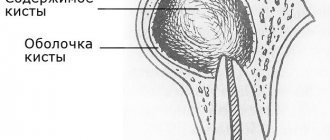
What is a cyst? Jaw cyst
A cyst is a cavity that is lined with epithelium and filled with fluid or soft material.
The formation of teeth (odontogenesis) is a complex process in which connective tissues and epithelium (enamel organ, dental follicle and dental papilla) participate.
The enamel organ refers to an epithelial structure derived from the oral ectoderm. The dental follicle and dental papilla are ectomesenchymal structures, because they are partly derived from neural crest cells.
For each tooth, odontogenesis begins with the apical (affecting the tip of the tooth root) proliferation of the epithelium of the oral mucosa, known as the dental lamina. The dental lamina gives rise to the enamel organ, a cap-shaped structure that subsequently takes on the shape of a bell. After the formation of the enamel organ, the dental lamina cord usually fragments and degenerates. However, small islands of dental lamina may remain after tooth formation. They are believed to be responsible for the development of some odontogenic cysts and tumors.
The enamel organ has four types of epithelium. The inner lining of the enamel organ is called the inner enamel epithelium and becomes the ameloblastic layer that forms tooth enamel. The second layer of cells adjacent to the inner enamel epithelium is the intermediate layer. Adjacent to this layer is the stellate reticulum, followed by the outer enamel epithelium. The enamel organ is surrounded by loose connective tissue known as the dental papilla. Contact with the epithelium of the enamel organ causes the dental papilla to produce odontoblasts, which form dentin. As odontoblasts lay down dentin, they induce ameloblasts to form enamel.
After initial crown formation, a thin layer of enamel organ epithelium, known as Hertwig's root sheath, grows at the apex of the tooth root. This epithelial expansion later becomes fragmented but leaves behind small nests of epithelial cells known as Malassez remnants in the space of the periodontal ligament. They are the source of epithelium for most periapical (radicular) cysts, but do not cause any odontogenic neoplasms, with the exception of squamous odontogenic tumor.
Causes of maxillary sinus cysts
Predisposing factors for the formation of a maxillary sinus cyst are:
- Individual anatomical and topographical features of the structure of the nasal cavity, which impede the free movement of air through the nasal canals (congenital deformities, deviated septum).
- Blockage of the excretory duct of the glands located in the mucous membrane of the paranasal sinus. This happens with frequent rhinitis, sinusitis, allergic rhinitis and polyposis.
- Inflammatory dental diseases with infection spreading to the root canals. The paranasal sinuses are separated from the roots of the molars and premolars by a thin septum, so infection from pulpitic or periodontitis teeth often causes an inflammatory process in the maxillary sinus.
- The formation of a dental cyst, which grows into the maxillary sinus and continues to grow inside it.
The maxillary cyst gradually grows larger, filling the sinus without pronounced clinical symptoms. The presence of a formation can be suspected during exacerbations caused by colds, acute respiratory viral infections, local hypothermia, and decreased immunity.
Why do jaw cysts form?
Odontogenic cysts (developing or inflammatory) get their name from the nature of their origin. Most jaw cysts are lined by epithelium, which is derived from odontogenic epithelium.
The occurrence of such cysts is usually associated with unerupted teeth (third molars of the lower or upper jaw, second premolars of the lower jaw, and canines of the upper jaw). They can also form near additional teeth and in combination with odontomas. The occurrence of odontogenic cysts is rarely associated with baby teeth.
Most often, jaw cysts are discovered between the ages of 10 and 30 years. Men, especially white-skinned men, suffer from them more often.
In most cases, dental cysts do not manifest themselves in any way and are an accidental finding obtained during an X-ray examination. But, in some cases, they can reach significant sizes, which lead to the expansion of bone tissue, but even they may not give pronounced symptoms until a secondary infection occurs.
Types of dental cysts
Tooth cysts have different classifications, each of which is formed according to certain characteristic parameters of the pathology.
According to the nature of the disease, they are distinguished:
- residual cysts – occur after tooth resection (removal) surgery; this is the most common type of cyst;
- retromolar – formed during severe eruption of wisdom teeth;
- radicular - cysts are located on or near the tooth root;
- follicular – at the heart of such cysts is the germ of a permanent tooth; follicular neoplasms arise as a result of poor quality care of baby teeth.
Classification of neoplasms according to their origin:
- odontogenic – arise as a result of the transition of the inflammatory process from other dental diseases;
- non-odontogenic - the causes of the development of such cysts include problems not related to the teeth and oral cavity.
Locations of cystic formation:
- anterior teeth;
- teeth that are adjacent to the nasal sinuses with their roots;
- wisdom teeth.
How do jaw cysts appear?
In most cases, the cyst does not cause significant symptoms. Its development can be provoked by incorrect treatment of dental diseases or caries.
Odontogenic cysts are usually distinguished by type of origin:
· dentofacial cysts – their occurrence is associated with the crown of a tooth that could not erupt;
· keratocysts – are a consequence of Nevoid basal cell carcinoma syndrome;
radicular or radicular cysts – are of inflammatory origin and most often result from a reaction to necrosis of the dental pulp;
· bifurcation buccal cyst – typical for children 5-10 years old, it is formed in the area of buccal bifurcation of the first molars of the lower jaw;
· primary cyst – in most cases it is a keratocystic odontogenic tumor;
· orthokeratinized cyst – also refers to a subtype of keratocystic odontogenic tumor;
· eruption cyst – usually formed from a degenerating dental follicle and forms in the gum when the tooth erupts;
· newborn gum cyst – formed from the remains of the dental plate on the gum of a newborn;
· adult gingival cyst – is a variant of the lateral periodontal cyst;
· lateral periodontal – a non-inflammatory cyst on the side of the tooth, formed from the remains of the dental plate;
· calcifying cyst is a rather rare pathology, which is characterized by cystic and neoplastic signs;
· glandular cyst is a formation with a respiratory epithelial lining and potential relapse; in its manifestations it is similar to the central variant of poorly differentiated mucoepidermoid carcinoma.
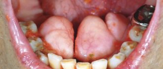
Odontogenic cysts are difficult to detect at an early stage. It gives virtually no symptoms. The patient may be alarmed by tooth displacement or a change in the color of the diseased tooth. If the cyst has reached a significant size, the patient may notice protrusion of the bone structures.
A long asymptomatic course can lead to the formation of inflammatory processes, which are dangerous due to the development of suppuration and can provoke pathological fractures of the jaw bones.
If a cyst has formed in the upper jaw, it can cause nosebleeds, headaches and impaired nasal breathing.
The main symptoms associated with the presence of an odontogenic cyst (pain, fever, inflammatory changes in the oral cavity) usually appear in the later stages of the disease.
Price
The cost of treatment or removal of a dental cyst consists, like many other surgical interventions, of a number of factors. The complex of diagnostic procedures performed, types and methods of treatment, conservative preparation of canals, preoperative preparation, the operation itself, the osteoplastic materials used, postoperative observation - all this affects the final cost of treatment in these clinical situations.
A dental cyst is a serious pathology. In all cases, even in cases of complete absence of any symptoms, it poses a health hazard. With regular visits to the dentist, you can be sure: a dental cyst, if there is one, will be detected in time, and subsequent treatment will allow it to be eliminated without leaving a trace, preserving the health of the tooth.
According to antiplagiat.ru, the uniqueness of the text as of October 16, 2018 is 99.9%.
Key words, tags: X-rays, tooth extraction, orthopantomogram, periodontitis
How are odontogenic cysts diagnosed?
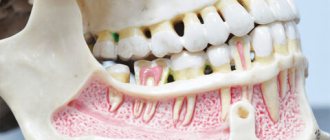
The leading method for identifying odontogenic cysts is radiography, which is capable of visualizing jaw cysts at an early stage of their development. On an x-ray image, the cyst is distinguished by the presence of clear boundaries, and the formation itself gives a characteristic shadow of a round or oval shape, immersed in the sinus of the tooth root.
Ultrasound examination also helps to recognize odontogenic cysts.
As already mentioned, pronounced symptoms are characteristic of the late stage of development of a pathological formation, therefore it is difficult to diagnose a cyst at the initial stage, relying only on symptoms.
The final diagnosis is made on the basis of histological examination. It is important to differentiate an odontogenic cyst from other pathologies (adenomatoid odontogenic tumor, ameloblastic fibroodontoma and calcifying epithelial odontogenic tumor).
The CT scan method is widely used in the diagnosis of jaw cysts to confirm the presence of calcifications along the wall of the cyst, as well as tiny spots that are usually not found on x-rays. In addition, computed tomography is necessary during the surgical planning stage.
What is the prognosis for the disease?
How successfully the situation in a patient with an odontogenic cyst will be resolved depends on at what stage the cyst was discovered, how severe the symptoms were and how it was treated.
As a rule, the use of surgical treatment gives a positive prognosis. Therapeutic treatment provides a positive prognosis only if it is started at the initial stage of the disease.
A negative prognosis may be associated with detection of the disease at a late stage: odontogenic cysts can provoke the development of serious pathologies that cause deformation of the maxillofacial tissues.
What treatment methods for jaw cysts exist?
The choice of treatment method for an odontogenic cyst directly depends on the symptoms it causes, as well as the results obtained during instrumental and laboratory diagnostics.
If surgical treatment is chosen (cystotomy or cystectomy), the maxillofacial surgeon performs complete removal of the cyst. In some cases, it is necessary to remove the cyst along with the affected parts of the tooth root. Treatment is carried out in a hospital setting.
If the choice falls on therapeutic treatment, the doctor will carry out procedures aimed at reducing inflammation. This is a long process, taking at least six months.
The first step is to drain the contents of the cyst using a special drainage tube, which is inserted into a small incision in the tumor. As the contents drain out and the tumor shrinks, the size of the tube is adjusted downward.
After removing the contents of the cyst, the dentist cleans the root canals and administers medications that destroy the tumor tissue. At the end of all procedures, the doctor uses a special solution aimed at accelerating healing.
Treatment is monitored radiographically.
Both after surgical and therapeutic treatment, the patient requires preventive measures that will help avoid the re-formation of an odontogenic cyst.
Diagnosis of tooth root cyst
To make a diagnosis and carry out appropriate treatment, the dentist collects and analyzes the medical history. During the initial diagnosis, many patients report the fact of endodontic treatment performed to eliminate periodontitis or pulpitis. Some patients indicate an exacerbation of the disease after intraoral dissection.
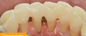
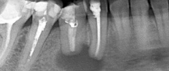
Radiography is used as the main diagnostic method. Below is a photo and x-ray of a dental cyst.
To obtain an x-ray, several methods are used, the first method is based on contact intraoral x-ray, the advantages of this technique:
- determining the degree of destruction of the jaw bones;
- assessment of the condition of the tooth root and tooth canal;
- assessment of the quality of canal filling;
- identifying the presence of perforations and fragments of instruments and materials in the tooth canal;
- determination of the relationship between the cyst and the roots located near the teeth.
The second method of performing radiography is an orthopantogram; the procedure is a panoramic photograph of both jaws and the maxillary sinuses of the upper jaw.
Another method of the procedure is a survey X-ray in the nasomental projection; the X-ray covers the bones of the skull from the nose to the chin; using the image, the doctor assesses the condition of the maxillary sinuses and detects cysts that have grown into the nasal cavity.
In addition to radiography, to detect a tumor, the patient may be prescribed an electroodontic diagnostic procedure. This technique helps to assess the degree of such an indicator as the electrical excitability of the teeth that are located next to the cystic tooth. If the value exceeds 60 microamps, the dentist prescribes endodontic treatment to the patient.
For diagnostic purposes, histological and cytological studies are used to determine whether the neoplasm is benign or malignant.
Diagnosing a dental cyst is not difficult, but only qualified dentists can carry it out in a hospital setting; under no circumstances try to independently determine the presence of a cyst and do not take therapeutic measures; strictly follow the doctor’s recommendations.