From this article you will learn:
- what is a dental cyst - symptoms, photos,
- why does it form?
- cyst on the root of a tooth - treatment.
The article was written by a dentist with more than 19 years of experience.
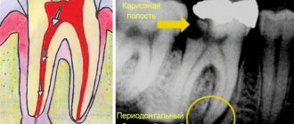
A dental cyst is a round-shaped inflammatory formation, which is a cavity in the bone tissue, which is lined from the inside with a fibrous capsule and filled with purulent contents (Fig. 1). In dentistry, the terms “root cyst of the tooth” or “radicular cyst” (from the word radix - root) are most often used to refer to this pathology. This is due to the fact that the most common place for its formation is the area of the apex of the tooth root.
The formation of a cyst on the root of a tooth is associated with an infection in the root canals. Most often it appears - 1) either in the absence of timely treatment of pulpitis or periodontitis of the tooth, 2) or is a consequence of poor-quality root canal filling. The formation of a cyst occurs gradually and over a long period of time. First, at the apex of the root, under the influence of infection in the root canals, a small apical granuloma is formed, which subsequently increases in size and transforms into a cyst filled with pus.
What does a dental cyst look like: photo
Pay attention to how a cyst on the root of a tooth looks on an x-ray - it looks like intense darkening at the apex of the tooth (for your convenience, we have limited this area with arrows). Such intense darkening at the apex of the tooth root indicates the presence in the bone tissue of a site of bone destruction in the form of a cavity. In Fig. 3 you can see what a cyst looks like at the apex of the root of an extracted tooth (like a sac filled with pus).
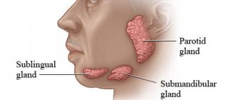
This article is written for patients and therefore will contain unnecessary medical details. But to complete the picture, it is worth adding that a dental cyst can be localized not only at the apex of the tooth root, but also on the lateral surface of the root, as well as between the roots of multi-rooted teeth - in the root bifurcation zone. But such localizations of root cysts are quite rare, and therefore we will examine in more detail the symptoms and treatment of a dental cyst localized at the apex of the root.
Type of cyst on the root of an extracted tooth (video 1) –
Tooth cyst: symptoms
If a dental cyst occurs, there may be no symptoms at all for a long time. In this case, jaw cysts are diagnosed accidentally, for example, during a routine X-ray examination before the start of prosthetics (usually a panoramic photograph of the teeth is taken for this - an orthopantomogram). But minor symptoms may still occur. For example, there may be periodic minor pain when biting on the causative tooth or slight pain when pressing on the gum in the projection of the tooth root.
Moreover, the patient will feel such symptoms inconsistently, but only periodically. For example, they can occur after hypothermia, against the background of decreased immunity and colds. In very rare cases, sharp pain may occur when biting on the causative tooth, against the background of which swelling of the gums and even soft tissues of the face may also be observed. Against the background of an exacerbation, slight mobility of the tooth with a cyst may also appear. Also, as a rule, such an exacerbation may be accompanied by a slight fever and weakness.
In some cases, a dental cyst can be suspected not only by these secondary signs. Sometimes you can even see it, of course, if it reaches a large size. If left untreated, the size of the cysts slowly increases, which is associated with the constant production of pus into the cyst cavity. An increase in the amount of pus in the cyst cavity leads to an increase in the pressure of the cyst on the walls of the bone cavity surrounding it. Under such pressure, the bone tissue is slowly resorbed and the cyst expands. I have operated on a number of patients whose cysts reached 3-4 cm in diameter.
On the upper jaw, root cysts increase in size much faster than on the upper jaw. This is due to the fact that the bone tissue of the upper jaw is softer and more porous in structure, and therefore its resorption always occurs faster. If a root cyst occurs in the premolars and molars of the upper jaw (teeth 4-5-6-7-8), the cyst may even occupy the entire volume of the maxillary sinus, and removal of the dental cyst in this case will require a maxillary sinus. In some cases, large root cysts may be visible during examination of the oral cavity, and not just on a targeted or panoramic x-ray.
Root cyst on the upper jaw (view in the oral cavity) –
Please note that, as in this case, the cyst deformed the hard palate. Such deformations can be observed not only in the palate, but also on the anterior side of the alveolar process of the upper and lower jaw. Very often, large cysts lead to complete destruction of bone tissue on the anterior surface of the alveolar process of the jaw (in the projection of the root of the causative tooth). And it turns out that under the mucous membrane in this area there will be a fibrous membrane of the cyst. In this case, during palpation you will not feel the hard bone; the finger will fall inward a little.
In what cases might you suspect you have a root cyst? –
Firstly, if you periodically experience discomfort or slight pain in one of your teeth when biting (in this case, gentle tapping on the tooth will also cause discomfort). But such symptoms are usually characteristic of apical periodontitis, which, by the way, also needs to be treated urgently. But what is not typical for ordinary apical periodontitis, but is typical for a root cyst, is pain on palpation of the gums in the projection of the root of the causative tooth.
Therefore, if there is discomfort in one of the teeth when biting, you can palpate (press) with your finger on the gum in the projection of the root of this tooth. Pain when pressed is highly likely to indicate the presence of a cyst at the root of the tooth (24stoma.ru). This diagnosis may also be supported by the presence of a dense or soft “bulging” of the gums in the projection of the root of the causative tooth, which can occur when the root cyst reaches a large size.
Main symptoms and signs
A cyst at the base of the root causes swelling of the lower part of the face and pain.
At the beginning of the pathological process, the patient does not experience any discomfort, but may notice a small bump or bump in the jaw area. Subsequently, the formation begins to increase, destroying bone tissue, causing facial asymmetry, and enlargement of the cheeks in the affected area. As the disease progresses, the following appears:
- pronounced swelling of the gums and cheeks;
- nasal congestion on the affected side;
- jaw deformation;
- pain syndrome resulting from compression of nerve endings.
When a cyst becomes infected, a person complains of sharp pain during chewing, swelling and redness of the affected gums, peeling of the skin in the area of inflammation, headache, drowsiness, and increased body temperature.
X-ray diagnostics –
As we said above, the final diagnosis can only be made using an x-ray, on which a cavity formation in bone tissue will always look like a rounded darkening.
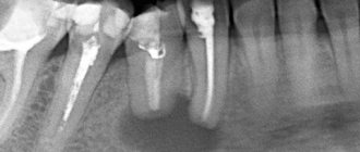
In the x-ray images below we have presented you with 3 options for localizing root cysts. The most common location is root cysts at the apex of the tooth root, but cysts on the lateral surface of the root or located between the roots of multi-rooted teeth are much less common. Radiographs of root cysts of different locations –
Cyst on the root of the tooth: causes
As we said above, the formation of a cyst on the root of a tooth is associated with an infection in the root canals. Root canals open at the tops of the roots with small holes, which means that during inflammation, bacteria and their toxins will travel outside the tooth, forming foci of inflammation at the tops of the roots. At the first stage, a so-called apical granuloma (up to 0.5 cm in size) is first formed at the root apex, which is a section of granulation tissue surrounded by a loose fibrous capsule.
Such a granuloma does not yet have a cavity with pus inside, but with a further increase in its size (over 0.5 cm), a cavity is formed inside it, lined from the inside with epithelial cells. Such a cavity formation already corresponds to the concept of “root cyst,” although in dentistry small root cysts ranging in size from 0.5 to 1.0 cm are often also called the transitional term “cystogranulomas.” And the root cyst itself is most often called a formation with a diameter of more than 1.0 cm. Inside the cavity of the cyst there is pus, which is produced by its inner membrane.
Learn more about the reasons for the development of a cyst at the apex of the tooth root -
Infection in the root canals does not occur on its own, and this is not some kind of psychosomatics and related problems, for example, bronchial asthma. Infection in the tooth cavity and root canals occurs in the following two situations:
- Untreated caries, pulpitis and periodontitis - tissues affected by caries contain a large number of cariogenic microorganisms. If caries is not treated, then bacteria gradually enter the dental pulp (neurovascular bundle), causing inflammation in it - pulpitis. If pulpitis is not treated in time, which should consist of removing the infected pulp and filling the root canals, the infection penetrates through the root canals outside the tooth. This leads to the development of a focus of inflammation at the apex of the tooth root, followed by the formation of a root cyst (Fig. 5).
- Poorly sealed root canals - imagine that you have cured your pulpitis in time or the inflammation that has just begun at the apex of the tooth, but after some time you still have a cyst formed at the apex of the root. This happens very often and is due to the fact that the root canals were filled poorly (according to official statistics, dentists fill root canals poorly in 60-70% of cases). The fact is that the infection develops in the unfilled part of the root canal or where the canal was not tightly filled with a filling substance.
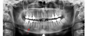
Normally, each root canal of a tooth should be filled to the apex of the root. If the canal is not filled to the top of the root, then an infection develops in the unsealed part of the canal, which penetrates beyond the tooth and also causes the formation of a cyst. In Fig. 6-7 you can see radiographs of teeth whose root canals were poorly filled, which in both cases caused the formation of a dental cyst. In Fig. 6, white arrows mark unfilled areas of the root canals. In Fig. 7, only traces of filling material are visible in the root canal, i.e. The root canal is not sealed tightly and not all the way.
Reasons for the development of root cysts in other locations –
The appearance of a root cyst on the side surface of a tooth is usually due to the fact that numerous small branches (opening with holes on the side surface of the root) always extend from the main root canal in the root of the tooth. Therefore, even with high-quality filling of the main root canal, there is a very small risk of a granuloma or cyst appearing on the lateral surface of the root.
A little more often you can find a cyst in another location - between the roots of multi-rooted teeth (in the bifurcation of the roots). Such a cyst usually occurs if the integrity of the hard tissues of the bottom of the tooth cavity is damaged, for example, as a result of a crack or perforation of the bottom of the tooth cavity, or perforation of the upper half of one of the root canals. Such complications are most often the result of dentist mistakes in the treatment of pulpitis and periodontitis.
Tooth cyst: treatment
If you have a cyst on the root of a tooth, treatment can be conservative (therapeutic) and surgical. Conservative treatment will consist of treating the cyst with a special medicinal paste based on calcium hydroxide, which will temporarily fill the root canals. Accordingly, if the root canals have not been filled, the tooth is opened and the root canals are mechanically treated. If the root canals have already been sealed, but poorly, they are first unsealed.
Below we will show how therapeutic treatment of a dental cyst is carried out using a specific example. It should be noted that good results with conservative therapy can be achieved even when treating fairly large cysts. However, there are situations where conservative therapy is less optimal than surgical removal of the cyst.
Surgical treatment is reasonable if...
- If the root canal is filled, but there is a pin in it, and there is a risk of fracture of the tooth root when removing the pin from the canal.
- If the root canal is filled, and the tooth has an artificial crown or is the support of a bridge (after therapeutic treatment of the cyst, the crowns will have to be redone, and this costs money).
- For large cysts.
- If the cyst arose as a result of perforation of the tooth root.
- If the cyst often suppurates, for example, every time you try to temporarily fill the root canals with medicinal paste.
A prerequisite (including in the presence of a pin and a crown) is that the root canal must be well sealed for at least 2/3 of its length. This will allow you to cut off the root apex with the unfilled part of the canal, removing it along with the cyst shell, and at the same time maintaining sufficient length of the root so that it can withstand the chewing load. A very important point is the convenience of surgical access, so surgical removal of a cyst is rarely performed, for example, in 6-7 teeth.
Therapeutic treatment of cysts using a specific example -
Let’s say right away that this method of treatment is very long (at least 2-3 months), it will require numerous visits to the dentist, and it is also the most financially expensive. The sequence of actions will be as follows (we will describe the main stages below):
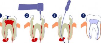
- Working with tooth root canals – if the root canals of the tooth have not been previously filled, then at the first stage the dead pulp is removed from the tooth and instrumental treatment of the root canals is carried out. If the root canals have already been filled, then they are first unsealed. In this case, the cyst arose due to insufficient filling of the root canal to the root apex (Fig. 8).
Because the canal was previously sealed, then it must first be unsealed, which was done (Fig. 9). In fact, in this situation, it was much easier and cheaper for the patient to perform a resection operation. It would last about 30 minutes, and you could forget about the cyst. But the patient refused surgical treatment due to fear.Example of treatment of a root cyst (Fig. 8-12) –
- Medicinal treatment of canals - the cyst contains pus, therefore, after unsealing the canals or removing the pulp, numerous rinsing of the root canal with antiseptics is required.
- Temporary filling of the canals with medicinal paste - first, using special instruments, a medicinal substance is removed from the apex of the root (directly into the cavity of the cyst), which has a powerful antiseptic effect. For this purpose, a paste based on calcium hydroxide is used. The same substance is then used to temporarily fill the root canal (Fig. 10).
- Repeating points 3 and 4 - if the cyst is large, then over the next few months you will need to make a couple of visits to the dentist. This is necessary to change the medicine, as well as to take a control x-ray to assess the dynamics of the reduction of the cyst.
- X-ray control – the effectiveness of treatment is assessed. A significant reduction in the size of the cyst on x-ray indicates the effectiveness of the therapy (the structure of the newly formed bone is even visible in the image), and in this case, you can proceed to the next stage - permanent filling of the root canals.
- Permanent filling of the root canals - if a decrease in the size of the cyst is noticed over the course of several months during treatment, then the root canals are finally filled, usually with gutta-percha (Fig. 11). Immediately after this, you can begin to restore the crown part of the tooth.
After the root canals have been completely filled and a filling has been placed, the patient must visit the doctor every few months and take a control x-ray. The image clearly shows the dynamics of cyst reduction and bone tissue restoration. Compare the X-ray image in Fig. 12 (taken 2.5 months after the start of treatment) with the image before the start of treatment in Fig. 8. The naked eye can see a reduction in the size of the cyst and restoration of the bone beams.
Emergency care for exacerbation of inflammation -
We have already said above that dental cysts are usually characterized by a chronic, sluggish course of the inflammatory process. However, in some cases, a sharp exacerbation of the process may occur - with the appearance of acute pain when biting on a tooth and/or swelling of the gums in the projection of the cyst. When a patient goes to the dentist due to an exacerbation, the tooth is immediately opened to allow the drainage of pus from the cyst cavity (through the root canals). You can see how this happens in video 1.
If there is swelling on the gum, the patient is sent to a dental surgeon to make an incision in the gum. You can see how the gum incision is made in video 2. After this, the tooth is left open, and the patient is prescribed antibiotics, antiseptic rinses, and non-steroidal anti-inflammatory drugs.
Well, in conclusion, I would like to say about how dangerous a dental cyst is if it is not treated. Root cysts slowly increase in size, and imagine that there is a 2-3 cm cavity filled with pus in your head. A growing cyst, for example, can engulf the roots of neighboring teeth, which will require filling the canals in them as well. In addition, the cyst can grow into the maxillary sinus and do many other bad things. We hope that our article on the topic: Dental cyst treatment was useful to you!
Sources:
1. Higher prof. the author’s education in therapeutic and surgical dentistry, 2. Based on personal experience as a dentist, 3. National Library of Medicine (USA), 4. “Outpatient surgical dentistry” (Bezrukov V.), 5. “Therapeutic dentistry: Textbook” (Borovsky E.).
Possible complications and prevention of pathology
When a cyst grows into the maxillary sinuses, pus may form.
To prevent the development of complications, the cystic cavity during the operation is filled with composite materials that help restore the original shape of the jaw. If treatment is not carried out in a timely manner, the risk of the following increases significantly:
- sinusitis, developing due to the germination of formations in the maxillary sinuses;
- loosening of teeth located near the problem area;
- infections of the oral cavity and internal organs due to a breakthrough of the purulent capsule;
- displacement of a number of teeth;
- formation of malocclusion in children;
- thinning of the jaw bones, which significantly increases the risk of fractures;
- blood clots that destroy the pulp.
A jaw cyst cannot be cured at home. Therefore, at the first alarming symptoms, it is necessary to visit the dentist and undergo appropriate treatment. To prevent a relapse, experts recommend following the rules of hygiene of both the oral cavity and the whole body, regularly brushing your teeth to remove stones, and following the recommendations of your doctor.
A jaw cyst is a benign formation that forms under the influence of a long-term inflammatory process that affects the oral cavity. This is a dangerous pathology, fraught with the development of sinusitis, bone deformation, loosening and loss of healthy teeth.