Root cysts or gingival granulomas are a common problem today. Neglect of regular examinations, hygiene rules, the development of caries or periodontitis contribute to the development of cystic formations. The disease affects all segments of the population, regardless of gender and age. In fact, it does not matter on which jaw a radicular cyst occurs, because the process of appearance, treatment, and consequences develop according to the same scenario. To eliminate pathology, there are a number of new techniques that can completely eliminate the disease with a guarantee of no relapses.
Radicular cyst as a disease
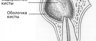
Cyst translated from Greek means “cavity”, “bubble”. In medicine, this bladder is often filled with blood, organic fluid or purulent exudate. The outer layer of the cystic component consists of soft connective tissue, and the cavity itself is filled with epithelium. Diagnosis of a radicular cyst is carried out specifically in the upper segments of the jaw bone. Detection of its signs in the lower jaw is quite rare. At the very beginning, the disease occurs in a latent stage and does not manifest itself in any way. If detection occurs, it is usually through an X-ray of a completely different dental area. Based on size, dentists distinguish two groups of cysts:
cystogranuloma (about 0.5 cm); cyst (more than 1 cm).
Radicular cysts of the jaws (lower or upper) are cavity neoplasms in the periapical dental area with cystic fluid, formed as a result of the inflammatory process of the periapical (root) part of the tooth. There are main forms of the disease:
acute process; chronic form.
Prolonged growth of the cyst can lead to gradual perforation or thinning of the jaw bone, which increases the risk of jaw fracture in the affected segment. A radicular cyst often requires surgical treatment, so if unpleasant symptoms are detected, a visit to the doctor is urgent.
Cyst on the root of the tooth: causes
As we said above, the formation of a cyst on the root of a tooth is associated with an infection in the root canals. Root canals open at the tops of the roots with small holes, which means that during inflammation, bacteria and their toxins will travel outside the tooth, forming foci of inflammation at the tops of the roots. At the first stage, a so-called apical granuloma (up to 0.5 cm in size) is first formed at the root apex, which is a section of granulation tissue surrounded by a loose fibrous capsule.
Such a granuloma does not yet have a cavity with pus inside, but with a further increase in its size (over 0.5 cm), a cavity is formed inside it, lined from the inside with epithelial cells. Such a cavity formation already corresponds to the concept of “root cyst,” although in dentistry small root cysts ranging in size from 0.5 to 1.0 cm are often also called the transitional term “cystogranulomas.” And the root cyst itself is most often called a formation with a diameter of more than 1.0 cm. Inside the cavity of the cyst there is pus, which is produced by its inner membrane.
Learn more about the reasons for the development of a cyst at the apex of the tooth root -
Infection in the root canals does not occur on its own, and this is not some kind of psychosomatics and related problems, for example, bronchial asthma. Infection in the tooth cavity and root canals occurs in the following two situations:
- Untreated caries, pulpitis and periodontitis - tissues affected by caries contain a large number of cariogenic microorganisms. If caries is not treated, then bacteria gradually enter the dental pulp (neurovascular bundle), causing inflammation in it - pulpitis. If pulpitis is not treated in time, which should consist of removing the infected pulp and filling the root canals, the infection penetrates through the root canals outside the tooth. This leads to the development of a focus of inflammation at the apex of the tooth root, followed by the formation of a root cyst (Fig. 5).
- Poorly sealed root canals - imagine that you have cured your pulpitis in time or the inflammation that has just begun at the apex of the tooth, but after some time you still have a cyst formed at the apex of the root. This happens very often and is due to the fact that the root canals were filled poorly (according to official statistics, dentists fill root canals poorly in 60-70% of cases). The fact is that the infection develops in the unfilled part of the root canal or where the canal was not tightly filled with a filling substance.
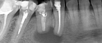
Normally, each root canal of a tooth should be filled to the apex of the root. If the canal is not filled to the top of the root, then an infection develops in the unsealed part of the canal, which penetrates beyond the tooth and also causes the formation of a cyst. In Fig. 6-7 you can see radiographs of teeth whose root canals were poorly filled, which in both cases caused the formation of a dental cyst. In Fig. 6, white arrows mark unfilled areas of the root canals. In Fig. 7, only traces of filling material are visible in the root canal, i.e. The root canal is not sealed tightly and not all the way.
Reasons for the development of root cysts in other locations –
The appearance of a root cyst on the side surface of a tooth is usually due to the fact that numerous small branches (opening with holes on the side surface of the root) always extend from the main root canal in the root of the tooth. Therefore, even with high-quality filling of the main root canal, there is a very small risk of a granuloma or cyst appearing on the lateral surface of the root.
A little more often you can find a cyst in another location - between the roots of multi-rooted teeth (in the bifurcation of the roots). Such a cyst usually occurs if the integrity of the hard tissues of the bottom of the tooth cavity is damaged, for example, as a result of a crack or perforation of the bottom of the tooth cavity, or perforation of the upper half of one of the root canals. Such complications are most often the result of dentist mistakes in the treatment of pulpitis and periodontitis.
Etiology and provoking factors
The appearance of a cystic component is the body’s response to the inflammatory process in the tooth. Cysts form in patients with a long course of carious destruction, when the lesion reaches the root part of the tooth. But there are other diseases or conditions that can serve as a trigger for the formation of cavity formations:
chronic periodontitis; severe pulpitis; trauma to the tooth and gums: previous illness of an infectious nature; chronic stomatitis; inflammation of the maxillary sinuses; abnormal bite; catarrhal otitis; diseases with a pronounced decrease in immunity; complication during the eruption of wisdom teeth.
A radicular cyst of the maxillary bone is most often formed under the influence of a long-term untreated carious tooth. Sometimes the cause of the appearance of a radicular cyst in the upper jaw is unprofessional treatment of caries, violation of canal filling technology, or lack of competence of a specialist in the treatment of chronic forms of periodontitis. Lack of treatment for a radicular cyst can lead to serious complications not only from the maxillofacial anatomical zone, but also from all organs and systems of the body as a whole.
Types of dental cysts
It is customary to distinguish between 2 types of dental cysts - follicular and radicular. Follicular cysts are less common than radicular cysts. In the presence of certain factors, it grows from the dental follicle - the membrane around the growing tooth. When it is large in size and involves adjacent teeth, it is called a follicular dentigerous cyst.
Radicular (odontogenic inflammatory) cysts form on the roots of teeth as a result of pathological processes spreading beyond the root apex. By location, radicular cysts can be apical (root), lateral (lateral), apicolateral and interradicular.
Clinical picture
The development of the cystic component can develop for about several years and does not manifest itself symptomatically. If unexpressed but unpleasant signs of a cyst appear, treatment should be carried out immediately, since treatment of the tumor after its significant growth will be quite difficult. As the cyst grows, it puts strong pressure on the surrounding connective tissue, so its growth is rapid. In the photographs, the cyst looks like a small spherical formation. The main manifestations are:
the appearance of severe throbbing pain; swelling and redness of the gums in the affected area; feeling of tooth mobility; feeling of fullness in the apical space; bad breath: prolonged persistence of low-grade body temperature.
An early cyst can be diagnosed by X-ray examination, and later stages of development are detected by visual examination of the patient’s oral cavity. Usually the suspicion of the presence of a cystic cavity is justified.
How dangerous is a cyst?
At first, when a cyst forms, the child may not complain of discomfort, so the cyst increases in size without making itself felt. But at one point, a cyst on the gum of a child’s baby tooth may fester—then, when the shell of the cyst ruptures, all its contents will spill into the bone tissue, opening access to infection.
The child may experience swelling in the jaw and even facial asymmetry. Further spread of pus is fraught with serious consequences - odontogenic periostitis, destruction of the jaw bone, diseases of the digestive system, kidneys, liver, heart, bone marrow damage and the subsequent development of osteomyelitis. The disease may also be accompanied by a fistula on the child’s gum.
Photo of a tooth cyst in a child
Diagnostic methods
After the patient contacts the dentist, a planned additional examination is prescribed, which will help identify the dental cyst in the upper jaw from other maxillofacial tumors and pathological changes. Among the main ones it is worth highlighting the following:
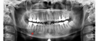
X-ray examination. On an x-ray, the cyst appears as a small oval or spherical shadow with clear outlines. The shadow is located at the top of the tooth root closer to its side. The periodontal gap is not displayed on the image, but significant destruction of the bone structure in the affected area is detected. Electroodontometry. The method is used when it is impossible to differentiate a radicular cyst on an x-ray. The excitability threshold of the affected tooth varies from 90 to 120 μA. Such indicators correspond to necrotic development of the pulp. Puncture. A puncture with a thick needle is performed to reliably exclude the malignancy of the cystic component. The contents of the cyst are sent for cytological examination.
A radicular cyst of the lower jaw bone is almost no different from the flow in the upper segments of the jaw. Often a radicular cyst is similar to osteoblastoma, but the main difference between a cyst is the absence of a cellular structure and the presence of clear contours of shadows in the projection image. Differential diagnosis allows you to reliably identify the disease and treat it according to accepted medical tactics.
Indications for tooth extraction with a cyst
As a rule, conservative treatment carried out on time or surgery to remove the cyst allows you to save the tooth. But this is not always possible. A tooth must be removed in the following cases:
— the presence of severe pain symptoms when drug treatment is ineffective; - purulent inflammatory process when it is impossible to drain it; — fracture of the coronal part of the tooth, without the possibility of restoration with anchor pins and/or stump inlays; — obstruction of root canals; — the presence of multiple damage to the roots of the tooth or large damage; the tooth is almost completely destroyed and orthopedic restoration is impossible; - no need for dental treatment due to the presence of a prosthetic plan agreed upon between the doctor and the patient.
Treatment process
Treatment of cysts can be therapeutic (conservative) or surgical. The treatment method is determined by the doctor after a visual examination of the oral cavity and the results of diagnostic studies.
Laser treatment
Laser beam treatment is gaining increasing popularity in dentistry. Under the influence of a laser, a tumor can be removed completely painlessly, without the risk of infection. In addition, the laser completely disinfects the affected channels and ensures rapid recovery. The laser treatment algorithm looks like this:
opening and expansion of dental canals; introduction of a laser beam into the canals: disinfection and removal of the cystic component.
The only disadvantages of the method are the relative high cost and the need for special high-precision equipment. Laser treatment also involves following the doctor’s recommendations immediately after the procedure (rinsing with an antiseptic and abstaining from food for up to 5 hours).
Conservative treatment
Therapy for a cystically altered tooth root requires disinfection, tooth cleaning and filling. An alternative treatment method is the introduction of a therapeutic suspension containing copper and calcium, followed by exposure of the tooth to low-power electrical discharges. The main indications for drug therapy are:
absence of fillings on root canals; poor filling in the root canals (not along the entire length); The size of the cyst barely reaches 8 mm.
In treatment, special drugs are used that negatively affect the cyst capsule and its contents. After which, the purulent exudate is completely removed, and instead of it, dental paste is injected into the cyst cavity to restore the bone structure. The manipulations are completed by filling the canal and crown. Cases of relapse of the disease are possible.
Operative method
Surgical treatment is carried out in some cases, since in 80% of cases a radicular cyst is an advanced process. The main indications for surgical intervention are:
presence of a pin in the root canal; early prosthetics of the causative tooth; the size of the cyst exceeds 8–8.5 mm; gum swelling and pain.
Until recently, the cystic component was removed along with the tooth. Alternative methods of surgical treatment are now being used to preserve the natural tooth. A tooth is removed only when the roots of the tooth have become part of a cystic structure or have been destroyed to the very root as a result of disease.
Main methods of surgical treatment:
| Cystectomy. | It is a complex but most effective method of cyst removal. The cavity is completely removed along with the damaged part of the root and the membrane. Indications for surgery are the rapid development of a tumor in the upper jaw to large sizes. |
| Cystotomy. | The procedure involves removing the anterior wall of the cystic cavity. The operation is performed in case of severe destruction of the bone floor of the nose, palatine plate, or with a large cyst. Cystotomy has the longest recovery period. |
| Hemisection. | The simplest method involves removing not only the cyst, but also part of the tooth root, the tooth itself or part of its crown. |
The postoperative period can take a long time. For the entire recovery period, antiseptic rinses are required, and sometimes antibiotics are required. You should not take aspirin, as it provokes bleeding. Recovery takes about a day, and swelling and slight soreness persist for several more days. If unpleasant symptoms persist or their intensity increases, it is important to immediately consult a doctor.
Traditional methods of treatment
Separately, it is worth mentioning the possibility of treating a tumor of the apical part of the upper jaw tooth with traditional healing recipes. Infusions of herbs, compresses and various poultices are good when the patient is in the postoperative period. Herbal treatment for tooth root cysts helps only as a local disinfectant along with traditional treatment methods. There are main herbs that heal gums faster:
Oak bark; pharmaceutical camomile; swampy cudweed; plantain and others.
If you start treatment only with herbs and compresses, you may not only fail to help your body cope with the cyst, but also lead to complications in the form of rupture of the cystic contents. Purulent exudate quickly spreads through the bloodstream and carries all bacterial microflora to vital organs.
Treatment
Radicular cysts are treated through surgery—cystotomy or cystectomy.
Cystotomy is used in the case of a large cyst that affects the roots of several adjacent teeth and destroys the walls of the maxillary sinus. The operation involves cleaning out the cyst cavity through a small hole made from the mouth, nose or paranasal sinus. Upon completion, the surgeon disinfects the cavity and inserts a swab with iodoform there. A week later, the tampon is replaced with a new one, and this continues until the doctor is convinced that the inflammatory process has stopped.
Cystectomy is the removal of a cyst by separating the fibrous membrane of the formation from the surrounding tissue. The operation ends with tamponade of the cavity or bringing together the edges of the mucous membrane that have been damaged. Cystectomy is done for small cysts.
Postoperative recovery will be successful if the patient takes oral hygiene instructions seriously: use antiseptic rinses and brush teeth carefully. In case of increased body temperature, as well as intense and prolonged pain, you should immediately consult a doctor.
Complications and prevention
Complications after surgery are possible if you do not follow the doctor’s special recommendations. A frequent cause of complications is the lack of self-discipline in the patient. The main complications include:
tissue infection during surgery; generalized sepsis; abscess syndrome around the affected tooth.
Rarely, the cause of postoperative complications is considered to be a lack of professionalism on the part of the dentist. Before treatment, it is important to inquire about the quality of services provided and the rating of the dental clinic. Complications include damage to molars in the maxillary sinuses and sepsis. If not the entire volume of purulent exudate is removed from the cyst, then the emergence of a secondary source of infection occurs almost immediately. Pus quickly spreads throughout the body and leads to dysfunction of many organs and systems. With the high-tech development of dentistry today, cases of complications as a result of cyst treatment are isolated episodes.
Preventive actions
When carrying out high-quality prevention, the patient must follow simple rules:
Carrying out regular hygienic teeth cleaning; sanitation of the oral cavity once a year; dental examination - 2 times a year; timely response to unpleasant symptoms in the oral cavity.
Treatment of cysts today is carried out successfully in a number of cases, so before visiting the dentist it is important to collect as much information as possible about the clinic and the professionalism of the specialist. You should not delay treatment, since in advanced cases, surgery is often the only possible way to eliminate the pathology.
For quick post-operative recovery, it is important for the patient to observe a hygienic regime, carry out preventive rinses of the mouth with herbal decoctions (chamomile, celandine, string, propolis) and medications (Miramistin, Iodinol). The patient’s discipline and attention to their own health are the key to oral health and the ability to treat any problems in the early stages using the most gentle methods.