Useful information for patients
The jaws are the basis of the facial skeleton. Not only the beauty of the profile depends on their anatomical structure, but also functional capabilities that are important for life - chewing, swallowing, breathing, speech, the formation of cavities for the sensory organs and much more.
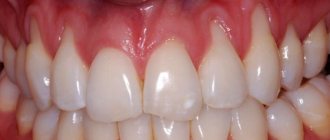
SYMPTOMS OF CORRECT BITE:
- There are no gaps between teeth.
- Chewing is not difficult.
- There is clear pronunciation.
- There are no cracks, chips or stains on the enamel.
- The teeth have the correct shape.
- There is no crowding of teeth.
- There is no pronounced inclination of the teeth in or out.
- The center of the incisors is located right in the middle of the face.
With improper development of bone tissue and the alveolar process, deformations such as microgenia, prognathia, overbite and others occur. In adults, the shape can also change, and this is mainly due to factors such as:
- Severe mechanical injuries.
- Consequence of surgical interventions.
- Various pathological processes (osteomyelitis, ankylosis, purulent inflammation and others).
- Incorrect orthodontic treatment or relapses after it.
- Partial or complete adentia (senile jaw).
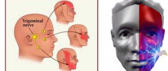
Changes in structure, various inflammatory and tumor processes lead to severe diseases of the jaw. The most common pathologies are osteitis, periostitis, osteomyelitis. At the first signs, such as swelling, redness, pain, fever, you should contact a dentist or surgeon.
ANATOMY OF THE JAW
UPPER JAW
The upper jaw is a fixed paired bone of the facial skeleton. It has an air cavity (sinus), 4 surfaces (orbital, anterior, posterior, internal), as well as 4 processes (frontal, zygomatic, palatine and alveolar).
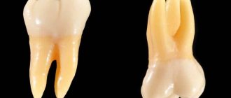
The dentition of the upper jaw is deviated slightly forward and outward. The crowns of the teeth increase in size from the incisor to the first molar, but the second molar is smaller than the first, the third is smaller than the second. The height of the crowns decreases successively from the incisor to the third molar, with the exception of the canines. The large roots of the teeth provide significant stability to the dentition of the upper jaw and to each tooth individually.
LOWER JAW
The lower jaw is a movable horseshoe-shaped bone of the facial skeleton. A large number of muscles are attached to it. The dentition of the lower jaw has a parabolic shape.
The teeth of the lower jaw are characterized by the fact that the incisors and canines are located perpendicular to the alveolar process, the chewing teeth are slightly inclined towards the tongue. The pattern of changes in the size and height of tooth crowns on the lower jaw is the same as on the upper jaw.
JAW DEVELOPMENT
The growth and formation of the jaw is closely related to the development of tooth germs and alveolar process. This process begins as early as the 7th week of intrauterine life. It is influenced by various endogenous factors (maternal toxicosis, hormonal imbalances, levels of vitamins and minerals), and after the birth of a child - also exogenous (external).
The jaw of a newborn is not yet completely ossified and fused; the lower part is shifted back in relation to the upper.
When a permanent dentition is formed (from the age of 6), intensive growth occurs due to the eruption of molars and incisors. Also, growth spurts are observed at 11–13 years of age; in boys, this usually occurs later. By the age of 18, the formation of bone tissue is completely completed.
CHANGES IN THE SHAPE OF THE LOWER JAW WITH AGE
| Jaw of a newborn |
| Jaw of a 4 year old child |
| Jaw of a 6 year old child |
| Jaw of an adult |
| Elderly man's jaw |
For patients who want to get a free consultation with a dentist in Krasnodar , we inform you that pre-registration for an appointment is being made at Dentistry on Gagarin. Call the numbers indicated on the website and sign up for a free consultation with a dentist with a diagnosis (consultation duration up to 15 minutes).
Topographic anatomy - what is it?
Topographic anatomy is a branch of science that studies the layer-by-layer structure of human body parts and internal organs, their location and interaction with each other.
Based on the knowledge of the discipline, it is possible to explain the symptoms that accompany organ damage, to understand the relationship and pattern of pathological processes.
This teaching received a second name - regional anatomy.
The topography of the jaw, in particular the teeth, contains a complex of knowledge about the structure and functions of bone formations in the mouth. Summarizes data on the structure of the jaw as a whole and its relationship with the oral cavity.
Information is necessary for doctors conducting medical practice in the field of dentistry and directly involved in the preparation of the jaw and bone formations: cleaning of root canals, filling, removal and adjustment of teeth.
The structure of the human upper jaw
The paired upper jaw is located in the middle of the facial segment of the skull and is motionlessly connected to its bones. The maxillary or air sinus, which opens into the nasal cavity, is part of it. The upper jaw is lighter than the lower jaw, as it has several sinuses (cavities), the largest of which has an average volume of 5 cubic centimeters.
The structure of the upper jaw is represented by a body with four surfaces:
The front surface , in the process of evolution, gradually changed its shape from flat to curved. The infraorbital margin separates it from the orbital surface in the upper segment. In the lower part, the anterior surface passes into the buccal alveolar process with small convexities corresponding to the location of the dental roots. On the medial edge is the nasal notch, which takes part in the formation of the anterior opening of the nasal cavity.
The infratemporal surface , which takes part in the formation of the septa of the pterygopalatine and infratemporal fossae, is fenced off from the anterior base - the zygomatic process. It contains the maxillary tubercle with several alveolar openings that lead into the canals of the same name.
The nasal surface takes part in the formation of the lateral septum of the nasal cavity. Its largest part is occupied by the maxillary cleft, leading into the maxillary sinus, located in the body of the maxillary bone. Anterior to the maxillary cleft lies the lacrimal groove, which helps in the formation of the nasolacrimal duct.
The orbital surface is similar in outline to a triangle. Takes part in the formation of the lower wall of the orbit. On its inner edge there is a lacrimal notch, which contains the lacrimal ossicle. In the posterior part, the infraorbital groove originates, developing into the canal of the same name.
In addition to the body, the organ includes four processes:
They differ in location, structure and direction.
The alveolar process is similar to a bony ridge extending down from the upper jaw. It is an arch on which eight recesses (alveoli) for tooth roots are located. The alveoli are separated from each other by interalveolar septa. The outer surface of the arch is called vestibular, the inner - palatine.
The zygomatic process extends in the direction of the zygomatic bone from the superolateral portion of the body of the maxilla. Between the alveolus of the first molar and the lower edge of the process there is a zygomaticalveolar ridge, which helps to redistribute the chewing load on the zygomatic bone.
The palatine process is a bony plate located horizontally that helps in the formation of the hard palate. On its lower rough side there are palatine grooves. The incisive canal lies in the anterior part of the process, and in the posterior part it connects with the plate of the palatine bone, located horizontally.
The frontal process extends upward from the body of the jaw, merging with the nasal segment of the frontal bone. The anterior lacrimal crest (located vertically on its lateral surface) initially limits the lacrimal trough. The ethmoidal ridge, located on the opposite - medial side, joins the middle turbinate.
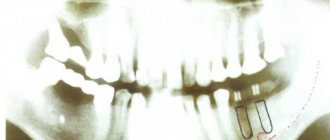
What is bone atrophy
Bone atrophy, or loss, is a result of tooth loss. Due to uneven load, the jaw loses volume - in the first year, tissue loss is 25%.
Atrophy can be stimulated by gum disease, endocrine, infectious diseases and the age factor. In patients, the blood supply to the jaw deteriorates, there is a lack of oxygen - the pressure on the tissue changes. The central or spongy layer, which has a porous structure, is subject to the process.
Jaw deformation has different intensity:
- I degree.
The blood supply is not impaired. It is possible to install a classic implant. - II degree.
The mucous membrane contracts. The operation is performed after plastic surgery. - III degree.
The contour is smoothed from the chin and in the oral cavity. Bone augmentation is a must.
The results of the deficiency are expressed in deterioration of speech, changes in facial proportions, and the appearance of wrinkles in the oral area. An advanced degree of atrophy causes displacement of the dentition, loss of adjacent or opposite units. Classical implantation is not possible with bone atrophy.
Functions of the upper jaw
The main roles are assistance in the functioning of the digestive system and speech apparatus. That is, the upper jaw is involved in the process of chewing (which is important for the primary processing of food) and the reproduction of sounds.
It forms the cavities of the nose, orbits and mouth, pterygopalatine fossae, outlines the correct location of the processes, and forms partitions between the mouth and nose. The jaw partly determines the oval of the face.
Osteoplasty
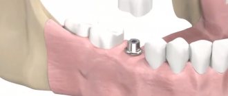
Classic dental implantation for bone tissue atrophy (height less than 10 mm) is preceded by bone augmentation surgery - osteoplasty. The procedure is carried out to ensure stable fixation of the implant and to avoid aesthetic complications during subsequent restoration of the element with prosthetics. For this purpose, the implantologist acts using one of the methods:
- Sinus lift.
The operation is performed on the upper jaw - the doctor lifts and displaces the maxillary sinus, making room for new bone. - Guided bone regeneration.
Bone material is added, covered with a membrane and sutured until it fuses with the jaw. - Replanting bone blocks.
The person's own bone material is used. It is removed from the lower jaw in the area of the wisdom teeth. The bone block is fixed with screws, bone granules are placed around it, and a membrane is attached. - Splitting of the alveolar ridge.
The dentist cuts the appendage and increases its thickness using a graft or artificial material.
It takes 3-6 months for the new bone to heal. It is possible to combine osteoplasty and implantation operations. The decision-making algorithm for performing osteoplasty simultaneously with implant fixation is as follows:
- Studying the possibility of positioning the artificial root in the correct position (using wax-up and surgical templates).
- Choice of osteoplasty technique. Depends on the degree of bone atrophy and its spread in height or width.
- If it is impossible to achieve stability of the artificial root with the existing bone volume, the extension is carried out in a separate stage.
Teeth size for prosthetics
It is especially important to preserve and, if necessary, restore the natural size of teeth during prosthetics, so as not to disrupt the bite and the full functioning of the dental system. The size of the crown is selected by the dental technician, focusing on neighboring or teeth of the same name, monitoring the correct closure of the rows and adjusting the prosthesis during installation.
Determining the size of artificial teeth with complete edentia is carried out by measuring the distance between the corners of the mouth using a special ruler - it corresponds to the width of the six front teeth. And the segment from the edge of the gum to the smile line is equal to the height of the crown. For example, during implantation it is necessary to correctly calculate not only the parameters of the orthopedic design, but also the size of the dental implant. It should be equal in length to the real root, but the choice of titanium rod parameters depends largely on the volume of the jaw bone tissue.
Development of tactics for correcting a child’s bite
Since the child is at an age when baby teeth are actively falling out and permanent teeth are erupting in their place (changeable bite), it is necessary first of all to create a place for the normal eruption of permanent teeth. Based on the current situation, it is necessary to increase the size of the upper jaw. Then you need to move the displaced teeth into place, allow all permanent teeth to erupt and align their position if necessary.
Commentary by speech therapist, myofunctional therapist T.B. Zukor: “When carrying out orthodontic treatment, it is not enough to just correct the position of the teeth; it is necessary to eliminate the cause of the malocclusion. In this case, mouth breathing was observed, as there were ENT problems. I, as a myofunctional therapist, can retrain a child from habitual mouth breathing to nose breathing, but only on condition that the nose is breathing! That is, the participation of an ENT specialist is required, who, for his part, will eliminate the existing problems. The stability of the result of bite correction directly depends on eliminating the causes of the disorder! If you don’t normalize breathing through your nose and restore the correct balance of the muscles of the tongue, cheeks and lips, you will have to wear retainers for a very long time, and maybe for life, since the teeth will constantly strive to get back into position incorrectly.”
Treatment plan:
Stage 1 - Expansion of the upper jaw.
A McNamara appliance will be used to expand the upper jaw. This is a fixed orthodontic plate appliance with a screw. The device is made individually based on impressions and is attached with rings or trays to the distant teeth. Using a screw, the device moves apart, stimulating the expansion of the upper jaw.
Stage 2 – Distalization (movement) of chewing teeth.
The chewing teeth that have been displaced due to the early removal of baby teeth must be moved to their place and pushed back. To move the teeth a sufficient distance, the Pendulum device will be used, which applies pressure to individual teeth, causing them to move in a given direction. The device is made individually using dental impressions and is fixed on the teeth for permanent wear.
Stage 3 – Eruption of permanent teeth.
At this stage, the child will not wear any orthodontic appliances, but will only be under the supervision of an orthodontist.
Stage 4 – Teeth straightening with braces.
Braces will be installed on permanent teeth, which will help set the teeth in the correct positions and create physiological interdental contacts.
Throughout all 4 stages of treatment.
It is recommended to undergo treatment by an ENT specialist (to eliminate the causes that interfere with breathing through the nose) and correction of the balance of the facial muscles by a speech therapist - myofunctional therapist to obtain a stable result of bite correction.
Commentary by orthodontist M.P. Sleptsova: “The child is still small, his teeth are only baby teeth, and the narrowing of the upper jaw is significant. Braces cannot correct this; they do not expand the jaw. In addition, sufficient distalization (movement back) of the distant teeth was necessary to prepare the site for the eruption of permanent teeth. Braces cannot cope with this task either. If the parents had not contacted the orthodontist in time, then it would have been possible to straighten the teeth only by removing some teeth or by surgically widening the jaw. It is also important to involve an ENT specialist and a speech therapist in the treatment in order to eliminate the incorrect type of breathing and get a stable result.”
Types of bone atrophy in the upper and lower jaw
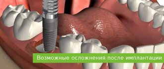
Destruction of the bone structure of the upper jaw is fraught with injury to the closely localized maxillary sinus. The cells in this area have a loose structure, the bone is thinner, and is actively resorbed in the area of the masticatory elements. When fixing long implants, there is a risk of injury to the sinus membrane, which can stimulate its rupture and the development of sinusitis or chronic runny nose.
The Schroeder classification identifies three types of atrophy of the upper jaws
:
- The jaw tubercles are pronounced, physical abnormalities are not visible, the mucosa is noticeably curved, the palate is deep. Implantation is possible without complications.
- The alveolar processes are not clearly expressed, the palate is of medium depth. Installing implants using the classical method is doubtful, but using a one-stage method is possible.
- The cells are seriously atrophied, the alveolar processes are smooth, the palate is flat, the fold at the level of the palate does not hold its shape. Fixation of classical implants is not possible; it can be done in one stage, depending on the location of the defect.
The tissue of the lower jaw is dense from below and does not decrease so rapidly, but as atrophy progresses, the dentist is faced with the problem of the nearby localized mandibular nerve (it is located under the roots of natural elements). Nerve injury can cause complete or partial loss of sensation in the lower facial part.
According to Keller, there are four types of mandibular atrophy
:
- The tubercles of the alveoli are visible on the jaw, a fold of the mucous membrane is noticeable. You can install classic or one-stage implants.
- The ridge becomes sharper, and muscles are attached at its base. Installation of a classic implant may cause discomfort, but a one-stage implant does not cause complications.
- The jaw of patients with early removed lateral teeth - the alveoli are thinned, the volume in the center does not decrease. It is not possible to implant classical artificial roots; one-stage ones are possible, but there is a fear of the implant shifting when chewing food in case of a single defect.
- In the area of the frontal incisors, the bone is clearly atrophied; the lateral row is not affected. Implantation for such bone tissue atrophy is possible using a combined method.
Orthodontist consultation
At the consultation, orthodontist M.P. Sleptsova conducted an examination and made a preliminary diagnosis, telling the parents how to correct a child’s malocclusion, approximately how long it would take for treatment and the approximate cost. Based on the results of the examination, we can say for sure that there will not be enough space for the eruption of permanent teeth and the teeth may begin to grow crooked. The fact is that permanent teeth are larger in size than baby teeth, and there is clearly not enough space in the jaw.
It is possible to say more precisely how much it will cost to correct a child’s bite after an orthodontic diagnosis, which includes taking impressions and calculating the impressions, performing a tele-roentgenogram of the head in frontal and lateral projection, as well as a panoramic image of the teeth and calculating x-ray images, consultation with an ENT specialist, speech therapist and osteopath. Diagnostics is needed to make an accurate diagnosis and develop the correct treatment tactics.
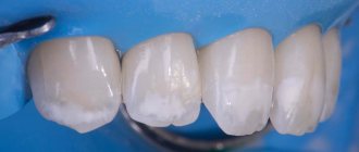
Photos of teeth before treatment:
Panoramic photo of teeth before treatment:
Diagnostics showed that there is a narrowing and shortening of the upper dentition, biretrusion (the upper front teeth are located behind the lower ones and should overlap them), displacement of the lateral teeth to the center, tortoanomaly (teeth rotated around their axis), hypertrophy of the tonsils, adenoids, infantile swallowing, mouth breathing.
Commentary by the chief physician of Dial-Dent S.V. Tsukora: “The causes of malocclusion in children may be improper swallowing, incorrect position of the tongue, the presence of a bad habit of constantly sucking a finger, pen, toy, etc., mouth breathing due to a constantly stuffy nose or due to habit, lack of solids in the diet food. The sooner parents show their child to an orthodontist, the easier it is to stop developing problems and prevent serious malocclusion. We have all the necessary specialists in our clinic - orthodontists, pediatric dentists, ENT specialists, speech therapists, osteopaths, so we can help in the most difficult cases, and the treatment result will be stable, since in addition to simply straightening teeth, we work with the cause of malocclusion.”