Tooth roots inevitably begin to become exposed in old age - this process is completely natural and is a reaction to the aging of the body. But old age is far from the only reason for this phenomenon, which spoils a beautiful smile and can lead to other serious illnesses in the future. Root exposure can occur at any age due to disease or pathology.
This article will talk about what to do in a situation where the gum has noticeably risen above the tooth, and you will also find answers to why this happens and what the consequences of refusing treatment may be.
When can a tooth be restored?
When the walls have completely crumbled, but the root of the tooth has not been destroyed, traditional dentistry sometimes suggests removing it. Today, using new protocols, it is possible to preserve the root and create a new stable unit - any of the functional ones, except for the “eights” (wisdom teeth). To do this, you need the root to be healthy:
- Not affected by caries, without fractures or cracks. If such defects exist, then only removal followed by implantation is indicated.
- No cyst. If there is one, then a method of treatment is selected. And only then can you begin to restore.
If a similar problem happens to you, do not rush to the surgeon. New technologies make it possible to accomplish the previously impossible - to preserve the root of a tooth. What to do in a particular case is decided at the appointment, after diagnosis. The vast majority of patients manage to save the tooth after treatment procedures and install a crown on the root of the tooth.
Examples when only the roots of the teeth in the upper jaw remain
Example 1. Restoration from one root of a front tooth
In this example (presented in full here), the situation was created literally by the patient’s hands. She tried to glue part of the tooth with superglue and, under the influence of dangerous substances contained in the glue, the crown of the tooth became completely unusable.
But the root of the tooth under the crown below remained intact and unharmed.
The length of the healthy part of this tooth root removed all doubts about the question - is it necessary to restore the root? Certainly! - it is possible and necessary. The tooth was restored in 1.5 hours using Cerec technology. And here in the photo is the happy owner of a new restored front tooth:
Example 2. Restoring 3 front teeth, of which only the roots remain
Here is a treatment example where I restored the aesthetics of her smile to a 22-year-old girl. The three front teeth on the upper jaw were completely destroyed. From the upper teeth 1.2, 1.1, 2.1 only
the roots
:
These three teeth were actually an aesthetically unsightly frame made from old fillings. Now we will not analyze the whole case in detail, I would like to draw your attention to the following points:
- the shape of the roots of the teeth and their condition made it possible to restore them
- There was no talk at all about any amputation - removal of the roots of the teeth of the upper jaw and implantation.
Our patient's smile was designed in a computer program:
So, in the photo below you see a general set for restoring our patient’s dentition, and among the modules there are 3 with a crown and a root.
The roots of the upper front teeth were quickly and, most importantly, effectively restored:
and our patient underwent a beautiful transformation and became the owner of a sweet and chic smile:
This is the enormous potential and aesthetic power of healthy tooth roots! Don't rush to delete them)
Example 3. Complete restoration of the smile zone on the front teeth of the upper jaw
This example is actually very popular on the Internet, since the patient is a TV presenter, and as a result of the treatment she found her new smile. Which is extremely important for her profession. I describe the detailed clinical case itself in another article, but here I would like to note the main points related specifically to the restoration of the roots of the front teeth with crowns.
In the picture below we have removed composite restorations and carious lesions. only the roots remain of the two front incisor teeth.
:
Crowns and “crown + root” modules were prepared in the laboratory:
After installation, the smile began to shine with bright colors:
You can see the entire chronology of our patient’s treatment in one large picture, step by step:
Why do tooth remains appear in the gums?
There are many reasons for this phenomenon:
- Perhaps the tooth has completely crumbled, but the root remains.
- The crown received a strong blow and a fracture occurred.
- An incomplete tooth extraction was performed. The remaining fragments cause inflammatory processes in the soft tissues, causing pain.
- Units with the neurovascular bundle removed are often destroyed. The so-called “dead teeth”. The nutrition of their walls is disrupted, the shell becomes fragile and, due to incorrect loading or due to increased fragility, the natural walls break off “at the root.”
If the process has already started, then urgent treatment of the tooth root is necessary. Otherwise, the remains of the unit will have to be removed to avoid infection of adjacent teeth and surrounding tissues.
The situation cannot be ignored: tooth fragments may not bother you at first, but then turn into a serious problem. When trouble occurs, immediately contact the dental clinic to promptly select treatment and find ways to restore the tooth.
Guarantees
In our Center, implants are installed with a lifetime warranty from the manufacturer - Nobel Biocare. We provide guarantees:
- lifetime for the installation of implants;
- 1 year for crowns.
The warranty program includes a complex for the implant, surgery, bone reconstruction and prosthetics.
The guarantee is valid provided that the patient follows the doctor’s recommendations, care rules and regularly visits the dentist.
Examples of canine restoration when only the root remains of the tooth
Example 4. The pin in the root of the lower tooth was removed, and the canine itself was restored
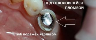
In this clinical case, I was restoring the lower canine, and the task was to save the root of the tooth and restore the coronal part of the canine, which was almost destroyed by caries. The anterior wall of the lower canine was made in the form of a filling, and it fell apart from the cutting edge.
As you can see in the photo, an anchor pin was installed inside the tooth - this is a pin that is screwed into the canal to strengthen the tooth. The tooth under the filling and the root of the lower canine were affected by caries. When the carious tissue was removed, only the root and a small part of the wall
:
You and I remember the main topic of our article, which tells us the following - if the tooth root remains, it can be completely restored. And this is not the first time that the unique Cerec technology has come to our aid. Using it and digital scanning, we restored the missing tooth module, which also included the root part:
Next, we restore the lower front tooth using a CEREC inlay and get an excellent result - the lower canine is completely restored:
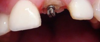
Example 5. The root of the tooth under the crown of the upper canine has not rotted, and therefore the tooth is 100% restored
In this case, there was a total restoration of the dentition (veneers and crowns) with the replacement of worn-out crowns on the front teeth. One of the upper canines under the crown was seriously affected by caries, but its root was practically healthy - only a small part of the tooth root was damaged, see the following photo:
The missing “crown + root” module and new crowns were manufactured in a laboratory. In general, everything is in a smile, it looks just great:
And the owner of a new beautiful smile herself cannot hide her admiration:
All the main tasks in this total work were successfully solved, but the main thing you should pay attention to is that we managed to save the tooth root here too.
Canal treatment under a microscope
In endodontics, the method of treating tooth canals under observation of the process through a microscope is widely used. This method allows you to see much more and provide better treatment. Root canal treatment under a microscope is a convenient method for the patient and the doctor. A magnification of 30-40 times under a microscope makes it possible to see all the branches of the canals, clean the canal perfectly and seal it.
The microscope allows you to see cracks in the canal, find all the branches of the canal, remove foreign objects, precisely treat hard-to-reach areas of inflammation, and remove the nerve. A microscope helps the dentist determine the condition of the filling, avoid damaging healthy tissue, and fill all the voids in the canal, leaving no room for infection to develop.
How to determine if a root is healthy
How to understand whether caries has affected the root of the tooth, whether the dentin of the tooth root is intact or whether there are defects? In order to have an objective picture of the process, it is necessary to perform an x-ray of the tooth root and a computed tomogram. A CT scan will determine the true condition of the root and show:
- The presence of inflammatory processes.
- It will determine whether there are neoplasms and what size they are (cyst or granuloma).
- The location of the roots in relation to the neighboring ones, the maxillary sinus.
This is the only way to see whether it will be possible to cure the tooth root using a tab.
Example 6. Restoration of teeth 4 and 5, when only one root remains
In the next total work I would like to highlight the 4th and 5th premolars. In general, the girl’s situation was quite complicated initially at the time she contacted me. Both the lower and upper jaws were restored. Let's look at only the top one.
Front 4 teeth
- these are 1.2, 1.1, 2.1 and 2.2 were entirely made of composite material, under which caries developed. And the teeth we were interested in, 1.4, 1.5, 2.4 and 2.5, were covered with metal-ceramic crowns under which there were metal inlays. Today, such structures are rarely used in advanced dentistry, since the same Cerec technology allows you to achieve excellent results with one module “crown + tooth root”, rather than breaking the structure into an inlay and a crown with an additional adhesive layer. In fact, we place a crown at the root of the tooth with the function of the missing root. There will be a separate example on metal tabs a little later.
So, in the photo below, the crowns were removed, the inlays were removed and carious tissue was removed from teeth 4 and 5, leaving healthy roots
:
If only the root remains of a tooth, this does not mean that it cannot be restored. And let this tooth root be pulpless, i.e. dead - such tooth roots feel great in bone tissue and orthopedic structures can be built on them. Using computer technology and 3D scanning, we first restored virtual teeth:
The following photo shows that on one side we place crowns with an inlay function in place of the 4th and 5th teeth, and on the other opposite side we place half-crowns also with an inlay function.
That is, these are single modular designs - veneers with a root part, which are currently the best for the patient
:
Installing veneers with the root part allows you to completely recreate an aesthetically beautiful dentition:
The stage of temporary prosthetics, which allows the patient to see his smile, plays an important role in the very process of its new formation, since the patient understands that the main problems with the restoration of the remaining teeth, and in fact the roots of the teeth, are left “far behind”:
After installing crowns and half-crowns and restoring the front teeth, our patient’s smile was transformed beyond recognition, the so-called. wow effect:
The clinical example described above can be viewed in detail HERE.
Canal filling methods
Canal filling
helps prevent the development of dental canal diseases. Canal treatment is a painstaking process, complicated by the fact that the dental canals themselves are narrow, the shape can be curved, which requires painstaking work to fill the entire canal. Today, dentistry has several methods for filling canals.
1. Heated gutta-percha
Gutta-percha is a hard material that becomes elastic when heated and ideally fills the canal cavity. Several methods are used to treat a tooth canal using gutta-percha:
1) liquid injectable gutta-percha;
2) continuous wave;
3) vertical condensation;
4) syringe administration of gutta-percha.
2. Lateral condensation - cold gutta-percha
A gutta-percha pin is inserted into a canal filled with sealer paste, compacted, and sealed.
3. Thermofil - volumetric filling with hot gutta-percha
A plastic rod is inserted into the canal and the canal cavity is filled with hot gutta-percha, penetrating into all branches, leaving no free space.
4. Depophoresis technology
It is used in cases of difficult access to a curved canal that was previously filled, as well as in cases where the canal contains a part of an instrument broken during treatment.
All methods are painless for the patient. After treatment, pain is possible for two weeks if the root is removed.
Why removing the root is not the solution to the problem
If there is no natural crown, and instead of it an empty space appears, especially when it is the roots of the upper front teeth, many people habitually rush to the surgeon. They want to remove the remains and quickly install a bridge, eliminating the aesthetic defect.
But first, it’s worth considering the situation with your doctor - there are certain risks during removal: damage to the mandibular nerve, excessive trauma to the gums, and others. In addition, tooth restoration based on its own root is always more gentle than any prosthetic method.
Dentists try to preserve every unit, since the absence of natural teeth leads to a decrease in the quality of life. If you simply remove the roots of permanent teeth, then immediate implantation is needed. In the absence of a unit, destructive processes begin in tissues, and bone volume decreases. Therefore, doctors do not recommend neglecting this problem. Even if there is no chewing molar, which is not visible.
Examples of restoration of fifth teeth on the upper and lower jaws
Example 7. A metal tab and caries left only the root of the 5th tooth
A patient came to me with a 3.5 tooth. It was a pulpless tooth - the lower five, it did not have any periapical changes or inflammation in the area of the root apex. The tooth was missing the upper half of the crown part of the tooth. This tooth 3.5 initially had a filling, a metal stump insert was installed in the tooth, quite deep, and a filling was already installed on top. Here's a “puff sandwich”:
As I said in the previous example No. 6, metal tabs are beginning to be used less and less in practice - they have been replaced by modern computer technologies, for example, Cerec. As a result of the atraumatic removal of the metal insert, the bone was preserved, nothing cracked: neither the root nor the remaining bone structure:
The restoration of the bottom five was carried out using Cerec technology in 1.5 hours
:
The “crown + root” module fell into place perfectly, the patient was very pleased with such a quick and effective solution to his problem:
Example 8. The top five were completely destroyed by caries. But the tooth root survived
With this example, I want to show you how deep caries can literally “eat” your teeth. And, of course, try to prevent it from developing like this. The patient is a fairly young man who has decided to radically improve his image. This is a laudable decision, but here are the teeth he came to me with:
Caries on almost all teeth. After removing the carious layer, the remains of the teeth appeared in this form:
Please note that in place of the upper tooth 5
There was practically only one root left with a small side wall. In this case, the same technology of tooth restoration using the “crown + root” module and crowns/half-crowns with an inlay function was used. The result of the treatment, as they say, is on the face:
The goal of treatment, according to the patient, has been fully achieved - a brutal Man
:
Possible complications after endodontic treatment
- The walls of the tooth cavity and the bottom are perforated, subject to the presence of dentin, if the instrument penetrates too deeply.
- The contents of the root canal are not completely removed in cases of obstruction, lateral branches, denticles or bleeding.
- The lumen is clogged with dentin filings, pulp residues, or an instrument that has broken in the canal.
- If the canal is bent, perforation of the root walls is possible
- The canal was not sealed thoroughly enough.
- The canal lumen was not expanded correctly.
- Filling falling out.
Complications may not appear immediately. After root canal treatment, the patient has slight sensitivity, pain, and discomfort in the area of the treated tooth and gums. If these sensations do not go away after two weeks and the pain intensifies, you should contact your doctor for an examination to determine the cause.
When and how can you restore a tooth with Cerec?
A common problem is the development of caries on the frontal units. The result is their complete destruction. The patient sometimes comes too late. Or the previous intervention does not bring the desired result. When several units in the smile area are damaged, and some of them only have tooth root tissue left, you can combine the installation of crowns, veneers and restoration of the upper visible part using special inlays in the root system. Sometimes large-scale intervention is needed - the smile area is corrected, damaged teeth are recreated. You can do a step-by-step restoration of 10 or even 20 teeth above and below.
Using Cerec technology, if there is a healthy root, it is easy to restore almost every tooth within a few hours.
- It will be necessary to determine the condition of the root and undergo a course of treatment.
- Then, after treating the upper roots, it is possible to install a module with the alignment of the tooth crown and the insert into the cavity in the tooth root.
A smile should always be charming. When restored, the new units are ideal, better than natural ones. In addition, natural teeth darken under the influence of pigments or turn yellow with age. Therefore, next to the restored ones they look unaesthetic.
To avoid such consequences, units can be corrected with veneers or crowns (when they are 50% destroyed and there is no stable support). To create an impeccable beauty zone thanks to Cerec, you only need a few visits to the doctor.
Alternatives
An alternative method for restoring sixes is a bridge prosthesis. This is a structure consisting of crowns tightly connected to each other. To restore one tooth, the bridge consists of 3 crowns. The two outer ones are fixed on the teeth on both sides of the defect. The supporting teeth are ground down to form the internal cavity of the outer crowns of the prosthesis. The middle crown is hinged and imitates the lost six.
This option is cheaper than implantation, but has disadvantages - grinding the enamel of living teeth leads to a reduction in their service life. Another important point is that the bone under the hinged crown does not receive stress during chewing, so bone tissue atrophy is inevitable.
Features of Cerec technology
Cerec root inserts and crowns are made from metal-free materials - it can be ceramic or zirconium dioxide. They can be installed even for allergy sufferers without fear. This technology has completely changed traditional approaches to dentistry.
Now the restoration process is comfortable and takes a minimum of time. In my clinic, the procedure is carried out in several stages.
I would also like to note that only one anesthesia is required and there are practically no errors when creating new teeth. Stages:
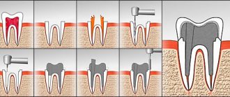
- An intraoral scanner determines the boundaries of the damage. This is especially important when only the roots of the front teeth or molars remain. The remaining tissues, antagonist teeth, and closed rows are scanned.
- The information is transferred to a computer, where all the images are combined into an image.
- The program creates a three-dimensional model of the remaining tooth, all defects are clearly visible. Then models of the inlay and crown are created that perfectly replicate the anatomical features of the tooth. Dimensions and shape are calculated accurately. It is determined what kind of restoration is needed.
- The design is made from blanks on a milling module. The elements are being tried on. In rare cases, correction is required. Shade discrepancies or other issues that displease the patient can be easily corrected within a few minutes.
- The completed root inlay fits hermetically to the remaining parts of the tooth, is securely fixed and exactly matches the shape.
Thus, it is possible to quickly restore units if only the roots of the teeth of the lower or upper jaw remain.
Associated symptoms
The tooth consists of three parts:
- Crown.
- Neck.
- Root.
In a healthy state, only the crown should be visible, the neck should be covered by the gum, and the root should be located in the alveolus (a special recess for the root). When the process of recession begins, the neck of the tooth is exposed, but that’s not all; along with this problem, the following symptoms usually appear:
- High tooth sensitivity when in contact with extremely cold, hot, sweet, salty and sour foods, because the tooth exposed the sensitive part of the neck, on which there is almost no protective enamel.
- Noticeable darkening of the enamel spoils the appearance; the situation is also aggravated when the neck of the tooth, which is yellow, is exposed.
- Visually, the tooth appears longer.
- Feeling of discomfort in the gums. When you try to press on it with your tongue, it does not fit tightly to the tooth, but moves away from it.
- When brushing your teeth, your gums become very swollen, painful and red.
- Wide interdental gaps appear at the base of the tooth.
It should be noted that any of the above symptoms are more acutely felt in adolescence and middle age, while older people may not notice or feel that anything is wrong. Therein lies a great danger, because adult patients most often come when the situation has reached a critical point and it is almost impossible to save the tooth.
Benefits of Cerec Restoration
The innovation has clear advantages compared to traditional prosthetics:
- The process does not take a week or several days, but only 1.5 hours
per tooth maximum. - Accuracy
. The computer minimizes errors and eliminates errors. - Biocompatibility
. Fabrics do not reject materials.
An inlay in the tooth root is created quickly, then the crown is modeled. The price is fully justified by the reliability, long service life of the structures and their aesthetics.
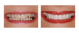
There is a striking difference between the smiles of patients when they only had the root of the tooth left, and the photo after treatment with Cerec dentures.
Is it worth getting an implant?
The absence of the sixth tooth threatens:
- displacement of neighboring units;
- advancement of antagonists on the opposite jaw.
Neighboring and antagonist teeth will slowly shift, which threatens to expose the roots, the appearance of wedge-shaped defects at the gum itself, exposure of the neck, which is sensitive to changes in temperature of water and food, and pain. In addition, there will be a change in the bite, since the teeth will be out of place. Therefore, it is necessary to place an implant to maintain healthy teeth and correct bite.
Example 9. Restoring the bottom six “from the root”
In this case, I would like to simply dwell on the moment of restoration itself. The patient is older, over 50 years old. And he felt the need to restore the chewing group of teeth. The picture shows the lower 6th tooth, of which practically only one root remains:
The tooth was restored in 1.5 hours
using Cerec technology. The very speed of tooth restoration in one visit to the doctor and the patient’s ability to immediately, as they say, use it in chewing gives a 100% head start on any existing method of tooth restoration. It works really well - you can see for yourself.
Example 10. Restoring the bottom seven
And I end my examples with the classic option of restoring the seventh lower tooth, of which only the root part remains. Removal of the lower 7th root of the tooth was not required; Cerec technology again proved to be excellent:
After modeling in 3D, manufacturing the module itself only took about 20 minutes, after which it was successfully installed on the remaining root:
Why do you need to build bone tissue?
Increasing bone volume (osteoplasty) is necessary for the primary stabilization of the titanium root. If the height and width of the alveolar ridge are insufficient, the implant simply will not stay in the bone. The method of osteoplastic intervention depends on the clinical picture.
The following methods are used to build bone in the lower jaw:
- Guided bone regeneration.
Increasing the height and width of the alveolar process by replanting osteoplastic material. The surgeon peels off the gum, fills the jaw bone with osteoplastic material, closes it with a barrier membrane and applies sutures. - Splitting of the alveolar process.
Increases bone width. The doctor makes a cut in the center of the ridge, alternately screws spreaders of different diameters into it (from smaller to larger), and fills the resulting space with bone granules. - Bone block transplantation.
An autogenous (taken from the patient) bone block is screwed to the bone, covered with a collagen membrane and sutured. The 6th tooth implant is installed after the block has healed. - Increasing the volume of the maxillary bone.
Because the maxillary structures are located close to the root system of the first molars, a sinus lift is required in 90% of cases. It can be closed or open. The essence of the operation is that the doctor carefully lifts the lower part of the maxillary sinus, fills the resulting space with osteoplastic material and installs a six implant.
Should the tooth root be removed? In what cases is this necessary?
Is it necessary to remove the root of a tooth? The root must be removed if:
- it is mobile, unstable or has a deep pocket;
- there are cracks;
- the root processes are “recessed” into the bone tissue and are located below their level;
- root tissue is softened or severely destroyed;
- There are chips in the tooth root.
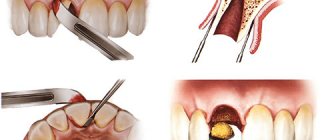
Modern surgery is as painless as possible. Doctors perform the procedure while maintaining the integrity of the surrounding tissue. In the absence of inflammatory processes, it is possible to remove the real tooth root with simultaneous implantation of an artificial one.
Price
Our Center has a case payment system, which means that the case includes all materials and necessary manipulations.
The cost of implant installation includes:
- implant and superstructures;
- work of an implantologist;
- anesthesia;
- basic bone building complex;
- primary and repeat CT.
The price of implants varies depending on the type of bone. Nobel Biocare PMC (cheaper) is intended for weak tissue, and Nobel Biocare Conical Parallel CC (more expensive) is intended for dense tissue.
The cost of the crown includes:
- production of a prosthesis by a technician;
- taking impressions;
- installation of a crown.
Tooth extraction (for simultaneous implantation), bone grafting or sinus lifting are paid separately. Prices for services can be found here.
CONCLUSIONS:
As you can see from the examples provided, I, orthopedic dentist Sergei Samsakov, an expert in the field of digital modeling and restoration of teeth using Cerec, manage to restore a beautiful smile even in almost the most hopeless cases. If you have similar problems, do not hesitate, contact us, and we will always find the optimal and, most importantly, beautiful solution to your situation.
And remember the simple truth: there are no hopeless situations!
Canal diseases
Canal diseases make themselves felt by inflammation and pain, so you need to see a dentist. If there is internal inflammation, but there is a possibility of soft tissue necrosis.
- Pulpitis
- inflammation of the dental pulp. With pulpitis, blood vessels collapse and die.
- Periodontitis
- inflammation of connective tissues resulting from complications of pulpitis.
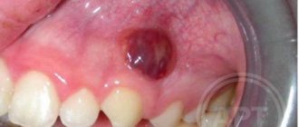
- Abscess
- the presence of pus in the gums, which arose as a complication of pulpitis
- Advanced caries
leads to inflammation of the tooth canals.