Not so long ago, a granuloma of a tooth was an indication for its removal... But today, thanks to modern treatment methods, it is possible to preserve the tooth and its functionality. This disease is characterized by a limited inflammatory process of periodontal tissues, which is accompanied by the formation of a rounded shape in the area of the tooth root. Its peculiarity is that it lasts for a long time without any clinical manifestations, but there comes a time when, under the influence of certain factors, the granuloma worsens. As a result, the symptoms worsen: pain, swelling and redness of the gum tissue.
The CELT Dentistry Department invites you to undergo treatment for dental granuloma in Moscow. We employ specialists who have decades of experience in scientific and practical work. They have modern diagnostic and treatment equipment in their arsenal, which allows them to correctly diagnose and treat patients in accordance with international standards. We have all the necessary licenses and certificates and work with the conclusion of an official contract. To find out the price of treatment for dental granuloma, go to the “Services and Prices” tab. We regularly update our price list, but we still ask you to check the numbers with our information line operators or at your doctor’s appointment.
Consultation with a dental surgeon - 1,000 rubles.
Resection of the root apex (frontal group) - 6,500 rubles.
Resection of the root apex (chewing group) - RUB 9,000.
Tooth extraction – from RUB 2,200.
At CELT you can get advice from a dental specialist.
- The cost of a consultation with a dentist-therapist is 1,000
Make an appointment
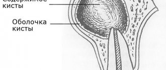
What is a dental cyst?
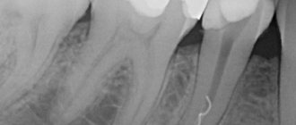
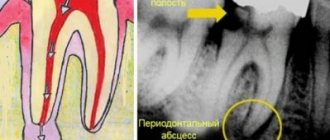
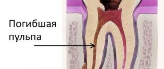
The inflammatory process - a dental cyst (from the Greek “kystis” - bubble) - is formed in the form of a small inflammatory ball that swells as part of a response to injury or infection. The inflamed area looks like a pus-filled bubble, sometimes reaching several centimeters in size. The body’s active work to restore order and remove slagged cells occurs constantly. At some point, a lot of them accumulate, and our excretory system selects the weakest area of the body through which they can be removed. In this case, the soft tissue around the tooth becomes the target, where a purulent neoplasm appears. An emerging problem can be diagnosed in the early stages using an orthopantomogram.
Why does a dental cyst appear?
What is the prerequisite for the appearance of such an unpleasant disease - dental cyst:
- physical trauma;
- periodontitis, peritonitis, pulpitis - inflammation of the soft tissues around the tooth;
- residual effects after diseases of the nasopharynx - ARVI, complications after influenza;
- decreased immunity;
- advanced caries process;
- poor oral hygiene;
- development of infection in the root canal;
- poorly placed crown, poor quality filling;
- the appearance of problems with the wisdom tooth.
Laser treatment of dental granuloma
Laser treatment is considered one of the most advanced in medicine and is also used in dentistry. It is believed that treatment of dental canal granuloma with a laser allows you to do without the use of drugs, since the beam destroys the accumulation of granulation tissue and destroys all pathogenic microorganisms. This impact has its supporters and opponents. Fans of laser treatment assure the maximum effectiveness of the technique and provide statistical data demonstrating the effectiveness of this method. On the other hand, the cost of treating dental granuloma with a laser is higher compared to classical endodontic manipulations; the use of certain medications is still required, and in case of non-standard structure of the root canals (in particular, with severe curvature), the use of a laser is less effective. It can also be used during direct surgical intervention in the process of processing and removing tissue. This reduces the invasiveness of the operation, but increases the final cost of treatment.
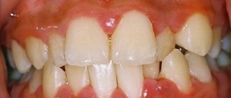
Symptoms of a dental cyst
It is practically impossible to recognize the disease at the stage of its inception. The epicenter of development is located deep in the tissues, so it is quite difficult to immediately make a diagnosis. The inflammatory process makes itself felt when external signs or pain symptoms appear:
- darkening of the enamel, redness of the gums;
- pain when biting, pressing;
- itching sensation near the gums;
- discomfort when chewing;
- the appearance of a tugging, sharp piercing pain in the area of the tumor.
During this period of time it is difficult to determine what is happening where. It seems to the patient that this or that tooth hurts - he may mistakenly indicate a healthy one, but in fact, “dead” cells accumulate, and the inflammatory process enters the next stage of development. A purulent formation is formed: the skin is stretched under the pressure of an enlarged, rapidly growing bladder. As the amount of pus increases, the blister grows in size, exceeding 1 cm in diameter. At this stage, the patient’s sensations and appearance make it possible to visually diagnose a dental cyst:
- swelling of the gums, face;
- severe aching pain;
- elevated temperature;
- lack of appetite, malaise;
- statement of the fact of pain in the lymph nodes, their enlargement;
- headache if a cyst appears in the maxillary sinus.
The problem cannot be ignored. The formation of a purulent capsule does not go unnoticed. The process must be kept under control to avoid side effects. We recommend that you consult a specialist when the first, even the slightest, signs of the disease appear. Timely medical care from a dentist helps save the tooth; its results have a positive effect and cause a cosmetic effect.
What is the difference between a cyst and a granuloma?
There are other diseases in the oral cavity that are symptomatically similar to a cyst. Specifically, a granuloma is an outwardly swollen round projection in the root zone of a tooth. The doctor must correctly diagnose the disease, since the sequence of exposure, removal techniques, and pharmacology have significant differences. Granuloma is eliminated with the help of medications, as well as accompanying mouth rinsing with phytocomponents. The cyst has deeper roots and does not go away without surgery.
Tooth granuloma: treatment of medium forms
To treat advanced forms of granuloma, endodontic techniques are used, which involve opening the root canal cavity. Treatment of diseases at this stage takes several stages and is carried out over a number of visits to the doctor.
Main stages:
- tooth treatment, opening and antiseptic treatment of dental canals;
- antibiotic-based medications are placed into the canal cavity;
- after the required period has expired, the canal cavity is opened again and filled with calcium-containing paste;
- At the final stage, permanent canal filling and tooth restoration are performed.
Separately, it is necessary to mention two modern physiotherapeutic technologies for the treatment of granuloma, which are successfully used in endodontics.
- Depophoresis.
This is cleaning the canal cavity from pathogenic microorganisms using a copper suspension. Under the influence of electric current, the smallest particles actively move throughout the entire canal cavity and destroy the source of inflammation. - Dental microscope.
A very useful and expensive device that allows for the most precise manipulations and provides a more predictable treatment result.
The main differences between granuloma and cyst
Indicators and signs for comparative analysis Granuloma Cyst External signs, structure Solid formation covered with connective tissue. Reddish color Cavity with liquid or pus, covered with stretched skin, transparent, dirty-white in color Overall dimensions, diameter of the abscess 40 – 80 mm 90 –300 mm Result of X-ray studies There are no contours of the tumor The image shows a clear border of the round capsule Clinical course of the disease Tooth stable and stands motionless in its place The tooth becomes mobile The condition of the gums and other periodontal soft tissues, lymph nodes Swelling appears in the oral cavity, and redness of the mucous membrane occurs. The effect on bone tissue is insignificant. The cyst develops locally, the inflammation is not transmitted to the mucous membrane. The tumor causes a negative effect by reducing the development of bone tissue, “corroding” it, so the picture shows the process when hard bone tissue decreases
Types of dental cysts
A molar cyst is distinguished by several characteristics:
- depending on the reasons for the appearance;
- according to location.
Distributing according to the reasons that provoke the appearance of a dental cyst, the following varieties are distinguished:
- A keratocyst appears on the lower jaw. Older people are susceptible to the disease. It can turn into a malignant form - it grows deeply into the body and destroys the bone.
- Odontogenic or radicular dental cyst is the most common type. After periapical inflammation and necrosis of the pulp, a granuloma of the dental root is formed in the upper part. The size of the purulent focus ranges from 3 mm to 3 cm. The process does not affect bone tissue and does not provoke tooth displacement.
- A cyst that forms at the site of teething is a retention cyst. It appears during the period of replacement of milk teeth with molars. A purulent fistula with bloody contents looks like a dark blue abscess. Education is formed if the process of eruption is slow. The purulent contents of the abscess should be opened and cleaned out.
- A calcifying odontogenic tumor occurs in the area of the support of the lower jaw. Experts do not have a consensus on the reasons for its formation - they have not yet been fully studied.
- A residual formation with pus occurs after tooth extraction as a result of careless work by the doctor. If a piece of root or a fragment of a tooth remains in the wound, a granuloma forms.
- A follicular cyst forms in place of soft tissues that resist the eruption of a new tooth. A cyst grows around a tooth hidden by the gum, enlarges and can spread to other teeth. The result of the development of a cyst can be tilting, tooth displacement or root resorption.
- A lateral periodontal cyst looks like a small formation, most often found on the lateral part of the root.
Depending on the location of the purulent tumor, there are:
- A cyst is formed on a tooth root. Three location options: basal, inter-root or peri-root zones. When a purulent capsule forms, the disease manifests itself well.
- The purulent focus is located in the maxillary sinus. Making a diagnosis is extremely complicated - you cannot do without an image of a certain area. In the advanced phase, the cyst provokes the appearance of sinusitis.
- At the initial stage, a cyst on the gum is asymptomatic. At the moment of filling with purulent contents, the bubble begins to grow rapidly. In treatment, medications are used, and they are also fought by puncturing the capsule, treating with saline solution and cleaning until complete healing.
- The formation of a cyst under the tooth crown occurs after unsuccessful disinfection during its installation after treatment. As a result of a loose fit, food debris gets under the crown, rots and creates a favorable environment for cyst growth. To treat it, you need to remove the crown.
- A cyst on the front teeth has practically no room for development and localization. It comes out at the initial stage of development.
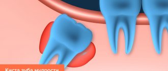
- A wisdom tooth cyst forms at the site of a long-erupting “figure eight.” As a treatment for this category of teeth, a proven effective method is recommended - removal of the cyst and wisdom tooth together.
Tooth granuloma: treatment of severe forms
Treatment of complex forms of granuloma, in particular large cysts, is possible only using surgical techniques. The same applies to the treatment of this disease after unsuccessful manipulations that caused numerous perforations of the tooth root. Below are the main granuloma removal techniques.
| Complex granuloma on the root of a tooth: treatment | Description of the technique |
| Cystectomy (apex resection) | The doctor removes the granuloma along with the root tip, after which the canals are filled. Before the procedure begins, it is often necessary to drain the pus, which requires an incision in the gum and a certain time for the fluid to completely drain out (on average 2-3 days). To restore bone tissue in the area of surgery, the use of osteoplastic materials is required. |
| Hemisection | The granuloma is removed along with part of the root and crown, after which the tooth is restored. This is often how granulomas are treated when a tooth canal is perforated. |
| Separation | The technique is applicable exclusively to molars. The tooth is divided into two parts, after which the doctor carries out all therapeutic and restorative manipulations through the resulting gap. |
| Removal of a tooth | An extreme measure that is resorted to when all other methods are impossible for a number of reasons. These include severe damage to the tooth, as well as numerous perforations or a vertical crack in the root. |
Important:
Treatment of tooth granuloma under the crown requires extraction and subsequent replacement of the orthopedic element. If after treatment of granuloma the tooth hurts and the severe pain does not subside after several days, you should consult a doctor.
What to do with a dental cyst?
Features of the development of purulent inflammation of various forms and localizations show that the formation occurs at a slow pace. When pronounced symptoms appear, the cyst emerges on the surface of the mouth, gum or root, inflamed and filled with pus. Patients complain of severe malaise, “fever,” experience pain and discomfort when chewing food.
A decade ago, the only solution to getting rid of the problem was the ability to remove the source of pain. Modern equipment, special instruments, and medications (painkillers, anti-inflammatory drugs) make it possible to save a tooth by treating a serious tumor. Depending on the stage of development, surgical or therapeutic methods are used.
The latter option involves mechanical cleaning of the canals, careful processing and filling. The surgical procedure involves removing the damaged surface while preserving the tooth. The missing root tissue is replaced with a specific material. Complete removal is considered only in the case of a wisdom tooth or when damaged tissues occupy most of the tooth and root. An exceptional situation when there is no need to treat a dental cyst is if it appears next to a baby tooth and develops slowly, and there is only a short time left before replacing a molar one.
Why is a dental cyst dangerous?
Despite the fact that being the result of a positive struggle of the body and a reaction to negative processes, reduced immunity, a dental cyst can lead to serious side effects and cause complications. The most dangerous and common of them:
- Flux is the concentration of a purulent process in the periosteal jaw area under the gum. A type of complex disease externally manifests itself in the form of a tumor, and a sharp throbbing pain is felt.
- Periodontitis is an advanced form of a purulent cyst that provokes the spread of infection to bone tissue.
- Tooth loss. A side effect that occurs extremely rarely, but occurs if the patient does not consult a dentist in a timely manner and does not receive drug treatment. The damaging effect extends to nearby hard areas of bone tissue, and there is a danger of losing several adjacent teeth.
Possible more complex exacerbations and consequences:
- inflammation of the lymph nodes;
- purulent abscess;
- fracture of the jaw due to decay of bone tissue and its complete destruction in a certain area;
- sepsis and osteomyelitis.
Even with minor signs of a dental cyst, treatment should be started. The sooner you see a dentist with a problem, the greater the chance of a complete recovery. While maintaining oral hygiene, make regular appointments with your dentist, and do not neglect the importance of medical examinations. You will not have to deal with the consequences of a dental cyst, since a qualified doctor will be able to recognize it at the initial stage. Take good care of your mouth at home and follow your dentist's advice.
Make an appointment with a therapist by phone+7(985)532-21-01
Retrograde pulpitis: diagnosis, causes, treatment, complications
Acute traumatic pulpitis treatment in the clinic on Tverskaya, Chekhovskaya
Tooth root caries treated or removed
Treatment of wisdom tooth caries if it hurts
Tetracycline teeth treatment, whitening, veneers
Treatment of caries between the front teeth
Do fissure dental caries need to be treated?
Cystotomy
The cystotomy procedure consists of several successive stages.
After drilling out the tooth, the dentist carefully cleans the root canal and removes the pulp. During a very deep cleaning of the canal, the doctor gets to the shell of the cyst and removes its upper part, after which the contents are pumped out of the neoplasm, and the walls of the cyst are treated with disinfectants. Then a special paste is placed into the canal and it is closed with a temporary filling. During subsequent dental visits, tissue healing is assessed. If the tissue heals quickly (which indicates that the infection has been destroyed), then the canals are sealed and a permanent filling is installed.