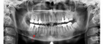
Diseases of the teeth and oral cavity not only cause pain and discomfort, but also lead to serious problems, such as cysts. This type of disease is quite rare and occurs due to infection in the root canals. Essentially, a cyst is a bubble filled with fluid or pus. The size of the tumor is usually a couple of millimeters, and sometimes can reach several centimeters. Most often, a cyst occurs on the root of the tooth, gum, and also under the crown, and it can be detected during an X-ray examination at the dentist.
Quite often, a cyst in the mouth does not show any symptoms at all: the initial stages proceed calmly and unnoticed. Depending on the type of cyst and the degree of its development, certain symptoms may be observed, but not necessarily all at once. Therefore, if you find any signs of a possible cyst, contact your dentist. Experienced specialists will conduct a full examination of the oral cavity, take x-rays and prescribe appropriate treatment.
Main types of oral cysts
Modern dentistry in Belarus identifies several varieties of this disease, each of which has its own reasons for its formation:
- Radicular (develops as a result of advanced caries)
- Keratocyst or primary cyst (formed at the stage of formation of our body, from the remnants of pharynx-forming tissue that occurs due to disturbances in the development of teeth)
- Follicular (occurs in cases where the tooth cannot erupt normally and forms under the gum)
- Retention (can occur in children during the period when baby teeth are replaced by permanent ones)
- Residual (formed in the bone as a result of complex tooth extraction)
The main reasons for the development of cysts are still considered to be untreated caries, injuries to the jaw skeleton, as well as improperly performed root canal treatment and various infections of the oral cavity.
Why does a dental cyst appear?
The main causes of cysts in teeth are:
- Development of infection in the root canal of a tooth
- Injury
- Chronic diseases of the oral cavity and nasopharynx
- Inflammation of periodontal tissues (periodontitis, periodontitis)
- Reduced immunity
- Caries
- Poorly installed filling or crown
- Difficult eruption of wisdom teeth
How to treat an oral cyst in Minsk
To get rid of such an unpleasant neoplasm, first of all it is necessary to unseal the canals, if they have been sealed, of course, then treat them with antiseptic agents and temporarily seal them with medicinal paste. If the treatment is successful and the cyst decreases in size, the final root canal filling is performed. If the cyst develops more severely, surgical intervention will be required. The doctor completely clears the cyst of pus, and also removes damaged tooth roots, or the tooth itself, if necessary. But there is no need to be afraid, any doctor will do everything necessary to save the tooth.
Specialists of the Family Dentistry Center in Minsk offer their clients high-quality dental treatment under anesthesia for children and adults using drug sedation.
You can calmly treat your teeth in your sleep, get rid of various problems and diseases of the oral cavity, including cysts, not only comfortably, but also painlessly. Modern equipment that meets European quality standards, highly qualified doctors, affordable prices and an individual approach to each patient - we know what the secret is to a beautiful smile and healthy teeth.
Why does a lump form on the lip?
As we said above, a retention cyst forms inside one of the small salivary glands, which are massively scattered throughout the mucous membrane of the entire oral cavity (24stoma.ru). Each such salivary gland has a thin duct that opens on the surface of the mucous membrane and allows the secretion of the salivary gland to enter the oral cavity. When this duct is blocked, the secretion of the salivary gland accumulates in its lumen, stretching the walls of the gland.
The cause of duct blockage is usually mechanical trauma to the mucous membrane. This is why patients note that as soon as they bit their lip, a lump formed. The secretion in the salivary gland is constantly being formed, and therefore the lump on the lip will constantly increase in size - until either spontaneous opening occurs, or you accidentally or deliberately bite through it. If the integrity of the cyst is violated, then you can see how a clear viscous liquid leaks out of it onto the surface of the mucous membrane.
After emptying, the cyst collapses, and for some time you do not notice it. Remember that you cannot deliberately bite or puncture such a cyst (for example, with a needle). The fact is that even if the secretion of the gland comes out, the cyst shell will still remain inside the tissue. This will result in the same cyst forming over and over again.
Treatment of a cyst without tooth extraction
The success of treatment directly depends on how early it was detected. Therefore, dentists recommend that patients undergo regular preventive examinations and seek help at the first signs of illness. More recently, therapy necessarily involved tooth extraction. Of course, this approach did not suit either the doctors or especially their patients. Today, dentists' methods have changed significantly, and it is possible to get rid of a cyst without losing the beauty of your smile.
Treatment of the cyst can be surgical or conservative. The choice of therapy method depends on: the type, age of the patient, personal wishes and is determined for each patient.
Treatment methods
Once a cyst is detected, it is necessary to begin treatment as soon as possible. Depending on the type and severity of the disease, the treatment method will be determined: therapeutic, surgical or laser.
The therapeutic method is used for small diameter cysts. First, the doctor makes a hole in the tooth, through which the contents of the cyst are pumped out and the medicine is injected. The canals are treated with a disinfectant, after which a temporary filling will be installed. The treatment period can take from 1 to 3 months.
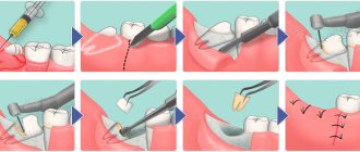
Most often, to treat a dental cyst, a surgical method is used with complete or partial preservation of the tooth. Less often, the tooth has to be removed completely.
There are three types of surgical treatment:
- Cystectomy. This is the most gentle type of treatment. To begin with, treatment and disinfection of the tooth canals is carried out, after which an incision is made in the gum, a hole is sawed out in the bone and the cyst is removed along with part of the root.
- Hemisection. This type of operation is similar to the previous one, only in addition to part of the root, part of the tooth crown is also removed, which makes this type of treatment more traumatic.
- Cystotomy. The simplest and least traumatic type of operation. In the area of the cyst on the gum, the doctor makes an incision through which the contents of the cyst are pumped out. Despite the simplicity of the operation, the recovery process will be long.
Laser cyst treatment is a new technique that allows you to quickly, sterilely and painlessly treat the affected tooth. During treatment, the fillings are removed and a laser is inserted into the canals to remove the cyst. Remaining liquid and other particles are removed using a pump.
Classification
Experts distinguish two types of tonsil cysts:
- Retention - formed after blockage of the ducts of the glands of lymphoid tissue, have a classic round shape, a thin capsule, filled with serous or purulent secretion. After emptying (evacuation of contents or spontaneous breakthrough), they are filled again.
- Dermoid or congenital - formed during intrauterine development, inclusions of embryonic tissue are always found in them. These true tumors, caused by genetic causes or harm suffered by the mother during pregnancy, are very rare. The capsule is usually dense and the contents are viscous.
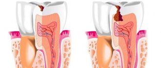
How does a tooth root cyst appear?
The causes of cysts are varied, and among them it is impossible to single out the prevailing ones. So, this may be the result of physical trauma to the maxillofacial bones, or a rare but common medical error, in which the tightness of the root canal was broken, which became the basis for the emergence of a source of infection. A similar situation occurs when a dental crown is installed incorrectly. But the reasons for cyst formation are not limited to this. Untreated infectious diseases (in particular, sinusitis or periodontitis) are no less a provoking factor.
Diagnosis of symptoms of tooth root cyst
The appearance of a cyst implies the formation of a cavity, which gradually grows while simultaneously filling with pus. This condition can last for a long time, and the patient does not experience pain. A sign suggesting the presence of a cyst is slight pain that occurs when pressing on the gum. Such a symptom only in rare cases becomes a reason to visit a dentist. Basically, it occurs in later stages, when the pain intensifies and becomes nagging, continuous, cannot be relieved with analgesics, and inflammation begins to additionally manifest itself in the form of swelling and swelling of the gums in combination with a persistent unpleasant odor in the oral cavity. Finally, a situation cannot be ruled out in which a fistula occurs - that is, a channel through which the contents of the cavity independently come out.
An effective way to detect a cyst in a timely manner is to conduct an X-ray examination followed by its opening and removal.
It should be noted that at the very beginning the disease poses minimal danger to surrounding tissues, since the cavity itself is reliably isolated by thick walls. However, as pus accumulates, the pressure on the walls increases, creating the possibility of a breakthrough, which can lead to blood poisoning and, in the long term, damage to the structure of the jaw bones. At the same time, the dynamics of cyst development are unpredictable and strictly individual. However, in the presence of other inflammatory processes or infections, in a weakened body the transition to the acute stage can occur quickly.
Particular attention should be paid to early diagnosis of the disease in pregnant women. This is due to the complexity of treatment: during pregnancy, only stabilization of the cyst is possible. If the inflammatory process worsens, the choice of treatment methods is strictly limited. That is why, at the planning stage of pregnancy, it is necessary to undergo a full examination by a dentist.
Our services in otorhinolaryngology
The administration of CELT JSC regularly updates the price list posted on the clinic’s website. However, in order to avoid possible misunderstandings, we ask you to clarify the cost of services by phone: +7
| Service name | Price in rubles |
| Taking ENT smears | 600 |
| Videoendoscopy of the upper respiratory tract | 2 400 |
| Radio wave removal of skin (mucous) neoplasms of ENT organs | 4 000 |
All services
Make an appointment through the application or by calling +7 +7 We work every day:
- Monday—Friday: 8.00—20.00
- Saturday: 8.00–18.00
- Sunday is a day off
The nearest metro and MCC stations to the clinic:
- Highway of Enthusiasts or Perovo
- Partisan
- Enthusiast Highway
Driving directions
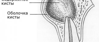
What is a cyst? Jaw cyst
A cyst is a cavity that is lined with epithelium and filled with fluid or soft material.
The formation of teeth (odontogenesis) is a complex process in which connective tissues and epithelium (enamel organ, dental follicle and dental papilla) participate.
The enamel organ refers to an epithelial structure derived from the oral ectoderm. The dental follicle and dental papilla are ectomesenchymal structures, because they are partly derived from neural crest cells.
For each tooth, odontogenesis begins with the apical (affecting the tip of the tooth root) proliferation of the epithelium of the oral mucosa, known as the dental lamina. The dental lamina gives rise to the enamel organ, a cap-shaped structure that subsequently takes on the shape of a bell. After the formation of the enamel organ, the dental lamina cord usually fragments and degenerates. However, small islands of dental lamina may remain after tooth formation. They are believed to be responsible for the development of some odontogenic cysts and tumors.
The enamel organ has four types of epithelium. The inner lining of the enamel organ is called the inner enamel epithelium and becomes the ameloblastic layer that forms tooth enamel. The second layer of cells adjacent to the inner enamel epithelium is the intermediate layer. Adjacent to this layer is the stellate reticulum, followed by the outer enamel epithelium. The enamel organ is surrounded by loose connective tissue known as the dental papilla. Contact with the epithelium of the enamel organ causes the dental papilla to produce odontoblasts, which form dentin. As odontoblasts lay down dentin, they induce ameloblasts to form enamel.
After initial crown formation, a thin layer of enamel organ epithelium, known as Hertwig's root sheath, grows at the apex of the tooth root. This epithelial expansion later becomes fragmented but leaves behind small nests of epithelial cells known as Malassez remnants in the space of the periodontal ligament. They are the source of epithelium for most periapical (radicular) cysts, but do not cause any odontogenic neoplasms, with the exception of squamous odontogenic tumor.
Diagnosis of a cyst
As a rule, detecting a cyst does not cause any particular difficulties, since the pharynx can be clearly seen even with the help of a backlit frontal mirror. The main thing is to contact an otolaryngologist for examination. ENT doctors use various instruments during examination - spatulas, elevators, and, if necessary, endoscopes.
The cyst is determined in different parts of the tonsils, on the surface or in the depths. It has the appearance of an opaque round formation, similar to a ball, whitish in color, elastic and mobile. There may be no signs of inflammation of surrounding tissues.
You cannot touch or put pressure on the cyst yourself - it can burst, and its contents are unknown. At best, the purulent contents will enter the stomach, and at worst, it will spread to other organs.
The doctor examines cysts larger than 1 cm very carefully, without damaging the surrounding tissues, trying not to touch the capsule.
Diagnosis of a cyst includes puncture (puncture and removal of contents). If necessary, the resulting material is examined in the laboratory to understand the nature of the disease. If a malignant process is suspected or blood is leaking from a cyst, treatment begins immediately, without waiting for the end of the examination.
Depending on age, general condition and concomitant diseases, the doctor may prescribe other clinical tests. The blood coagulation system, the functioning of the heart and lungs are required to be examined.
Cyst in a child’s mouth on the gum
The development of cysts in the mouth in childhood occurs frequently, which is explained by improper dental care, injuries received from a fall, as well as untimely treatment of caries. A cyst in a child’s mouth on the gum may also appear during teething.
Treatment of cysts in childhood should be carried out by a dentist, who will determine the type of formation and the reasons for its development. The capabilities of modern medicine make it possible to painlessly treat cysts in children at the initial stage of development.
However, some parents, after a cyst in the child’s mouth was discovered, look at photos, study thematic forums and use traditional methods of treatment. Self-medication for this disease can lead not only to tooth loss, but also to further spread of the source of infection and the formation of fistulas.
The Yusupov Hospital treats patients over 18 years of age. If a cyst is found in a child’s mouth on the gum, experts recommend seeking help from a pediatric dentist.
Symptoms of a dental cyst
The difficulty in diagnosing a cyst lies in the absence of symptoms. In some cases, a person may pay attention to such signs of a dental cyst as darkening of the enamel, slight displacement of the tooth and discomfort when chewing solid food, but most do not attach any importance to this. By the time pain from a dental cyst begins to appear, the granule can already grow up to 1 cm in diameter. From this moment on, the patient begins to feel other quite noticeable signs. Here are the symptoms of a dental cyst that may appear at this stage:
- aching pain in the area of inflammation;
- swelling, which can spread to the face;
- enlarged and painful lymph nodes;
- elevated temperature;
- headaches - with the development of a cyst in the maxillary sinus;
- general malaise.
Under no circumstances should these symptoms be ignored, otherwise serious consequences will arise, and removing a tooth with a cyst is not the worst of them. At the first signs of the development of the disease, you should consult a specialist.
Tonsil function
The tonsil is a lymphoid tissue located in clusters in the pharynx. The tonsils are one of the organs of the immune system that protect the internal environment of the body from harmful influences. There are six tonsils in the human pharynx: two paired and two unpaired; together they form the so-called Pirogov-Waldeyer protective ring. The palatine tonsils (popularly called tonsils) are located closest to the entrance of the pharynx, and these tonsils suffer more often than others. The lymphoid tissue of the tonsils supports general and local immunity. Lymphocytes mature in it and antibodies are produced.
The palatine tonsils play a vital role. They are the largest and differ in their structural features. Upon careful examination, it is clear that the surface has depressions or lacunae, smoothly turning into crypts or deep folds lined with mucous membrane. Only the lateral surfaces are covered with a capsule of connective tissue, and all the rest are in contact with everything that enters the pharynx.
At CELT you can consult an otorhinolaryngologist.
- Initial consultation – 3,000
- Repeated consultation – 2,000
Make an appointment
Tooth root cyst in children
The disease has features and varieties that are characteristic strictly for children. First of all, we are talking about formations that arise and go away on their own, without complications: in the form of a rash that appears on the gums, as well as the so-called “Epstein’s pearl”. Both varieties do not pose a danger due to the absence of purulent masses, which means that the cavity is infected. In addition, they may not occur at all, being only some of the phenomena accompanying the development of the child.
The situation is different with the appearance of a cyst during the growth of teeth (both milk and molars). In this case, the resulting cavities are infected, and given the previously described asymptomatic nature of the disease in the early stages, the most reliable method of determining its presence is regular visits to the doctor. This allows for successful treatment at the very beginning of the development of the cyst, saving the tooth in almost 100 percent of cases.