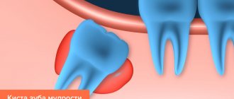
Tooth root granuloma is a limited inflammation of the periodontium, which is a small round formation located in the area of the tooth root. The disease manifests itself in most cases in response to a complicated course of periodontal inflammation. The peculiarity of the disease is the latent symptoms in the initial period and the manifestation of severe pain with the progression of the inflammatory process.
Granuloma is presented in the form of a compacted node at the tooth root, in the periodontium. This name is given to formations up to 5 mm in size. Nodes that are larger (up to 1 cm) are classified as cystogranulomas.
Make an appointment with a therapist by phone+7(985)532-21-01
Education is the pathological epicenter. Starting from a tooth root granuloma, inflammation spreads, gradually destroying the tooth. The disease requires treatment or tooth extraction. Otherwise, the process continues to progress and will lead to complications in other organs. Most often, the pathology spreads to the muscles of the face, neck, and heart area.
How does a dental root granuloma form?
The process of development of pathological formation includes 3 stages:
- Development of dental disease leading to complications. Progression of inflammation with the accumulation of pathogenic microorganisms in the dental pulp. The pathological process, developing in connective tissues, leads to cell death over time.
- Pathology continues to develop. The infectious environment ends up in bone tissue. The result of the process is the formation of a neoplasm that transforms into the described granuloma.
- Against the background of bone tissue detachment from the infectious focus, a compacted connective tissue capsule is formed in the local area. In the resulting environment, complicated inflammation occurs, which leads to the rapid proliferation of bacterial microorganisms. At this time, the tissue grows rapidly, bacteria turn into purulent mucus. When visiting a doctor at the current stage, the specialist diagnoses acute granuloma.
The disease significantly undermines the human immune system. As the pathology develops, the tooth begins to loosen - the roots of the element are exposed.
Periodontal pockets, pathological formations between the gum and dental tissue, can also lead to the development of the disease. The cause of cavities is hard tartar.
The pocket holds a lot of bacteria, which leads to the formation of a gap between the gum and the socket. It becomes a “transport” for infection that penetrates into the root of the tooth. Subsequent growth of tissue at the base of the tooth root and filling of the cavity with pus is a granuloma.
Computed tomography in dentistry
Diagnostics using the latest tomograph in is your opportunity to conduct a high-quality CT scan of teeth in Kurkino, in a place convenient for you, where you can subsequently undergo the necessary treatment. We have installed advanced equipment Planmeca ProMax 3D with ProFace function. This 3D scanning system provides maximum information content of the obtained data, creating images of bone tissue layer by layer and with high clarity. If you need a dental CT scan in Khimki, our clinic in Kurkino, a neighboring district of Moscow, will offer you every opportunity to conduct a thorough analysis of the condition of the oral cavity in any projections.
Causes of dental granuloma
Experts divide all the causes of granuloma into two groups: primary and secondary.
Main provoking factors
We are talking about factors that are key conditions for the development of the pathological process. Among them:
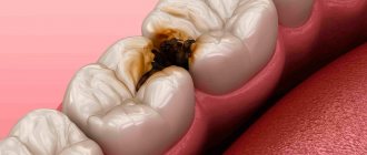
- advanced caries;
- untreated pulpitis formed as a result of inflammation of the connective tissue;
- fracture of the tooth, leading to the penetration of infection into the element of the dentition;
- periodontal inflammation;
- inappropriate antiseptic treatment of the tooth following removal of the element or therapy.
Minor provoking factors
The group of secondary causes includes factors that can, but are not required to lead to the formation of granuloma. Among them:
- constant stress;
- physical stress;
- sudden change in climatic conditions;
- hypothermia of the body;
- serious cold.
The development of the disease can also be triggered by poor-quality therapy (canal filling, etc.). Often the appearance of inflammation is preceded by tooth extraction and a complete lack of infection prevention after manipulation.
The area of the removed element tightens the tissue, and after microbes enter the cavity, the inflammatory process of the periodontium begins to develop. In the absence of preventive measures, the granuloma quickly grows, filling with pus. If treatment is neglected, the formation moves along the gum - infective endocarditis occurs, often leading to death.
It is possible that granulomas may appear when primary teeth are removed in children. Accordingly, the development of the disease in childhood cannot be ruled out.
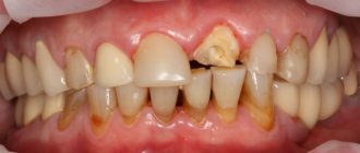
Symptoms of granuloma on the root of the tooth
In the initial stage of the disease, the patient does not notice specific symptoms. The first sign that indicates a pathological phenomenon is a sharp pain syndrome.
An additional symptom of the disease is the sensation of a foreign object in the mouth. The role of a foreign object in the oral cavity is played by overgrown tissue. It can be easily felt with your tongue.
If the patient can see a granuloma, you should prepare for the following symptoms of pathology:
- redness, swelling of the gums;
- inflammatory process in the mouth, accompanied by unstable hyperthermia;
- change in enamel color (darkens);
- discharge of pus from under the gums;
- feeling unwell, migraine;
- flux.
Swelling and redness can be observed on several sides of the tooth (internal, palatal, behind the lips). If you press on the affected area, the pain increases sharply, the pain is bursting, and progresses over time.
To summarize: the basic symptom is acute pain observed against the background of tissue swelling. Then the patient notes significant changes in body temperature.
How is granuloma on a tooth diagnosed?
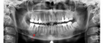
The dentist makes the diagnosis. The patient undergoes an x-ray. With a granuloma on an x-ray, a specialist notes darkening at the tooth root.
In a hospital, the patient may be prescribed radiovisiography, a type of x-ray that provides a reduced dose of radiation. In this case, the situation is assessed not on an x-ray, but on a monitor. Accordingly, the diagnostic method is classified as digital.
The most favorable outcome is diagnosing granuloma at an early stage of development. Pathology is often determined during the treatment of other dental diseases. The specialist notes swelling of the gums, which is very painful. A doctor may also suspect a granuloma based on the protruding bone at the apex of the tooth.
Particular attention is paid to patients whose oral cavity has crowns and pulpless teeth, since the diagnosis of “granuloma” in these categories of patients is made much more often.
How harmful is CT scanning to health?
The second most popular question among patients after “what is a dental CT scan” is “is it harmful?” People are wary of any method based on x-rays, and that's normal. Of course, the radiation dose is also present in computed tomography, but the decisive factor is its level, and it is minimal!
X-ray radiation standards are strictly limited by the requirements of SanPiN 2.6.1.1192-03. They indicate that in one CT session the patient receives only 40-50 μSv (microsievert), and the permissible dose is 3500 μSv per year. Even a large 3D dental CT scan emits 10 times less radiation than conventional radiography. It turns out that with such a minimal load it is possible to perform almost 100 CT procedures per year without the slightest harm to health.
If your doctor has prescribed you to have a CT scan of your teeth before implantation, extraction or other intervention, you should not refuse. The radiation received during the study is negligible, but the harm from neglecting the best diagnostic method may be too great.
How to cure dental granuloma
For a patient with granuloma, therapy is indicated at the first stage of pathology development. He visits the dentist, undergoes x-rays, then follows all the specialist’s recommendations and remains calm during the necessary manipulations.
The treatment course for this type of pathology may include:
- Conservative treatment.
- Surgical intervention.
- Traditional therapy.
The first two methods are most effective in the fight against granuloma. They are held within the walls of the clinic. Traditional methods of therapy are applicable only as supportive measures.
Conservative method
The therapeutic approach involves the prescription of antibacterial agents, sulfa drugs, and tooth filling. This approach makes it possible to stop inflammation and preserve the element of the dentition.
Antibiotics are designed to quickly stop the progress of inflammation, eliminate the pathogenic environment, and cleanse the tooth of infection. At the same time, the patient rinses the mouth with antiseptic agents. They help relieve pain.
In case of deep caries, an inflammatory phenomenon is observed in the pulp - the doctor carries out canal cleaning and removal of the infectious focus. Then the doctor puts the drug into the tooth cavity and performs a temporary filling. After a certain period, a permanent filling is installed in this place.
In severe cases of the disease, therapeutic methods of treatment are powerless - surgical therapy is indicated for the patient.
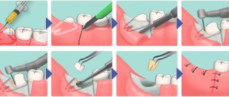
Surgery
The essence of the operation is to open the gums and remove purulent discharge through drainage. This method eliminates inflammation. As an additional measure, they resort to prescribing antibacterial drugs, analgesics and antiseptics. The prognosis after surgery is favorable.
The surgical treatment of dental granuloma involves one of the following procedures:
- Gum opening and drainage.
- Removal of the apex of the tooth root.
- Hemisection of the tooth.
- Removal of an element of the dentition.
If there is a pocket on the gum or a gap on the tooth, the cyst is dissected and the accumulated substance is removed from the cavity.
Root resection
Medical manipulation consists of the following stages:
- Violation of the integrity of the shell.
- Cleaning the channel.
- Filling the cavity with the drug.
- Elimination of formation (granuloma) and the apex of the tooth.
- Replacement of unsuitable fabric with artificial material.
- Installation of a seal.
Hemisection of tooth
A therapeutic measure is resorted to if there are a large number of tooth roots and the disease is extremely neglected - it is not possible to preserve the root part. The process consists of 4 stages:
- Removal of the root and the crown part of the tooth in contact with it.
- Filling the cavity with material.
- Placement of the crown.
- Monitoring the clinical picture after surgery using radiography.
Removal of a tooth
If it is not possible to save the tooth, they resort to removing it. Indications for manipulation are:
- prolonged pain;
- formed gum canal;
- dental crack (vertical);
- complete destruction of the element or its crown;
- visualization of root perforation;
- obstruction of the root canals.
Traditional methods of treatment
Traditional therapy can alleviate the patient’s condition by reducing the manifestation of concomitant symptoms: Below are several effective traditional medicine recipes:
- Tincture of propolis and calamus. Prepare calamus root and dry propolis (30 g each). Pour the components with alcohol or vodka and leave in a dark, cool place for 14 days. Use as a rinse.
- Herbal decoctions. Sage, chamomile, and eucalyptus can be used as raw materials. Use for mouth rinsing.
Pulp is the general name for the “internal organs” of a tooth. To understand what root canal treatment is, why it is needed, and what the criteria are, let’s look at the structure of the tooth.
A tooth has two main parts: the crown and the root. Inside each tooth there are cavities that are filled with loose fibrous connective tissue - pulp. In the past, root canal treatment was called “removing the nerve.” But the nerve is only one of the components of the pulp.
tooth structure
A tooth is a whole living organ. Through the apex of the tooth root – the apex – a bundle of blood and lymphatic vessels and nerve endings enter the tooth. When, due to advanced caries or as a result of injury, the enamel and dentin of a tooth are damaged, the infection penetrates inside, into the pulp, and inflammation of the internal tissues of the tooth occurs - pulpitis.
Depulpation or treatment of pulpitis includes two main points: cleaning and filling . This is significantly different from installing a regular filling. During cleaning, the doctor removes all the inflamed pulp from the coronal part and all the root pulp. In order to do this efficiently, there are modern diagnostic methods, including computed tomography (CT) and work with a microscope. They allow you to accurately assess the number of root canals, their location, shape and degree of destruction.
depulpation
Until some point, it was believed that the upper sixth teeth had three roots, but now in 80% of cases there is an additional fourth root canal. The lower incisors have one canal, but additional branches can often be seen on a CT scan or under a dental microscope. Previously, they went unnoticed, which means they were infected. The infection spread, complicated inflammation occurred, and the tooth was destroyed. Modern dental techniques, such as the use of CT scans and microscopes, give doctors more opportunities for accurate diagnosis.
The first important criterion in the treatment of pulpitis is maximum cleaning of the canals. They need to be passed all the way to the apex, to the very apex, where the neurovascular bundle enters the tooth. This is done mechanically using special tools, so-called files. After this, the root canal is expanded, removing all the inflamed pulp, and a taper is created so that it can be properly filled with filling material.
Channels are often curved, have micro-branches, and their mechanical processing is limited. On the first visit, after mechanical cleaning, a preparation is left in the tooth, which chemically sterilizes the branches of the canals that are inaccessible for mechanical treatment. After a while, the canals are filled and the tooth crown is restored.
We always try to keep teeth alive so that they perform their functions well. A pulpless tooth is a dead organ. It is like a prosthesis that is neither capable of regeneration nor of perceiving too cold or hot to protect itself. But there are situations when, due to caries or injury, the pulp is damaged, becomes inflamed, and that very familiar toothache occurs, which, due to a natural change in the blood supply to the body, intensifies by night. Previously, arsenic was applied under a temporary gluten filling, and then the nerve was mechanically removed. For such manipulations, dentists are feared at the genetic level. But today in modern clinics, treatment of any pulpitis is accompanied by anesthesia, and patients do not experience any pain.
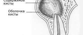
What are the criteria for treating teeth with pulpitis? They often think that it is enough that the tooth does not hurt and there is a filling. But even if the tooth hurts for 3-4 days or a week without treatment, the pulp dies. Due to infection, the nerve, blood vessels and tooth become dead. But if the pain goes away, this does not mean that the tooth has recovered! It becomes a hidden source of infection, and the next stage may develop when the infection progresses further. The tooth is secured in its bed by a system of ligaments. The space between it and the jaw is filled with connective tissue - periodontium. If the pulp in the tooth has already died, it will become a source of infection, which passes into the periodontium and then we get periodontitis - inflammation of the root tips and tissues around the tooth. In this case, the body tries to localize the source of inflammation, and a cyst may appear (from the Greek κύστις “bubble”). This is a cavity that fills with exudate (pus), and from which bone destruction can begin.
In a living tooth, a possible reaction to cold/hot due to enamel defects quickly passes because the tooth is restored. But if the pulp is inflamed, then under the influence of chemical and mechanical irritants it will hurt for a long time. Depending on the stage of inflammation, the duration of pain increases. As soon as the inflammatory process moves to the periodontium, we begin to feel pain when biting. This happens because the ligaments on which the tooth is suspended in its socket normally absorb pressure. But if the tooth is sick, then the inflamed receptors perceive any pressure as an excessive load and signal about it.
The immune system can hold back the development of such hidden inflammatory processes for some time, but as soon as the immune system is weakened, due to banal hypothermia, the amount of exudate can begin to increase sharply, and then we see gumboil and facial asymmetry. This is already inflammation of the periosteum. There is also a milder course of periodontitis, when a fistulous opening is formed on the gum, and during exacerbation, pus comes out of it. In patients, it happens that something swells, then goes away, an abscess appears and goes away. The presence of a fistula may be an indication that there is a problem with the roots of the teeth.
It is important for patients to know that even high-quality root canal treatment involves depulping the tooth. The tooth becomes dead, and this has much more difficulties than treating caries.
Timely diagnosis, prevention and treatment will save you from complex root canal treatment. But if it has come to this, it is important to understand that this is a responsible operation. It is not quick, it is not cheap, but it is necessary. Root canal treatment can take a long time – two to three visits. This depends on the number, shape of the root canals and the degree of infection.
Complications with dental granuloma
Lack of appropriate treatment for granuloma can lead to the following complications:
- Complete tooth loss. The root is destroyed, the inflammatory process is transferred to the soft tissues.
- Osteomyelitis of the jaw.
- Formation of a cyst-like formation.
- Malignant tumors.
- Involvement of other organs and systems in the infectious process (sinusitis, pyelonephritis, etc.).
- Penetration of purulent discharge into the skull (meningitis, encephalitis, etc. develops).
- Migrating granuloma. The skin on the patient's face bulges, abscesses and fistulas form.
Preventive measures
Prevention of the disease is complex. The actions of a person who wants to prevent the formation of granulomas require compliance with the following medical recommendations:
- Following the rules of personal hygiene (daily brushing of teeth, rinsing the mouth).
- Timely treatment of bleeding gums.
- Regular visits to the dentist (at least 2 times a year).
- Timely change of toothbrushes to prevent infection in the mouth.
- Attentive attitude to your own health - reaction to pain, discomfort in the oral cavity, timely contact with a specialist.
- Complete cure of dental diseases.
- Use of medicated toothpastes for preventive purposes.
- Regular rinsing of the mouth with herbal decoctions.
- Including foods that are sources of calcium and vitamins in your daily diet.
Is there an alternative to CT
There are many other diagnostic imaging methods (x-ray, orthopantomogram, ultrasound, etc.), but only CT provides the possibility of highly accurate, separate images of all types of tissue at different angles and to different depths. Although a panoramic dental photograph remains an equally important diagnostic tool for a dentist today, it can only provide a general overview. In turn, a 3D tomogram allows you to obtain not a single flat image of the jaw, but a whole series of sequential multiplanar images in different projections and without the distortions inherent in a panoramic image.
Example:
due to the different density of bone structures exposed to X-ray radiation, it is impossible to see less dense bone in a 2-dimensional image; accurate information is provided by a 3- D image of the teeth.
Is it possible to cure dental granuloma?
The prognosis for treatment of granuloma depends on the stage of development of the pathology and the chosen therapeutic strategy. When organizing treatment at the first stage of the clinical picture, the prognosis is always favorable. Positive results are obtained with timely treatment of granuloma in children.
If treatment is started at the stage of formation of purulent discharge, the success of therapy depends on the location of the inflammatory process. If it is the root part, the prognosis is rarely favorable - the tooth is removed. If pus collects only in the gums, after surgery and a course of antibiotics the patient makes a full recovery.
Lack of timely treatment can cause the patient's death. The pus, bypassing the muscle tissue, reaches the heart area - sepsis appears, leading to death.
To summarize, it is worth emphasizing that granuloma is a truly serious pathology. The appearance of the first signs of the disease is a reason to visit the dentist and begin treatment. Otherwise, the progression of inflammation will lead to irreversible complications.
Make an appointment with a therapist by phone+7(985)532-21-01