CLASSIFICATION
Odontogenic infections (infections of the oral cavity), depending on the anatomical location, are divided into truly odontogenic
associated with damage to tooth tissue (caries, pulpitis);
periodontal
, associated with damage to the periodontium (periodontitis) and gums (gingivitis, pericoronitis), surrounding tissues (periosteum, bone, soft tissues of the face and neck, maxillary sinus, lymph nodes);
non-odontogenic
, associated with damage to the mucous membranes (stomatitis) and inflammation of the major salivary glands.
These types of infections can cause serious life-threatening complications from the cranial cavity, retropharyngeal, mediastinal and other localizations, as well as hematogenously disseminated lesions of the heart valve apparatus, sepsis.
Purulent infection of the face and neck
may be of non-odontogenic origin and includes folliculitis, furuncle, carbuncle, lymphadenitis, erysipelas, hematogenous osteomyelitis of the jaws.
Specific infections (actinomycosis, tuberculosis, syphilis, HIV) can also be observed in the maxillofacial area.
MAIN PATIENTS
Infections of the oral cavity are associated with the microflora that is constantly present here. Usually this is a mixed flora, including more than 3-5 microorganisms.
With truly odontogenic
infections, along with facultative bacteria, primarily viridans streptococci, in particular (
S.mutans, S.milleri
), anaerobic flora is released:
Peptostreptococcus
spp.,
Fusobacterium
spp.,
Actinomyces
spp.
In case of periodontal infection, five main pathogens are most often isolated: P.gingivalis, P.intermedia, E.corrodens, F.nucleatum, A.actinomycetemcomitans
, less commonly
Capnocytophaga
spp.
Depending on the location and severity of the infection, the patient’s age and concomitant pathology, changes in the microbial spectrum of pathogens are possible. Thus, severe purulent lesions are associated with facultative gram-negative flora ( Enterobacteriaceae
spp.) and
S.aureus
.
In elderly patients and hospitalized patients, Enterobacteriaceae
spp also predominate.
In the conditions of domestic bacteriological laboratories, it is quite difficult to isolate a specific pathogen of a certain odontogenic infection. However, it seems possible to localize pathogens that play a major role in the development of oral infections in supragingival and subgingival plaque.
When to take an antibiotic for teeth
Antibiotics are drugs that are used to treat bacterial infections.
It is important to note that we are talking specifically about bacterial diseases, not viral or fungal ones. Antibiotics are powerless against viruses or fungi. Therefore, if we are talking about a viral or fungal disease, then in such cases it is appropriate to use antiviral and antifungal drugs, respectively. Antibiotics are also used in dental practice when a tooth hurts. At the same time, it is important to understand in which cases the pain is of infectious (bacterial) origin, and in which – non-infectious.
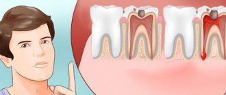
A common type of toothache is due to pulpitis. It occurs when caries has already eroded the top layer of enamel. The resulting hole reaches the pulp of the tooth, which is why the latter begins to hurt when exposed to various irritants - thermal, chemical or mechanical. In this case, antibiotics are useless. In order to relieve pain due to pulpitis, anti-inflammatory drugs are used.
In cases where a dead tooth hurts, there is a high probability that the cause is bacteria. As you know, dead teeth (with filled canals) cannot hurt on their own. If such pain occurs, it means that bacteria have grown in the cracks or cavities of the tooth. Microorganisms multiply, infect the gums and lead to the accumulation of pus. The latter exerts pressure, which leads to pain. The above pathology is nothing more than a gumboil. The antibiotic for flux is selected by the doctor after diagnosis. After all, it is necessary to establish the type of pathogenic bacteria so that the antibiotic for flux will work for sure.
Antibiotics in dental practice are also used in the following situations:
- After surgical operations . Tooth extraction and other surgical interventions are sometimes complicated by an infectious process. To avoid bacterial infection, surgical treatment is accompanied by antibiotic therapy.
- Periodontal diseases . Advanced variants of periodontal pathologies are sometimes complicated by purulent periostitis (flux). To avoid such developments, treatment of periodontal diseases is sometimes accompanied by the use of antibiotics.
The above cases are not the only ones in which antibacterial drugs are used. These medications are prescribed by the doctor based on the clinical picture, as well as the individual characteristics of the patient. In some cases, antibiotics are also prescribed for preventive purposes - when there is a possibility of a bacterial infection. These risks are assessed by a doctor.
Indications for the use of antibiotics in dentistry
The need for treatment with antibiotics depends on the nature of the infection and the body's ability to withstand the course. The main reasons for prescribing antibiotics include:
- When advanced caries threatens pulpitis, the dentist may prescribe antibiotics to limit the spread of the pathological process. The patient is prescribed antihistamines to complement the effectiveness of antibiotics.
- With the development of an inflammatory process of periodontal tissue (periodontitis), antibiotic therapy allows the destruction of protozoa, gram-negative anaerobes in the oral cavity. Various dosage forms of drugs are used for treatment: gels, ointments, intramuscular and intravenous injections, tablets.
- The proliferation of pathogens, poor immunity, caries and dense plaque can lead to the development of gingivitis. After laboratory detection of the sensitivity of microbes to the antibiotic, a course of treatment is prescribed. Antibiotic drugs are mainly used for catarrhal gingivitis.
- The appearance of purulent accumulations inside the oral mucosa provokes the appearance of a fistula. The process occurs due to the proliferation of anaerobic gram-negative bacteria, streptococci, staphylococci, Pseudomonas aeruginosa or Escherichia coli. A doctor-prescribed course of antibiotics, which are also used for dental implantation, will help cope with the infection.
- Inflammation of the connective tissue around the root of the tooth is called periodontitis. The occurrence of the disease is a consequence of dental trauma, a complication of pulpitis, caries, or an error in dental treatment. If the process is not stopped, pus may appear. The effectiveness of antibiotics for periodontal disease is felt after preliminary cleaning of the periodontal tissue.
- The result of inflammation of various origins can be a granuloma - a cavity of granulation tissue filled with fluid. Location: on the gum near the root of the tooth. It is important to start treating granuloma in the early stages. The use of antibiotics facilitates the opening of the granulosa vesicle and suppresses the infection accumulated in it, and serves to prevent infection. Self-medication with antibacterial drugs is unacceptable.
Antibiotics for gumboil and toothache: types of drugs
Today in dental practice various antibiotics are used for gumboils and toothache. Let us remember that antibiotics are usually divided into classes. Let's look at the most popular types of antibiotics for purulent gum disease.
Nitroimidazoles
Nitroimidazoles are synthetic antibacterial drugs that exhibit high activity against anaerobic microorganisms (bacteria living in conditions without oxygen). The most popular drug in this group is metronidazole, which has been used since the 60s of the last century.
The mechanism of action of nitroimidazoles is associated with disruption of DNA replication and protein synthesis of microbial cells. Such violations are incompatible with the life of the bacterium.
Representatives of this group of antibiotics are active against most anaerobic bacteria (both gram-positive and gram-negative). Some types of protozoan microorganisms and the bacterium Helicobacter pylori are also sensitive to nitroimidazoles. The latter is the cause of the development of gastritis and gastric ulcers (as well as tumor lesions of the mucous organs of the gastrointestinal tract).
Fluoroquinolones
This is a group of drugs that exhibit antibacterial activity against a wide range of bacteria. It is noteworthy that fluoroquinolones are not considered antibiotics in the classical sense of the word. The fact is that antibiotics (even synthetic ones) always have analogues of natural origin. Fluoroquinolones have no natural analogue. The most popular representatives of fluoroquinolones include ciprofloxacin, ofloxacin, moxifloxacin, lomefloxacin, levofloxacin and others.
The mechanism of action of fluoroquinolones is reduced to inhibition of a number of important microbial enzymes. This leads to disruption of DNA synthesis and death of the bacterial cell.
Lincosamides
Lincosamides are a group of antibacterial drugs that includes antibiotics such as lincomycin and clindamycin. The mechanism of action of lincosamides is reduced to suppressing protein synthesis in bacterial walls.
The role of the doctor in excluding the possibility of alveolitis
Actions of the dental surgeon to exclude alveolitis:
- Thoroughly clean the socket so as not to leave a tooth fragment or cyst in it. After extracting a tooth, especially a damaged one, a control x-ray of the socket is often taken;
- Be careful and avoid injury. The tooth extraction was accompanied by bone injury due to the inept actions of the doctor. Usually the bone is injured during complex extraction, when tooth extraction cannot be carried out using conventional instruments. Difficult procedures include the removal of unerupted and incorrectly positioned wisdom teeth, multi-rooted teeth, and teeth with curved or destroyed roots. An inexperienced doctor may not understand the situation and damage the bone;
- Control during anesthesia. An overdose of anesthetic leads to a sharp decrease in the amount of blood released after tooth extraction and insufficient formation of a blood clot;
- Prescribing appropriate antibacterial drugs, especially if the tooth extraction was difficult or was carried out against the background of purulent inflammation.
Therefore, it is very important to seek help from competent and experienced specialists who are fully familiar with all the intricacies of the tooth extraction procedure.
Sometimes alveolitis can develop despite preventive measures taken due to certain reasons. These reasons include:
- Reduced immunity of the patient, inability of tissues to regenerate;
- Tendency to bleed, resulting in a blood clot not forming;
- An increase in the amount of the hormone estrogen in women when taking hormonal contraceptives or certain diseases, which leads to the destruction of a blood clot.
A timely visit to the dentist if alveolitis develops is the key to a quick cure. Patients of our clinic who have undergone tooth extraction surgery receive detailed recommendations on oral care; if they are strictly followed, the likelihood of developing alveolitis is excluded.
Antibiotics for tooth root inflammation
For toothache caused by inflammation of the tooth root, doctors also prescribe fluoroquinolones (as for gumboil). In addition, for inflammatory pathologies of the tooth root, antibiotics of the penicillin and tetracycline series are also used. Let's take a closer look at them.
Penicillins
These are the first antibiotics that were discovered by Alexander Fleming in 1928. The main mechanism of action of penicillins is bactericidal. These drugs disrupt the synthesis of peptidoglycan, a component of the bacterial cell wall. This leads to the death of the bacterium.
Currently, different types of penicillin antibiotics are used in dental practice. The most common are amoxicillin and ampicillin.
Tetracyclines
Another class of antibiotics that was discovered a long time ago - back in the 40s of the last century. Tetracyclines act against a wide range of bacteria, both gram-negative and gram-positive microorganisms. The mechanism of action of tetracyclines is inhibition of protein synthesis in bacterial cells. Microorganisms stop synthesizing vital proteins, which is why they die.
Complications of alveolitis
If alveolitis is not treated in a timely manner, the following complications may develop:
- Odontogenic sinusitis is an inflammation of the maxillary sinus caused by the spread of infection from the inflamed sockets after the removal of premolars or molars of the upper jaw;
- Phlegmon - purulent inflammation spreads to the surrounding soft tissues;
- Acute periostitis - pus accumulates in the periosteum area;
- Odontogenic osteomyelitis is a purulent-necrotic lesion of the jaw bone;
- Sepsis - an infection enters the bloodstream, causing it to become infected.
Alveolitis itself is not so dangerous, but its complications are life-threatening. Therefore, if you notice symptoms of alveolitis, contact your doctor immediately.
How long do antibiotics work for toothache?
The therapeutic effect of antibiotics occurs within the first three days. If relief does not occur during this time, then a decision is often made to prescribe another drug.
The minimum course of antibiotics is 5 days. In this case, an individual course of therapy is established for each patient depending on a number of factors.
Sometimes antibiotics do not work and the patient does not feel a therapeutic effect from taking them. Let's consider typical cases when antibacterial drugs do not work:
- Insufficient dose of medication . When carrying out antibiotic therapy, strict adherence to the dosage is an important condition. If you take less than what your doctor prescribed, there will be no effect. In other words, there was not enough active substance to suppress pathogenic flora. Please note that the dosage of the antibiotic also depends on the age and weight of the patient. The heavier the patient, the more drug is required to achieve the required drug concentration in the blood.
- Insufficient duration of therapy . Strictly adhere to the regimen and duration of taking the drug. Some patients, feeling relief, decide to interrupt the course, which makes a mistake. Premature interruption of the course of antibiotic therapy is fraught with the development of antibiotic resistance - resistance of pathogenic and opportunistic flora to antibacterial drugs.
- Some concomitant diseases . Before prescribing medications, the doctor must have comprehensive information regarding the patient’s health. It is important to inform your doctor about all chronic and recent acute diseases that may affect the effectiveness of antibiotic therapy.
- Antibiotic intolerance . The patient may have individual intolerance to some types of antibiotics. Don't despair. If one antibiotic is not suitable, then there is always an alternative antibacterial drug. Sometimes a patient has problems taking an antibiotic orally (in the form of tablets, capsules or solutions). In such cases, a decision is made to inject antibiotics, bypassing the gastrointestinal tract.
- Interaction with other active substances . The doctor must be informed about the medications the patient is taking. Some drugs cannot be taken together with antibiotics, as they neutralize the effect of the latter. In addition, drinking alcoholic beverages is prohibited during antibiotic therapy. Otherwise, the treatment will not bring the desired effect.
Additional physiotherapy treatment (click to expand)
Physiotherapeutic treatment is an important addition to drug therapy; with the help of physiotherapy, the intensity of inflammation can be significantly reduced and healing time can be accelerated. For alveolitis, the following techniques are used:
- UV therapy - the hole is irradiated with short-wave ultraviolet light, which kills pathogenic microorganisms and reduces the level of inflammation.
- SMV therapy is a method of treatment with an electromagnetic field, based on the effect of centimeter waves on the area of inflammation. The procedure helps improve blood circulation and metabolism, due to which toxic substances are removed from tissues faster and regeneration processes are accelerated. SMV therapy also has an analgesic effect.
- UHF therapy – the body is exposed to a high-frequency electromagnetic field. For alveolitis, UHF therapy is used if the patient's regional lymph nodes are enlarged.
- Electrophoresis – medications are injected into inflamed tissue using electrical impulses. For post-extraction alveolitis, electrophoresis is used to reduce pain. For this purpose, solutions of novocaine, lidocaine, trimecaine are used.
- Fluctuarization is a treatment technique with pulsed currents of a sinusoidal shape with a low frequency. As a result of the procedure, blood circulation and lymph flow improves, swelling resolves, and the level of inflammation decreases.
- Laser therapy - the hole is exposed to infrared laser radiation, which has an anti-inflammatory effect, reduces swelling and redness of soft tissues, and accelerates healing.
What you need to know when taking antibiotics
Let's consider some facts about antibiotics that will help the patient avoid mistakes during antibiotic therapy:
- Taking antibiotics should not be combined with alcohol . Doctors prohibit drinking alcohol because alcohol increases the likelihood of side effects. It has been established that drinking alcohol during antibiotic therapy increases the likelihood of headaches, abdominal pain, tachycardia, and increased blood pressure. In addition, alcohol is an additional burden on the liver, which is already having a hard time coping with medications. Thus, drinking alcoholic beverages increases recovery time.
- Antibiotics, orange juice and milk . Few people know about this, but it is not advisable to take antibiotics with fruit juices (in particular, orange, grapefruit, apple or pineapple), as well as milk or dairy products. This is due to the fact that the above drinks impair the absorption of antibiotics. For this reason, the effectiveness of antibiotic treatment deteriorates. Therefore, after taking medications, refrain from drinking juices and milk for at least 3 hours.
- Antibiotics with meals . Some types of antibiotics should be taken on an empty stomach. For others there are no strict restrictions, and they are allowed to be taken with food. Read the medication instructions carefully and consult your doctor regarding your medication regimen.
- Medicines and antibiotics . The instructions for the antibiotic contain a list of drugs with which it is undesirable to combine it. Some combinations may weaken the effect of the antibiotic or increase the likelihood and intensity of side effects.
- Proper distribution of the antibiotic dose . The frequency of antibiotic intake per day is directly related to its effective concentration in the blood. For example, if the instructions say that the drug should be taken once a day, this means that after administration it remains effective for 24 hours. Thus, you need to take the antibiotic a second time the next day, at about the same time. If the instructions say that you need to take the drug 2 times a day, then the interval between doses should be 12 hours; if 3 times a day, the interval should be 8 hours.
The use of antibiotic therapy during pregnancy and lactation
Most antimicrobial agents are prohibited for use by expectant mothers and during breastfeeding. When prescribing antibiotics to suppress the purulent-inflammatory process in the gums and periosteum, the doctor takes into account the benefits for the woman and the possible risk for the developing fetus.
With the active spread of infection, suppuration, development of sepsis, severe complications against the background of the pathological process, it is possible to take certain groups of antibacterial agents. The new generation of antibiotics is less toxic to the fetus.
Under strict medical supervision, in the minimum effective dose, the following is prescribed:
- Ornidazole;
- Metronidazole;
- Josamycin;
- Azithromycin;
- Clarithromycin;
- Clindamycin;
- Cefepime;
- Ceftaroline.
The list of approved drugs is indicated by the dentist. The best option is to additionally consult with a gynecologist managing the pregnancy.
Anti-tuberculosis antibiotics are allowed to be used by pregnant women in combination with other drugs according to a specific regimen for a long period.
Adverse reactions from antibiotics
Antibiotics, like other medications, have side effects. Let's look at the most common problems that patients encounter from taking these drugs:
- Digestive problems . This is due to a disruption of the normal microflora. Antibiotics affect not only pathogenic and opportunistic flora, but also normal flora. A change in the balance of microflora leads to the development of gastrointestinal disorders. In particular, this is diarrhea, bloating, and sometimes constipation.
- Allergic reactions . After taking antibiotics, some patients experience allergic reactions. These include skin rash, runny nose, sneezing, watery eyes and swelling. In severe cases, severe allergic reactions, including anaphylactic shock, are possible. It is noteworthy that disruptions in the functioning of the immune system (and associated allergic reactions) are often a consequence of intestinal disorders. Let us remember that a huge number of intestinal bacteria are directly involved in the functioning of the immune system.
- Headache . This is a relatively common side effect of taking antibiotics. Some patients who have never previously complained of headaches experience this symptom while taking antibiotics.
- Sensitivity to the sun . While taking antibiotics, it is not recommended to sunbathe or visit solariums. This is due to the fact that some antibacterial drugs act as photosensitizers - photosensitive components. This significantly increases the likelihood of getting a burn.
- Fungal infections . Since antibiotics also suppress the normal flora of the mucous membranes, this increases the likelihood of developing fungal infections. For example, oral candidiasis (thrush) is often caused by taking antibiotics.
- Teeth staining . Another problem caused by antibiotics is tooth pigmentation. Most often, this problem is encountered by patients taking tetracycline antibiotics. In some cases, such staining is irreversible, especially in childhood.
- Other side effects . The range of adverse reactions from antibiotics is not limited to the above effects. While taking these medications, the patient may experience joint pain, double vision, problems with the liver and kidneys (due to intoxication), and even depressed mood and depression.
The patient's role in the prevention of alveolitis
According to statistics, in most cases, the cause of the development of post-extraction alveolitis is the patient’s incorrect actions after tooth extraction, ignoring the recommendations of the attending physician. After tooth extraction it is prohibited:
- Remove the blood clot from the socket. The blood clot that forms in the hole after tooth extraction prevents microbes from entering the wound, and the healing process under it proceeds quickly and without complications. Patients can remove the clot by touching it with the tongue, with fingers, by vigorously rinsing the mouth immediately after tooth extraction, and also while eating solid foods.
- Do hard physical work, sports, take hot baths, go to the sauna. Intense loads and elevated temperatures provoke the opening of the wound, and bleeding resumes; pathogenic microorganisms can enter the wound.
- Smoking, drinking alcohol. Bad habits lead to excessive irritation of the mucous membranes, healing of the hole is much slower, and inflammation may occur.
To speed up healing and prevent the development of inflammation in the socket, you need to:
- In the first few days after tooth extraction, review your menu, excluding spicy, too salty, sour, and hot dishes. All food should be soft, pre-chopped;
- Maintain careful oral hygiene. The presence of chronic inflammation of the gums and carious teeth in the mouth can cause infection of the socket, so after tooth extraction you need to carry out antiseptic baths (they should be prescribed by a doctor);
- After each meal, rinse your mouth with clean water to remove any remaining food; this must be done very carefully so as not to remove the blood clot covering the hole;
- Brush your teeth carefully, trying not to touch the socket with the brush.
Other ways to treat dental and oral infections
The advisability of antibiotic therapy is determined solely by the doctor after diagnosis. In some cases, it is impossible to do without antibiotics, since it is not possible to get rid of the pathogenic infection in other ways.
In some cases, for inflammatory pathologies of the teeth and oral cavity, antiseptic drugs and anti-inflammatory drugs can be used. Folk remedies, for example, rinsing with herbal decoctions, have also proven themselves to be quite effective in the treatment and prevention of inflammatory diseases of the oral cavity. However, remember that traditional medicine cannot be used as the main type of treatment. They can be used as an additional measure to the main treatment.
Symptoms
Acute periodontitis is characterized by an attack of severe pain, during which swelling and swelling of the gums is observed, but the root top of the tooth is not yet destroyed. The chronic course of periodontitis practically does not manifest itself and is often asymptomatic; occasionally there is pain when biting. This stage of the disease can only be identified by contacting a dentist, who will make an accurate diagnosis using x-rays.
The acute form of the disease will not show any significant changes in the tooth area, but the chronic form will manifest itself in the appearance of a purulent sac, an abscess at the top of the root. In order not to cause damage to the oral cavity and save the tooth, long-term treatment will be required, which will take quite a lot of time, but it is also worth noting the fact that with modern medicine this process will be painless.
How to treat inflammation of the periosteum of a tooth before visiting a doctor?
Self-medication for any purulent processes is very dangerous, so it is recommended to consult a dentist at the first symptoms of the disease. However, before visiting a doctor, the patient can alleviate his condition somewhat by applying cold to the cheek on the affected side and rinsing his mouth with an antiseptic solution at room temperature (chlorhexidine soda-saline solution, chamomile or sage decoctions). Here's what you absolutely can't do:
- Apply warm compresses and drink hot drinks.
- Apply any bandages yourself or use medications without a doctor’s prescription.
- It is better not to take analgesics before visiting the dentist.
- If you are undergoing surgery (opening an abscess), you should not take aspirin, since it changes the rheological properties of the blood and can cause bleeding.
Features of the treatment of periodontitis with fistula
The inflammatory process can occur with complications, one of them is odontogenic fistula. More often it occurs with granulating periodontitis. These are holes in the mucous membrane, formed as a result of the proliferation of granulations and destruction of the tissues surrounding the tooth. Through the resulting fistula, pus is released, which is formed during inflammation under the root of the tooth.
To some extent, the resulting fistula helps to reduce the pain experienced by a person: the purulent contents do not linger in the tissues, but come out, easing the course of the disease. But you should not delay the examination by the dentist and the appointment of treatment - this is fraught with tooth loss.
The fistula will not disappear on its own; it is necessary to remove the cause that caused it – inflammation in the periodontal tissues. The treatment regimen for root inflammation is standard in all cases:
- thorough mechanical cleaning of the canals
- carrying out disinfection
- temporary filling with a drug until inflammation in the periodontium disappears
In particularly advanced cases, overgrown granulations are surgically removed.
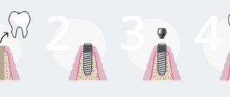
Prosthetics with crowns
Installing a crown on a tooth is a procedure to restore its anatomical shape, color, chewing function and aesthetics.
In practice, any tooth under a crown (with or without a preserved nerve) can begin to hurt. In this case, only a specialist can provide assistance. Lack of professional treatment or late contact with a doctor aggravates the situation; removal of the causative tooth may be necessary.
After professionally performed prosthetics, pain should not occur. At first, you may experience some discomfort associated with getting used to the new product in your mouth. If you have a crown installed and your tooth hurts, you should immediately tell your doctor about it.
Why does a tooth hurt under a crown: reasons
Thus, if you have a toothache under the crown or the gums under the crown are inflamed, the reason almost always lies in poor-quality therapeutic preparation of the tooth for prosthetics. Of course, in most cases, dental therapists take control photographs after filling the canals, but even if they see mistakes, most doctors simply will not waste time and redo something.
Unfortunately, this approach in Russia is the norm rather than the exception. And if your tooth hurts under the crown, then the first thing you should do is an x-ray, which will show one of the main mistakes that dentists make when filling root canals. The image will show whether the tooth can be treated or whether it needs to be removed. The most common mistakes made by doctors include...
Root canals are not filled to the root apex –
Let us remind you that in most cases, teeth must be depulped before prosthetics. Depulping a tooth means that the dental pulp (neurovascular bundle) is removed from it and the root canals are filled. When filling root canals, there are certain standards, the implementation of which helps prevent subsequent inflammation in the area of the tooth roots.
In Fig. 1,2 you can see how well-filled root canals look on radiographs. However, when the doctor does not work and fills the canals not to the top of the root, conditions are created in the unsealed part of the canal for the spread of infection. Failure to fill the canal by just 1-2 mm can already cause inflammation at the apex of the tooth root (Fig. 3).
On X-ray 3 you can clearly see the unfilled part of the root canal (it is shown by a white arrow). Black arrows show the boundaries of a periodontal abscess, which on an x-ray looks like intense darkening in the area of the root apex. The reason for its formation is the development of infection in the unfilled part of the root canal. This dental disease is called periodontitis.
What does inflammation of a tooth look like under the crown (Fig. 4) –
- "Gutta-percha" is a material for canal filling,
- “periapical abscess” is a focus of purulent inflammation in the form of a purulent sac at the apex of the root (depending on the size of the purulent focus, the latter is called either a granuloma or a radicular cyst).
Poor obturation of root canals –
Inflammation can also be caused by poor obturation of the root canal with filling substances (gutta-percha and sealer). Those. the canal can even be sealed to the top of the root, but it is not sealed tightly, with many pores and empty spaces.
This could also be the reason why your tooth hurts under the crown, because... Such poor-quality canal filling also leads to the development of inflammation at the apex of the tooth roots. Poor canal obturation can also be easily determined by a targeted photograph of the tooth.
Perforation of the walls of the root canal –
Perforation is literally “a non-physiological hole.” In other words, this is a hole in the root of the tooth that is created artificially. The only physiological hole in the tooth is at the top of each root. The most common perforations that occur are:
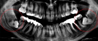
- During instrumental treatment of root canals - using tools for mechanical expansion of the root canal, the doctor can make a number of mistakes. For example, instead of expanding the root canal along its course (from the mouth of the canal to the apex of the root), the doctor will direct the instrument perpendicularly through the canal wall, which will lead to the appearance of a “hole” in the root wall (Fig. 5, 6).
- During fixation of the pin in the root canal, doctors also very often allow perforations if the technique for fixing the pins in the root canal is not followed. Such perforations are also determined by radiographs and corresponding symptoms (Fig. 7).
Fracture of an instrument in the lumen of the root canal –
This happens quite often, but in most cases it is again due to the fault of the doctor. Below you will read about the main reasons for instrument failure in the canal. The only good reason for a breakdown that cannot be blamed on the dentist is if your root canals have a very severe curvature.
- Violation of the technique of rotating the instrument in the root canal - instruments for treating root canals are quite thin and require strict adherence to a certain technique of use. For example, most instruments cannot be rotated more than 120 degrees in the root canal. If the instrument is rotated 360 degrees in the root canal, this can naturally cause a fracture of the instrument, which is associated with the curvature of the root canals.
- Reuse of instruments—breakage of instruments can also occur due to the fact that the doctor uses “old instruments.”
Instruments for mechanical expansion of root canals are made of special metal. Any metal gets microcracks during loading, which is called “metal fatigue.” Repeated use of an instrument for root canal treatment greatly increases the risk of instrument breakage (24stoma.ru). Tools for canal treatment come in different sizes and differ in thickness. The thinnest instruments have sizes No. 6,8,10, 15. In Europe and the USA, such instruments are generally disposable and their reuse is not allowed. Instruments of other sizes can be reused after sterilization. But in Russia, in the vast majority of clinics, in order to save money, no one throws away such instruments, and they work with them “to the limit.” What affects the incidence of tool breakage. - When working in highly curved, difficult-to-pass channels - In this case, instrument failure may occur through no fault of the doctor, because
Instrumentation in such root canals is itself risky. But you cannot refuse and not do it. The presence of a foreign body (piece of instrument) in the root canal or beyond can be determined using an x-ray (Fig. 8, 9). And the problem here is that in most cases it is not possible to remove the instrument fragment from the canal. This prevents high-quality filling of the root canal, which in the vast majority of cases leads to inflammation developing, the tooth under the crown aching, or the gums under the crown hurting.