01.12.2019
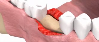
Penetration of bones and tooth roots into the maxillary sinus indicates perforation of the latter. A similar complication may be accompanied by treatment at the dentist. Perforation of the maxillary sinus occurs quite often. From this article you will learn the causes of this pathology, signs of perforation, methods of diagnosis and treatment, as well as prevention. Photos of tooth roots in the maxillary sinus can also be seen in the sections of the article.
Reasons for the breakup
The sinus, called the sinus or maxillary sinus, is not characterized by tightness. It interacts with the nasal cavity through a small gap. As a rule, a sinus rupture occurs in the area of its lower part.
This happens due to the characteristic structural features and against the background of certain diseases, namely:
- The roots of molars and premolars are located very close to the sinus. In some cases, the bone layer is quite thick and reaches 1 cm. However, sometimes the barrier can be less than 1 mm in thickness.
- The first and second molars in the root part can be located directly in the sinus cavity and are separated only by the mucous membrane.
- Rapid thinning of the bone layer due to inflammatory diseases in acute or chronic form, including cysts, periodontitis and periodontitis.
- Thin trabeculae of bone in the maxillary tissues.
All these factors can cause a sinus rupture during dental treatment. In this case, there may be no violation of the technology of performing dental procedures. This may be a result of each patient's individual anatomy. Let's figure out why the tooth root penetrates the maxillary sinus.
Why you should entrust treatment to the ENT department of dentistry
ENT dentistry is a symbiosis of two medical areas, a multidisciplinary approach to the treatment of inflammation of the maxillary sinuses of odontogenic origin. Only an experienced maxillofacial surgeon with ENT training can make an accurate diagnosis and create a sound rehabilitation plan.
As a rule, odontogenic causes of sinusitis are simply ignored during routine examination by an otolaryngologist. Treatment in a city clinic without high-quality diagnostics or in the absence of it at all turns into a multi-part series with monthly visits to an ENT doctor, dragging on for many years, causing inconvenience and worsening the quality of life.
Unified drug therapy or traumatic sinus punctures are prescribed, which, if they bring relief, are for a short time. Inflammation from the acute stage becomes chronic with periodic exacerbations. A person runs from one doctor to another to no avail, but without identifying and eliminating the cause, odontogenic sinusitis cannot be cured !
Frequent cases
Perforations of the maxillary sinus occur, as a rule, as a result of the dentist’s work during dental treatment. Tissue ruptures are typical for the following situations:
- Tooth extraction.
- Dental implantation.
- Endodontic therapy.
- Tooth root resection.
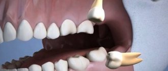
If perforation occurred during a tooth extraction operation, this may be a consequence of unskilled and excessively rough work of the dentist, as well as the individual characteristics of the structure of the maxillary sinus of the patient himself. If the dental roots are located directly in the sinus cavity, perforations during the removal of molars are almost inevitable.
One of the complications during endodontic therapy can also be perforation of the tooth root, which in most cases is associated with perforation of the sinus floor. This situation can occur as a result of excessive expansion of the root canals, as well as when using brute force when installing pins and compacting cement for fillings. In this case, most often not only the root of the tooth may be in the maxillary sinus, but also its fragments and particles of filling material. And this is dangerous for the patient.
When a rupture occurs during installation of an implant or during filling a root canal, as well as as a result of inserting pins into the root of a tooth, this in the vast majority of cases is a therapeutic error by a specialist. Sometimes you can find the roots of a wisdom tooth in the maxillary sinus.
What factors are taken into account by the dentist when performing an intervention?
The likelihood of sinus perforation increases when the tooth root is located in close proximity to it at the time of extraction. Therefore, surgical treatment should be carried out with extreme care and subsequent X-ray monitoring of the tissue condition after surgery. The dentist also monitors the condition of tissues when using pins for implantation or when treating root canals for filling purposes, since there is a high risk of filling material and root fragments penetrating into the maxillary sinus.
If the process occurs at the time of implantation into bone tissue or when filling canals, then this is a direct mistake that is made by the doctor during his therapeutic tactics. The only way to control manipulations is X-ray control and the experience of the dentist, who, when working, must take into account the anatomical structure of the patient’s upper jaw. In this case, damage to the bottom of the sinus is fraught with serious complications that are difficult to eliminate. Especially if this happens during the implantation of artificial roots. The bone tissue in the upper jaw quickly undergoes degenerative changes, and this leads to a decrease in the height of the alveolar process.
When performing root resection to eliminate cystic formations, as in the above cases, the dentist must fully examine the patient. If the history is insufficient, perforation may occur in a situation where the doctor does not know the exact size of the bone plate that separates the bottom of the sinus from the wall of the cyst itself. In this case, the prognosis worsens if it is necessary to remove a large volume of bone tissue.
Tooth root cyst in the maxillary sinus
Removal of the tooth root is considered the most effective method of treating cysts localized in the apex area. When the patient has not been carefully examined, and the dentist does not have information about the thickness of the bone tissue that separates the cystic wall from the sinus floor, and also in the case when it is necessary to remove a large amount of jaw bone, rupture of the sinus tissue quite often occurs.
Recovery after surgery
No hospitalization
A hospital stay for 2-3 days after surgery (as happens in city hospitals) is not required.
Gentle surgical protocols, the use of modern functional equipment and a microscope allow treatment to be carried out as delicately as possible .
All operations are performed in a controlled, drug-induced sleep - this is not general anesthesia ! Recovery from the state is easy, without dizziness, memory loss or clouding of consciousness. The drugs are absolutely safe for the body and are eliminated naturally 40 minutes after stopping the supply.
For patients with vascular and cardiac problems, a day hospital , where you can calmly recover and recover under the supervision of our anesthesiologist-resuscitator.
Accelerated rehabilitation
Recovery after the intervention occurs within a week. For those who want to speed up the process, our Center provides a set of procedures to reduce pain, resolve hematomas and swelling - carried out on the day of surgery.
Home care
Medications are prescribed - antibiotics, painkillers and decongestants. To prevent the patient from running to pharmacies in a postoperative state and to avoid purchasing counterfeit products “on the side,” the entire package of drugs is collected and given out free of charge .
The package contains all the necessary medications for taking and caring for the surgical area.
Please follow the recommendations and do not skip taking medications to avoid complications. The instructions are in the medicine package.
If you suspect a worsening condition, contact the clinic immediately. The telephone number for the 24-hour patient support service is listed in the memo.
Symptoms
When perforation and entry of the tooth root into the maxillary sinus occurs during dental resection procedures, characteristic signs of rupture appear, including:
- Bleeding from the tooth socket with small air bubbles, the number of which increases when you try to exhale sharply through your nose.
- Bloody nasal discharge from the side of the ruptured sinus.
- A sharp change in the timbre of the injured patient’s voice, characterized by nasal sound.
If there is a tooth root in the maxillary sinus, the symptoms cannot go unnoticed. Sometimes, after resection, the patient begins to complain about difficult passage of air through the resulting hole, as well as pressure and heaviness in the projection of the maxillary sinus.
When perforation occurs during implantation or endodontic therapy, signs of such a complication include:
- Specific failure of the instrument or material for implantation after some effort to move it into the jaw.
- Changing the position of the instrument used in the resulting wound.
- The appearance of small air bubbles and blood discharge from the hole.
Perforation may not be detected and repaired immediately after a sinus rupture, which leads to infection of the sinus cavity and is accompanied by signs of acute sinusitis or sinusitis.
The following symptoms are typical for such pathologies:
- Acute intense pain in the sinus area of the upper jaw.
- Swelling of the nasal mucosa on the side of the injury, accompanied by difficulty in nasal breathing.
- Discharge of purulent secretion from the nose.
Systemic complications and symptoms of intoxication appear, which are characterized by chills, headaches, weakness and increased body temperature. But how to detect the root of a tooth in the maxillary sinus?
Accelerated rehabilitation
After the operation, you will receive free medications and instructions with rules of behavior during the rehabilitation period. We have collected the necessary set of drugs to improve your well-being at home. Please take them as prescribed by your doctor and follow the recommendations to avoid unpleasant consequences.
For quick recovery after surgery, our Center offers its own method of rehabilitation in 1-2 days. The procedures allow us to minimize the formation of possible hematomas, swelling, and eliminate pain. The program uses:
Microcurrent therapy
A weak pulse current normalizes metabolic processes, activates ATP production, and improves tissue nutrition. Regeneration starts, healing accelerates, swelling decreases. Muscle spasm goes away.
PRP plasma therapy
Injections of purified and platelet-enriched plasma from one’s own blood trigger cellular regeneration processes using the body’s internal reserves. The manifestation of edema and hematomas is reduced.
Biostimulation of the face
Lymphatic drainage drugs D-NUCLEO and MesoSculpt C71 help reduce swelling. Active anti-inflammatory components improve blood flow, increase tissue nutrition, and improve healing.
Diagnostics
For an experienced dentist, specific testing may not be necessary to confirm a maxillary sinus perforation.
If we are talking about tooth resection, then such a complication as soft tissue rupture is accompanied by a typical clinical picture. If the dentist is in doubt when perforation occurs during endodontic treatment, the following additional examinations may be required:
- Probing the hole formed after tooth extraction, as well as the root canal. For this purpose, a thin probe is used. In this way, it is possible to establish the absence of a bone bottom in the wound. During the study, the instrument passes through soft tissues without hindrance.
- Carrying out an X-ray examination of the sinuses. The resulting images will show darkening in the sinus cavity, indicating the accumulation of blood in this area, as well as filling material and fragments of teeth and implant. In some cases, contrast radiography may be required with the introduction of a special substance into the perforation cavity.
- CT scan. This diagnostic method makes it possible to detect ruptures and the presence of foreign bodies. In this case, the image is as accurate and informative as possible.
- If the perforation is old, it is recommended to donate blood for a general examination, based on the results of which it will be possible to draw a conclusion about the presence or absence of an infectious focus in the body.
After carrying out the necessary diagnostic measures and confirming the fact of a rupture of the maxillary sinus, the dentist will prescribe appropriate treatment. So, a person has a tooth root in the maxillary sinus - what to do?
Can a dentist diagnose sinusitis?
In case of pulpitis, the patient finally comes to the dentist. The doctor fills the canals, removes the nerve, but part of the infection still remains in the apical delta - after all, it has penetrated there a long time ago!
The apical delta is a branching at the tips of the roots that can never be adequately sterilized. And if a tooth has many roots, then, naturally, the entire system of root canals is one, like the root of one tree.
Therefore, if an infection gets into a tooth, it is already inside.
And it’s normal that the body fights it, and the sinus mucosa begins to thicken. In the process, some people develop granulomas, others - cysts. But cysts are changes in the periodontium, and the mucous membrane itself begins to thicken and degenerate.
Sometimes it happens that polyps are formed - spherical thickenings on a stalk. Hypertrophy of the columnar epithelium may occur. That is, as a result of chronic inflammation, many, many layers are formed.
The dentist can conduct a computed tomography scan, but it can only identify changes in the mucous membrane in volume - simply establish the presence of this fact.
And only an otolaryngologist can say what exactly happened there. To do this, you need to conduct an endoscopic examination.
But in most cases, it is the dentist who is the first doctor to detect sinusitis on a CT scan.
When a person realizes from various symptoms that he has inflammation in the sinus, he also learns about this from the dentist.
Treatment
The choice of treatment for maxillary sinus perforation depends on the changes that occur as a result of the rupture. Non-surgical treatment seems possible in case of violation of the integrity of the tissues during tooth extraction, when the pathology was detected immediately, and according to the results of the radiographic examination it is clear that there is no infection of the sinus cavity and there are no foreign bodies in it. With such a clinical picture, the doctor, as a rule, tries to preserve the blood clot formed in the hole as much as possible. In addition, preventative measures are taken to prevent wound infection.
For this purpose, a small gauze swab soaked in an iodine solution is inserted into the hole. As a rule, the tampon is self-fixed in the wound cavity. However, sometimes it may be necessary to place a stitch in the gum. Treatment with an iodide solution should be carried out for 6-7 days until the defect disappears and full-fledged granulations are formed. It is not recommended to remove the tampon from the socket during treatment, as this can damage the clot and lead to infection of the wound.
When a tooth root is found in the maxillary sinus, everyone should know what to do. In some cases, the doctor decides to close the defect with a special plastic plate, which is secured to adjacent teeth with clasps. Thus, it is possible to achieve separation of the sinus and oral cavity.
No hospitalization required
Operations in our Center are performed by operating teams of experienced maxillofacial surgeons with ENT training in a sterile operating room. The treatment is as gentle and minimally traumatic as possible; a 24-hour hospital stay is not required; you will go home the same day .
The intervention lasts about an hour, under sedation. After completion, the patient quickly returns to clear consciousness without unpleasant consequences or risk of complications. After 30-40 minutes you can safely go home. For patients with concomitant cardiovascular diseases, a day hospital is provided . You can lie down for the time necessary for recovery under the supervision of our anesthesiologist-resuscitator.
Preventing inflammation
In addition to preserving the wound, preventive therapy is carried out aimed at preventing the development of the inflammatory process. Most often, antibacterial drugs and anti-inflammatory drugs are prescribed. In addition, drops are prescribed for instillation into the nasal passages, which have a vasoconstrictor effect. Treatment can be carried out both on an outpatient basis and while staying at home.
If the examination reveals the presence of a foreign body in the maxillary sinus, then treatment is carried out only in a hospital setting. Therapy consists of surgical intervention to open the cavity and remove the tooth root from the maxillary sinus.
What are the dangers of sinus injury?
Since the complication does not always manifest itself immediately, symptoms of sinusitis that arise after a few years force the patient to consult a regular ENT doctor. Treatment, as a rule, does not bring results - in most cases the cause is not found. You start going from one doctor to another, and precious time is lost. The longer a foreign body is in the sinus, the higher the risk of tumor formation. If sinus perforation is left unattended, it can lead to:
- chronic sinusitis and sinusitis
- inflammation of the roots of adjacent teeth
- osteomyelitis of the jaw
- encephalitis and meningitis
Complications
Rupture of the maxillary sinus is a serious consequence of dental procedures, the treatment of which most often occurs in an inpatient setting. What is the risk if the tooth root has grown into the maxillary sinus?
Self-treatment of the problem using traditional medicine methods can lead to even more serious complications, including:
- Severe inflammatory process in the sinus cavity with further infection of adjacent tissues. Then osteomyelitis develops in the upper jaw.
- Transition of the inflammatory process to other cranial sinuses, including the sphenoid, frontal and ethmoid.
- Loss of healthy teeth located in the perforation area.
- Formation of foci of pus, phlegmon and abscesses.
Due to the immediate proximity of the brain, and the fact that the tooth root has gone into the maxillary sinus and the latter has ruptured, the occurrence of an infectious lesion of the meninges cannot be ruled out. The next stage of serious complications may be meningitis or meningoencephalitis, which are life-threatening for the patient.
What dangerous consequences does a foreign body lead to?
A foreign object in the maxillary sinuses can lead to the following complications:
- bacterial or fungal sinusitis;
- encapsulation in the form of a cyst or other benign disease;
- granuloma formation.
In more complex cases, suppurative processes in the maxillary sinuses cause the development of the following complications:
- frequent colds and inflammation of the nasal cavity and sinuses;
- meningitis;
- hypertrophy of the mucous membrane;
- damage by coccal flora to other organs and systems (for example, heart muscle).
A particularly dangerous complication is inflammation of the meninges. When meningitis develops, the patient develops the following symptoms:
- significant increase in temperature;
- headache in the forehead;
- frequent vomiting that does not bring relief;
- rash on palms and soles.
If these symptoms appear, you should immediately consult a doctor!
And the possible consequences of getting filling materials can only be prevented by timely consultation with a doctor and regular preventive examinations in the mode prescribed by the doctor.
Prevention
As for preventive measures regarding perforation of the maxillary sinus, they consist of following the following rules:
- A thorough and comprehensive examination of the patient before undergoing serious dental procedures, including tooth resection or implantation.
- Correct assessment of the anatomical features of the jaw structure of each patient.
- Strict compliance with all technological requirements for complex dental procedures.
Old perforations
If the perforation of the maxillary sinus was not promptly identified and eliminated, then after 2-4 weeks the stage of acute manifestations will subside, and a fistula will form in the area of the defect, connecting the sinus cavity with the surface of the gum.
This process is simultaneously accompanied by symptoms of chronic sinusitis:
- constant dull pain in the sinus area radiating to the orbit and temple;
- nasal congestion on the affected side;
- purulent discharge from the nasal cavity, as well as from the fistula;
- Sometimes patients have swelling of the cheek on the side of the damaged sinus.
Most patients also complain of a sensation of air moving through the fistula when talking or sneezing, difficulty pronouncing certain sounds, and liquid food entering the nasal cavity from the mouth.
Treatment of such chronic perforations with fistulas presents some difficulties, since the presence of a chronic focus of inflammation in the maxillary sinus significantly reduces the effectiveness of therapy and quite often leads to relapse and re-formation of the fistula canal.
Such patients are indicated for surgical intervention, which includes opening the maxillary sinus with removal of all non-viable tissues and foreign bodies from its cavity, excision of the fistula and plastic closure of the defect. Antibiotics after removal of the fistula are prescribed for a course lasting 10-14 days with the simultaneous use of anti-inflammatory and antihistamine drugs, and the use of physiotherapeutic methods of treatment.
Consequences of perforation
Perforation of the maxillary sinus is a fairly serious pathology that often has to be treated in a hospital. Attempts to independently treat it with folk remedies at home without medical assistance can lead to the development of serious and dangerous consequences:
- The development of a pronounced inflammatory reaction in the sinus cavity with the spread of infection to the surrounding bone tissue and the formation of foci of osteomyelitis of the upper jaw.
- Spread of inflammation to other sinuses of the skull (frontal, sphenoid and ethmoid).
- Loss of healthy teeth located in the area of untreated perforation.
- Formation of purulent foci (abscesses, phlegmons).
Due to the close location of the maxillary sinus and the brain, after perforation, infection may spread to the meninges with the development of meningitis or meningoencephalitis, which threatens the patient’s life.
Possible complications when removing a tooth in the upper jaw
Perforation or perforation of the maxillary sinus is one of the most common complications that threatens the doctor when removing maxillary molars.
The maxillary sinus, also known as the maxillary sinus and also the maxillary sinus, is the largest of the peranasal sinuses. And the roots of the molars, or rather their tops, are located very close to its bottom, which is also the lower wall. Most often, perforation of the lower wall of the maxillary sinus occurs if the root of the tooth being removed enters the sinus cavity, and the mucous membrane lining the sinus only covers it.
If tooth extraction under such conditions is performed without any complications, the tooth is removed entirely and without root fracture (“normal extraction”), then simply perforation occurs, one of the most “successful” options. However, if the removal process is complex and is accompanied by root fracture, then when extracting a root fragment there is a great danger of it being pushed into the maxillary sinus.
This presents quite a challenge, which is exacerbated by the limited amount of dental appointment time. The next patient, who arrived exactly at the appointed time, is waiting outside the doctor’s door, and if we are talking about a private clinic, then once you make a client wait, you can lose him altogether, and with him, your earnings.
The patient wants to choose one dentist for the rest of his life and be satisfied with the quality of service. And the doctor must respond with efficient and competent service, as well as good service, including fast, timely appointments and polite treatment.
Rule one: don’t panic and get lost. The patient will immediately notice the doctor's embarrassment. The dentist is in the palm of his hand. You need to be completely confident in yourself.
Rule two: check for symptoms characteristic of maxillary sinus perforation. These include: – Patient difficulty in puffing out the cheeks. If there is a perforation, it will be difficult for him to do it, or he will do it at all. – Presence of nosebleeds from the nostril (from the side of the extracted tooth). – When breathing through the nose with the mouth closed, the patient indicates that air is entering the oral cavity. – Change in voice resonance – nasality appears. – Nasal discharge enters the mouth, the so-called “naso-oral discharge”.
If checking the symptoms has confirmed the doctor’s fears, the fact of perforation is confirmed, then it is necessary to perform an x-ray examination, which includes: 1. High-quality intraoral x-ray; 2. Occipito-mental radiograph at an angle of fifteen degrees; 3.Panoramic radiograph.
By taking any of these pictures, you can see confirmation of perforation. If no complications arose during tooth extraction, this means that the apex of the root did not break off and the dentist did not push its fragment into the maxillary sinus, then the image will only show a violation of the integrity of the lower wall of the sinus. Simply put, a hole in the bone tissue that communicates with the socket of the extracted tooth.
Uncomplicated perforation, in which there are no foreign bodies in the maxillary sinus, is treated by closing the perforation and suturing the defect. Please note that it is not enough to simply tighten the edges of the socket with sutures. To completely close the anastomosis, mobilize the mucoperiosteal flap.
It is important to make an incision through the mucosa, through the periosteum to the bone. In this case, the mucoperiosteal flap is separated, and not just the mucous membrane. If only the mucous membrane is separated, then the incision is not made all the way to the bone, and this risks bleeding.
Excessive tension of the mucosa leads to its rupture, which increases the healing time of the postoperative wound.
Let's consider the surgical technique. We highlight the stages of the operation: – It is necessary to create a wide base of the mucoperiosteal flap to ensure good blood supply. – The flap is separated from the bone. – Resection of the vestibular and palatal walls of the socket is performed with a minimum height of 3-5 mm, the sharp edges of the bone must be smoothed. Bayonet pliers can be used for this. – To increase the mobility of the flap, we cut the periosteum from the distal to the proximal edge of the flap, but the mucous membrane cannot be cut. This manipulation will increase the mobility of the flap so that it can be moved to the palatal side without tension. – The edge of the flap is grabbed with tweezers and carefully moved towards the palate until it touches the edge of the mucous membrane of the alveolar process from the palatal surface. Accuracy is important! – The socket is irrigated using a 0.2% chlorhexidine solution, then the flap is moved to the palatal side and sutures are applied. – It is recommended to apply two mattress vertical sutures to close the wound, after which the vertical incisions that were made on the mucous membrane are closed with ordinary interrupted sutures. – Vicryl, Silk, Nylon (No. 3) can be used as suture material. - Attention! You can't use catgut. It will swell and will not hold the edges of the wound, in addition, it will not allow the edges of the wound to be sutured to be accurately aligned. – To ensure that the underlying bone supports the sutures, they are placed on the palatal surface of the alveolar process. – Management of the postoperative period. The operation is completed.
We prescribe antibiotics, up to 5-6 rinses of the mouth with a 0.2% chlorhexidine solution per day, as well as nasal drops. Recommendations for the patient are to avoid pressure in the nasal cavity: you should not blow your nose, puff out your cheeks, or cough with your mouth closed. After five to six days, the stitches are removed.
This is what a simple operation looks like, which is performed if there are no fragments of the root of the extracted tooth left in the maxillary sinus. If an x-ray shows the presence of a foreign body in the sinus, it is recommended to use a vacuum pump, which will extract a root fragment from the sinus.
The manipulation is complex, its successful results are rare. If the root is not extracted, an antibiotic should be prescribed and the patient should be referred to the hospital for surgical treatment using Caldwel-Luc. This operation will be discussed later.
I would like to draw attention to some features of the operation described in detail above. When performing an outpatient maxillary sinusotomy, tuberal and infiltration anesthesia is performed.
During manipulations in the maxillary sinus, a tampon moistened with lidocaine is inserted into it. In the future, you can use it to try to remove the root from the sinus, while the tampon needs to be unrolled. If the perforation is small, it is enough to sew an iodoform swab onto the hole.
You cannot pack the hole - this leads to the formation of a fistula in 100% of cases. If the hole is not plugged, but sutured, then there is a higher probability that the resulting clot will organize itself, closing the hole.
It is not uncommon for a maxillary sinus fistula to be closed 3 weeks after perforation. Then many polyps are formed in the sinus, which can be easily removed with a thick swab.
A dental mirror helps you see what is happening in your sinus. To do this, you need to direct the beam through the perforation, which requires forming a flap. In general, the manipulation is considered simple, and any surgeon can perform a maxillary sinusotomy.