For many people, as practice shows, panic is caused by the mere fact that it is necessary to remove a wisdom tooth. This is due to the fact that patients imagine the procedure itself as painful. But the days when a dentist spent more than 2-3 hours chiseling out roots piece by piece with a hammer and chisel are long gone.
Modern clinics use new technologies and techniques, thanks to which the removal of a wisdom tooth in the upper jaw is quick, in a comfortable environment and absolutely painless.
Features and difficulties during removal
Extraction of the upper wisdom tooth is complicated by its specific anatomical features.
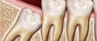
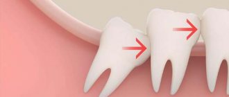
“Eights” erupt late, when the dentition is already formed. Therefore, very often they are incorrectly located, grow together with the roots of neighboring teeth, grow towards the cheek or even horizontally. Each of these factors complicates the extraction process placed on top of the 8th tooth. But if we compare wisdom tooth removal from above and below, then in the first case the surgical procedure is much simpler.
In the upper dentition, the bone is less dense and massive, there are much fewer blood vessels in the soft tissues and the risks of damage to the trigeminal nerve are lower. Therefore, the operation to remove the G8 is easier and safer.
Possible complications
The structural features of wisdom teeth determine the possibility of surgical and postoperative complications - this is another reason for patients to be concerned about the removal of wisdom teeth. Fortunately, they are quite rare and in most cases are associated with insufficient professionalism of the doctor. If the surgeon acts carefully, then unpleasant consequences can be avoided. However, it is worth knowing what you might encounter.
- Tooth destruction during the extraction process.
This can happen if the integrity of the enamel is damaged by caries, and the doctor presses too hard on the forceps. It is accompanied by a characteristic cracking sound and can greatly frighten the patient. Requires careful removal of fragments from the hole. - Fracture of the jaw bone.
This is also a consequence of the rough work of the surgeon, who, by applying forceps, can capture part of the alveolar process along with the tooth. - Alveolitis.
This is inflammation of the socket, accompanied by swelling, pain, fever and bad breath. Requires immediate sanitation of the wound surface and antibiotics. - Damage to the maxillary sinus.
A rare but very dangerous complication when perforation of the wall of the maxillary sinus occurs. To avoid this, the doctor should carefully study the x-ray and act as carefully as possible.
Features of tooth extraction in the upper jaw
The structure of the upper and lower jaws is significantly different. They also vary in density and location. Therefore, the method of tooth extraction will differ:
- on the upper jaw the operation is faster;
- the recovery period is shorter;
- pain relief is simpler and more effective.
Complications during tooth extraction in the upper jaw occur less frequently than in the lower jaw.
Anesthesia
The secret of high-quality pain relief is the correct delivery of the drug to the nerve. The choice of anesthesia method takes into account the patient’s age, his general condition, and the complexity of the tooth being removed.
Anesthesia of the upper jaw
The upper jaw is less dense than the lower jaw. It is covered with a cortical plate with many holes. Nerves and blood vessels pass through them. Pain relief is easier and faster than in the lower jaw
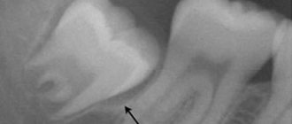
Sinus damage
Above the upper jaw is the nasal sinus, also known as the maxillary sinus. Its bottom may be close to the root of the tooth being removed. Then, during removal, sinus perforation may occur. This complication is typical for surgery on molars. This pattern is explained by the fact that the jaw in the area of the incisors is longer, and the nasal cavity is further away relative to them.
The main sign of sinus perforation is the escape of air, food and fluid through the nose. There is foam in the hole. Hospitalization in a hospital may be indicated for treatment.
Entry of part of the root into the sinus
Due to perforation of the sinus, part of the root can get inside it. This is caused by thinning of the bone plate or errors during the removal operation.
The symptoms when part of the root gets into the sinus are the same as when perforation occurs: air and contents of the oral cavity escape through the nose. But at the same time there is pain and increased body temperature. Treatment requires hospitalization.
Clinical researches
Repeated clinical studies have proven that the two-component mouth rinse ASEPTA ACTIVE more effectively combats the causes of inflammation and bleeding compared to single-component rinses - it reduces inflammation by 41% and reduces bleeding gums by 43%.
Sources:
- The role of anti-inflammatory rinse in the treatment of periodontal diseases (L.Yu. Orekhova, A.A. Leontyev, S.B. Ulitovsky) L.Yu. OREKHOVA, Doctor of Medical Sciences, Prof., Head of Department; A.A. LEONTIEV, dentist; S.B. ULITOVSKY, Doctor of Medical Sciences, Prof. Department of Therapeutic Dentistry of St. Petersburg State Medical University named after. acad. I. P. Pavlova
- The role of hygiene products in the treatment of periodontal diseases (S.B. Ulitovsky Honored Doctor of the Russian Federation, Honored Dentist StAR Prof., Doctor of Medical Sciences, Department of Preventive Dentistry of Pavlov Pavlov State Medical University, St. Petersburg) S.B. Ulitovsky - Honored Doctor of the Russian Federation, Honored Dentist of StAR, Prof., Doctor of Medical Sciences; E.S. Alekseeva - associate professor, candidate of medical sciences; A.A. Vasyanina - associate professor, candidate of medical sciences; V.A. Grigoriev - Associate Professor, Ph.D.
- The use of drugs from the Asepta line in the complex treatment of inflammatory periodontal diseases (N.V. Berezina E.N. Silantyeva S.M. Krivonos, Kazan State Medical Academy. Kazan.) N.V. BEREZINA, E.N. SILANTIEVA, S.M. KRIVONOS Kazan State Medical Academy
Preparing for surgery
Before removing a wisdom tooth in the upper jaw, the doctor prepares the patient for the upcoming operation. The preparatory process consists of:
- X-ray examination of the problematic tooth;
- clinical examination of the patient for the presence of pathologies and other concomitant diseases that may affect the course of the operation;
- removal of subgingival and supragingival deposits from the tooth being removed and adjacent teeth to prevent the penetration of infected plaque into the alveolus.
The surgeon also assesses the patient’s psycho-emotional state and, if necessary, recommends taking a sedative.
Rinse
Special rinses will help relieve pain and swelling at home. However, such treatment is symptomatic. If inflammation progresses, in no case should you limit yourself to rinsing only.
The treatment for inflammation recommended by dentists is ASEPTA Active mouth rinse. This unique two-component product with a combination of “chlorhexidine + benzydamine” has an antimicrobial, anti-inflammatory effect and provides an immediate analgesic effect.
To eliminate inflammation and relieve discomfort during wisdom tooth pericoronitis, it is possible to use such medicinal herbs as:
- chamomile;
- sage;
- calendula.
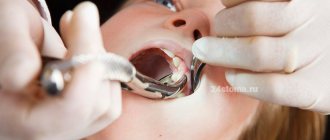
Easy removal
When nothing prevents you from removing the tooth from the socket and cutting the gums with a scalpel is not required, then such removal is considered simple. The procedure is carried out in the following order:
- the doctor orders an x-ray or recommends a CT scan;
- the problem area is numbed with local anesthetics;
- Using forceps, the surgeon first rocks the tooth and then pulls it out of the socket.
After the tooth is removed, the hole is treated with an antiseptic solution, after which a sterile swab is applied to it. If necessary, the wound is sutured. The entire procedure lasts about 10 minutes.
Difficult removal
Such an operation is considered difficult when the upper molar has to be extracted in parts with an incision in the gum and sawing of the crown. The procedure occurs in stages:
- X-ray or CT scan;
- gum pain relief. In case of a complex clinical picture, it is possible to perform the operation under general anesthesia;
- dissection of the gum is performed to open access to the crown;
- the surgeon pulls out the molar. It is often necessary to first remove the crown with a drill to ensure free access to the roots;
- the hole is carefully checked for the presence or absence of bone fragments.
In cases of bone resection, the hole is filled with special bone chips, which subsequently form new bone tissue.
Since the operation is complex, the rehabilitation period is longer and can last for several weeks.
Pain after removal of upper wisdom tooth
It should be understood that removing an upper wisdom tooth is much more difficult than removing any other unit in the dentition. The patient does not feel any pain during the operation. But when the anesthesia wears off, pain is inevitable.
To relieve pain, you should take painkillers recommended by your doctor. Cold compresses applied to the cheek relieve pain.
If within 3-5 days the pain does not disappear, but becomes more intense, then you should seek help from a dentist to find out the cause of this condition.
CAUSES OF IMPROPER TUNING
Third molars are the medical name for wisdom teeth. They do not have milk precursors, so they do not have ready-made conductive channels for exit. But their formation begins at the age of 5-6 at a speed that is individual for each person.
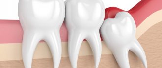
What causes the incorrect “crooked” cutting of eights? Despite the individual reasons for the appearance of eruption pathology, the most common ones can be identified:
- hereditary jaw structure;
- incorrect tooth position;
- non-standard size or unusual figure eight shape;
- metabolic and hormonal disorders of the patient;
- excessive number of teeth in a row;
- removal of several teeth to straighten the back, which created free space.
In each specific case, the process of eruption is accompanied by numerous sensations that do not allow one to independently determine the main reasons for their appearance and the nature of the pathology.
What to do if the bleeding does not stop
Immediately after removing a molar, the surgeon places a tampon on the socket to stop capillary bleeding. The patient needs to clench his jaw and not remove the tampon for 20 minutes. During this time, the blood coagulates and a clot forms in the hole, protecting the fresh wound from the penetration of bacteria into it.
If the patient has high blood pressure or poor blood clotting, the tampon should be kept in place for 40-60 minutes.
When a person strictly adheres to medical recommendations, but the bleeding does not stop, he should contact a dentist for help.
Safe and high-quality removal of wisdom teeth in Balashikha
The Berezka dental clinic offers qualified assistance to anyone who, for certain reasons, requires the removal of a wisdom tooth from above or below. The operations are performed by experienced surgeons using safe anesthesia and modern instruments.
Before the procedure, the dentist collects the patient’s medical history, conducts all the necessary examinations, and only if the clinical picture is present is it possible to extract the wisdom tooth.
The operation is performed in a comfortable environment for the patient. Innovative equipment and professional dentists ensure successful and safe removal of the most complex teeth.
CONSEQUENCES OF THE PROCESS
A long-term process of improper eruption of third molars can cause serious complications, the elimination of which will require long-term treatment and material costs:
- destruction of the roots of adjacent seventh teeth, which leads to their premature loss;
- the appearance of caries and destruction of the enamel of “sevens”;
- the appearance of a focus of inflammation on the cheek;
- the likelihood of inflammatory processes occurring in the bone part, in the gum area and base of the tooth (periostitis, ostomyelitis, phlegmon, pericoronitis);
- compaction of the soft tissues of the gums or cheeks, leading to their modification and degeneration, developing into benign or malignant neoplasms.
To avoid complications, you should contact a dental clinic as soon as possible. Only a comprehensive examination can predict the development of the eruption process and give recommendations on possible removal or treatment of a wisdom tooth.
In many countries, modern dentistry offers the removal of “eights” at the formation stage at a young age in order to save people from possible pain and complications.