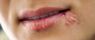
Cheilitis is a disease and inflammation that is expressed by damage to the lips (“ cheilitis on the lips ”), more precisely, inflammation of the red border, the mucous membrane of the lips and (in some cases) the skin of the lips. According to medical classification, there are different types of cheilitis. For example, simple cheilitis (Cheilitis simplex, the most common form), angular cheilitis (Cheilitis angularis, or angular cheilitis , or seizure, or inflammation of the corners of the mouth). Cheilitis can be either an independent disease (for example, exfoliative cheilitis, glandular cheilitis, actinic cheilitis), or a symptom of other diseases (for example, atopic cheilitis , eczematous cheilitis ). There are more than twenty types or types of cheilitis.
Causes of burns
Experts identify several types of lip damage, differing in the reasons for their occurrence. So, the most common causes of a burn on the lip are:
- Eating hot food;
- Eating hot spices or hot peppers;
- Smoking. Those who smoke unfiltered cigarettes are especially often affected;
- Unskilled waxing – injuries appear above the upper lip;
- Sunburn;
- Action by electric current;
- Injuries can occur during dental procedures;
- Careless self-treatment with iodine, alcohol or other chemicals.
The cause of the labia is varicose veins
Varicose veins usually appear in pregnant women. The pathology is found to one degree or another in 40% of pregnant women and affects the veins of the lower extremities and anus. The cause of the pathological condition is the pressure of the enlarging uterus on large venous vessels - the inferior vena cava and iliac veins. This causes blood stagnation in the pelvic area, vasodilation, and ultimately the appearance of varicose veins.
Common symptoms of varicose veins include:
- dilated, visible veins in the upper thighs and buttocks, spreading around the periphery;
- hyperalgesia, pain in the vulva and labia;
- hyperemia and swelling of the labia;
- discomfort during sexual intercourse;
- constipation;
- problems with urination.
Other risk factors for developing labial varicose veins are obesity, sedentary lifestyle, chronic constipation, lack of exercise and abdominal hypertension.
Diagnosis of the disease is based on ultrasound with Doppler of the pelvic venous vessels. Treatment of varicose veins of the vulva consists of embolization or sclerotherapy of diseased veins, which involves closing the lumen of the vessel. In pregnant women, the disease often goes away on its own a few months after birth.
Common types of injuries
There are many reasons for a lip injury. All of them are divided into several types. Their differences are in the aggressor. Let's list the varieties:
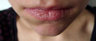
- Chemical burn of the lip - formed as a result of exposure to cleaning agents or detergents, acid, alkali, or iodine solutions. The longer the aggressor exerts influence, the more serious the injury will be. Timely intervention and medical care are required. As a result of such exposure, microcracks appear on the lips, which bleed, and damaged and dead skin creates a crust. The surface of the lips becomes very dehydrated and dry, which makes it even painful to open your mouth. Healing of such an injury takes a long time - from 1 to 2 months;
- Thermal burn is the most common injury. The cause may be exposure to boiling water or the application of hot objects. Often the injury is accompanied by damage to the palate of the mouth. Thermal burns have three degrees of severity. The first is mild, when slight pain is felt and medical attention is not required. You can cope with the problem with herbal compresses and special ointments. The second degree is of moderate severity, when the victim feels a sharp pain, blisters and small ulcers appear on the lip. The problem can be solved with medication. The third is severe, when pus is released from the blisters. As a result, deep deformation and tissue destruction can occur. Treatment can only take place in a medical setting;
- Sunburn - it appears if a person is in the sun for a long time. The peculiarity of injury is that it is not immediately detected. As a result, the lips peel, you can feel the skin tightness and burning. In rare cases, blisters appear. First aid for such an injury is to apply a cloth moistened with cold water to the lips for 15 minutes. Then apply medicinal ointment.
If you have a problem similar to that described in this article, be sure to contact our specialists. Don't diagnose yourself!
Why you should call us now:
- We will answer all your questions in 3 minutes
- Free consultation
- The average work experience of doctors is 12 years
- Convenient location of clinics
Single contact phone number: +7
Make an appointment
Lip tumors
Of all the anatomical parts of the oral cavity, the red border of the lower lip is most often affected. Lower lip cancer accounts for approximately 12 - 25% of the total number of tumors in this location. For reasons that are not entirely clear, tumor lesions of the red border of the upper lip are an extremely rare occurrence. The disease most often occurs in people over 60 years of age. Men are more often affected, the male/female ratio approaches 13/1. Predisposing factors are solar radiation, smoking, and alcohol abuse.
Clinically, the disease manifests itself as an exophytic (i.e., protruding) tumor or, much more often, an ulcerative bleeding surface. Since it is very difficult not to notice this lesion, the vast majority of patients seek medical help at an early stage of the disease. This circumstance (treatment of the early stages of any malignant tumors always brings better results) allows us to count on high effectiveness of treatment. In addition, lower lip cancer rarely metastasizes to regional (cervical) lymph nodes. At the time of initial treatment, involvement of the cervical lymph nodes is diagnosed in only 2–12% of patients. Damage to regional lymph nodes significantly worsens the prognosis of the disease. Thus, the 5-year survival rate in the presence of this lesion drops from 90 to 50% and forces one to resort to more aggressive methods of treatment.
Treatment for lower lip cancer is determined by the stage of the disease. Early stages can be cured with surgery or radiation treatment. There are 2 types of radiation treatment: remote and contact therapy (so-called brachytherapy).
The lip has a rather complex anatomical structure, which includes muscles with different directions of muscle fibers, skin, salivary glands, and subcutaneous tissue. Naturally, the goal of surgical treatment is not only radical removal of the tumor, but also restoration of the structure and function of the lower lip. Modern methods and techniques of plastic surgery can achieve good cosmetic and functional results. This is illustrated by the photographs below of patients who underwent surgical treatment for cancer of the lower lip.
Develops against the background of obligate (warty precancer, limited dyskeratosis, Manganotti cheilitis, Bowen's disease) and facultative (leukoplakia verrucous, keratoacanthoma, cutaneous horn, papilloma with malignancy, erosive-ulcerative and hyperkeratotic form of lupus erythematosus and lichen planus, post-radiation cheilitis) precancerous processes .
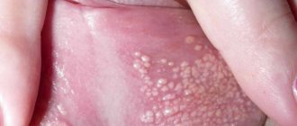
Warty precancer is characterized by the appearance on the red border of the lip of a sharply demarcated hemispherical lesion with a diameter of 4 mm to 1 cm, which protrudes above the surrounding tissues and has a dense consistency. Its color varies from an almost normal red border to a stagnant red. The nodule may be covered with thin scales and resemble a wart or keratinizing papilloma. Malignancy can occur 1-2 months after the onset of the disease.
The productive form of focal dyskeratosis is characterized by excessive keratinization of the epithelium and looks like a white spot with flat protrusions on a red border or an area of hyperkeratosis with stylized horny protrusions. The destructive form (erythroplakia) is distinguished by the fact that limited defects of the epithelial cover appear on the red border of the lip - erosions, ulcers, cracks. Malignancy can occur 6 months after the onset of the disease.
Abrasive pre-cancer cheilitis Manga-Notty is characterized by the formation of an oval or irregularly shaped erosion on the lip, often with a smooth, polished surface of a deep red color. Crusts may form on the surface of the erosion, which may cause slight bleeding when removed. There is no tissue compaction at the base and around the erosion. Spontaneous epithelization with subsequent recurrence is possible. Malignancy occurs 3 months to 30 years after the onset of the disease.
Lip papilloma is a clearly circumscribed tumor from 1-2 mm to 2 cm in diameter on a wide base with a villous or rough surface. As keratinization increases, the tumor acquires a whitish tint. Lip papilloma is considered a progressive precancerous disease. Compaction of the base of the papilloma and the appearance of pain are signs of its degeneration.
Treatment of diffuse lesions of the lower lip (cheilitis, leukoplakia, etc.) is predominantly conservative. Focal lesions are treated surgically, laser and cryodestruction.
Lip cancer
The term “lip cancer” refers to malignant tumors that arise in the mucous membrane of the red border of the lip. Neoplasms that have developed on the skin near the lip or mucous membrane of the vestibule of the mouth are not included in this group of tumors.
In the structure of cancer diseases in Russia (1997), lower lip cancer accounted for 1.4% of the total number of malignant neoplasms with an incidence of 2.8 per 100,000 population. The disease is most often observed in men over 70 years of age. In rural areas, lip cancer occurs 1.5-2 times more often than in urban populations. In recent decades, there has been a gradual decline in incidence. Approximately 90% of tumors of the lower lip develop due to exposure to known carcinogens, the main of which are tobacco and tobacco-containing substances. Alcohol is comparable in action to tobacco and is part of a group of agents that can potentiate tobacco-induced carcinogenesis. Other important etiological factors are chronic injuries and inflammation of the lip, ultraviolet radiation, changes in weather conditions, and occupation (work in the oil refining, coal, textile industries).
International classification according to the TNM system
Anatomical areas and parts:
1.Upper lip, red border. 2.Lower lip, red border. 3. Angles of the lips (commissures). T - primary tumor: Tx - insufficient data to evaluate the primary tumor, T0 - primary tumor not determined, Tis - preinvasive carcinoma (carcinoma in situ), T1 - tumor up to 2 cm in greatest dimension, T2 - tumor up to 4 cm in greatest dimension , T3 - tumor more than 4 cm in greatest dimension, T4 - tumor spreads to adjacent structures - bone, tongue, facial skin.
N/pN - regional lymph nodes. Regional lymph nodes for all parts of the head and neck, with the exception of the nasopharynx and thyroid gland, are the lymph nodes of the neck, including: submental, submandibular, nodes at the base of the skull near the great vessels (deep cervical), in the bifurcation zone of the common carotid artery (deep cervical) , posterior cervical (superficial cervical), located along the accessory nerve, supraclavicular, preglottic and paratracheal, retropharyngeal, parotid, buccal, postauricular and occipital.
N/pNx - there is insufficient data to assess the condition of the regional lymph nodes, N/pN0 - there are no signs of metastatic lesions of the regional lymph nodes. pN0 - histological examination of material from a selected area of neck tissue includes 6 or more lymph nodes; histological examination of the material obtained using cervical lymphadenectomy includes 10 or more lymph nodes, N/pN1 - metastases in one lymph node on the affected side, up to 3 cm or less in the greatest dimension, N/pN2 - metastases in one or more lymph nodes on the affected side, up to 6 cm in greatest dimension; or metastases in the lymph nodes of the neck on both sides or the opposite side, up to 6 cm in greatest dimension, N/pN2a - metastases in one lymph node on the affected side, up to 6 cm in greatest dimension, N/pN2b - metastases in several lymph nodes on the affected side, up to 6 cm in the greatest dimension, N/pN2c - metastases in the lymph nodes on both sides or on the opposite side, up to 6 cm in the greatest dimension, N/pN3 - metastasis in the lymph node more than 6 cm in the greatest dimension.
Note. The lymph nodes on the affected side include the lymph nodes located in the midline.
M - distant metastases: Mx - the presence of distant metastases cannot be assessed, M0 - no distant metastases, M1 - distant metastases.
The requirements for determining the рТ category correspond to those for determining the T category.
Grouping by stages
Stage 0 TisN0М0 Stage I Т1N0М0 Stage II Т2 N0М0 Stage III Т3N0М0 Т1-3N1М0 Stage IVA Т4N0-1М0 Any ТN2М0 Stage IVB Any ТN3М0 Stage IVC Any Т Any NM1
Clinic . Cancer most often affects the lower lip midway between the midline and the commissure. The main morphological form is squamous cell carcinoma (80-95%). Metastasis later, mainly to regional lymph nodes (5-9.2%). In the clinical course of cancer of the lower lip, two groups are distinguished - exophytic and endophytic. There are transitional forms between them. Exophytic cancers of the lower lip include papillary and warty forms. The papillary form develops from papilloma or against the background of focal productive dyskeratosis. Exophytic cancer visually represents a tumor of irregular shape (round, lumpy, flat), rising above the surface of the red border. There is infiltration of the underlying tissues without clear boundaries. The warty (fungous) form develops against the background of productive dyskeratosis and is characterized by the fusion of multiple small growths on the lip (“cauliflower appearance”), a gradual increase in infiltration of the underlying tissues and tumor disintegration. Endophytic cancer of the lower lip is represented by ulcerative and ulcerative-infiltrative forms. The ulcerative form is characterized by the formation of an ulcerative defect on the lip with a gradual deepening of the ulcerative surface, its bottom becomes uneven, its shape is irregular, the edges rise above the level of the red border, everted. The ulcerative-infiltrative form is characterized by infiltrative growth, which makes it difficult to determine the boundaries of the tumor. Endophytic forms of cancer are more malignant.
Diagnosis is based on anamnesis, examination, palpation of regional lymph nodes (submental and submandibular lymph nodes are examined bimanually), cytological and/or histological examination data. When making a differential diagnosis, it is necessary to keep in mind tuberculosis and syphilitic lesions.
Treatment . In stages I and II, the standard treatment method is radiation therapy (short-focus radiotherapy, electron therapy, contact radiation, interstitial or combined radiation therapy) with a total focal dose of 60 Gy.
The treatment of choice for stage I lip cancer is cryodestruction. Block resection of the lip with simultaneous plastic surgery according to Bruns is rarely used due to the poor cosmetic effect. Surgery is more indicated in the presence of residual tumor after completion of a radical course of radiation therapy; it is performed within 3-6 weeks after the end of irradiation.
For stage II tumors, 2-3 weeks after completion of treatment of the primary lesion, prophylactic upper fascial-sheath excision of the neck tissue on both sides is performed. The block of tissue removed includes the tissue of the submental and submandibular region and the lymph nodes of the fork of the common carotid artery, the lower pole of the parotid salivary gland.
For stage III lower lip cancer, combined radiation therapy is used (external gamma or electron therapy + interstitial or short-focus radiotherapy). The presence of clinically detectable metastases in the lymph nodes is an indication for preoperative irradiation of the cervical lymphatic collector on the affected side. Preventive or therapeutic fascial-sheath removal of cervical tissue (on both sides) is carried out after complete subsidence of radiation reactions and completion of regression of the primary tumor focus. The method of choice for treatment of stage III lower lip cancer, especially when the tumor has spread to the corner of the mouth, is a combined one. In this case, lymphadenectomy is performed simultaneously with removal of the primary tumor. To eliminate lip tissue defects formed after treatment, primary reconstruction is performed using local or regional flaps.
Treatment for stage IV lower lip cancer is combined. For multiple metastases in the lymph nodes of the neck, treatment begins with preoperative external beam radiation therapy. 2-3 weeks after completion of the first stage of treatment, surgery is performed on the primary lesion with simultaneous cervical lymphadenectomy. Fascial-sheath excision of the cervical tissue is performed for single, dislocated metastatic lymph nodes that are not fused with adjacent anatomical structures of the neck. The presence of multiple displaced metastases in regional lymph nodes or single, but limitedly displaced, fused with the internal jugular vein and the sternocleidomastoid muscle is an indication for performing the Crail operation. In case of bilateral metastatic lesions of the lymph nodes, cervical lymphadenectomy is performed simultaneously on both sides. In elderly and weakened patients, this operation can be done first on one side, and after 2-3 weeks on the other.
In case of unresectable tumors and the presence of contraindications to combined treatment, a course of palliative remote gamma therapy is carried out according to a radical program (60-70 Gy). Chemotherapy is used as part of palliative chemoradiation treatment (see Cancer of the mucous membrane of the floor of the mouth).
Recurrent lower lip cancer is treated surgically and in combination. For late limited relapses (developing 1 year or more after completion of short-focus radiotherapy), a course of radiation therapy (60-70 Gy) can be repeated.
Forecast . According to the summary data of various authors, the five-year survival rate for stage I is 91.2-96.9%, for stage II - 82.8-83.6%, for stage III - 63.5-75.4%, for stage IV - no more thirty%.
Prevention of lip cancer consists of timely treatment of precancerous diseases, quitting smoking, eliminating unfavorable external factors (increased insolation, etc.), and using protective lip creams.
SIGN UP FOR A CONSULTATION by phone +7 (921) 951 - 7 - 951
Share link:
Chemical burn - how to treat?
The most dangerous injuries occur after exposure to chemicals. First of all, you need to use the medical services of experienced specialists. But before they arrive, first aid should be provided. Many people face a problem when they have a burn on their lips and don’t know what to do. You need to do the following:
- Rinse the wound with water. If the aggressor gets into your mouth, rinse it with boiled water.
- If quicklime is the aggressor, water should not be used. You need to treat the burn with a sugar solution.
- It is necessary to reduce pain. To do this, you should make a compress with an anesthetic - novocaine or lidocaine.
After providing assistance, the help of medical workers and doctors is needed. Only he prescribes further treatment. If you do not consult a specialist, improper treatment can lead to dead skin.
How to treat thermal burning?
This type of damage is the most common. A common problem is lip burns from boiling water. If this happens, first aid must be provided. The healing process depends on the severity of the injury.
For a minor burn, follow these steps:
- Rinse the injury with cool water. This will relieve swelling, inflammation and pain.
- Apply manganese applications to the surface.
- Lubricate the wounds with carrot or potato juice.
- Freshly brewed tea bags help relieve swelling.
- Lubricate your lips with cream, ointment or sea buckthorn oil.
How to treat a moderate lip burn? You should apply the above steps, except for applying appliqués to your lips. Then it is recommended to seek help from a doctor. Sometimes surgical opening of the blisters is required.
If there is a third degree burn, you should immediately consult a doctor. Only he can minimize the consequences of injury and relieve pain.
Pain in the vulva due to infection - vulvitis
Inflammation is one of the most common diseases of the vulva and labia. May have a bacterial, viral or fungal etiology. The most common symptoms of inflammation:
- pain in the vulva and labia;
- burning and itching in the vulva area;
- swelling and redness of the labia;
- Due to itching, scratches form, which only increases the discomfort.
Vulvitis usually occurs as a result of sexually transmitted diseases - gonorrhea, chlamydia, syphilis, venereal ulcers, trichomoniasis and others.
Sunburn - what to do?
Burns are a common problem in hot weather. When exposed to the sun, in addition to the main symptoms, other signs of injury are noted: body temperature rises, severe itching is noted, chills or fever appear.
In this case, many do not know how to treat a burn on the lip. If you encounter a problem, do the following:
- Apply a thick layer of sour cream or kefir to the surface of your lips.
- Raw egg whites should be used to relieve pain. It must be applied to the wound.
- To relieve irritation and begin the anti-inflammatory process, you need to apply aloe leaf. It will need to be cut across and the cut applied to the injury.
- Then lubricate your lips with sea buckthorn oil.
Vulvar pain and Bartholin gland abscess
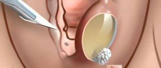
Bartholin's glands are large vestibular glands located at the back of the labia minora, right at the entrance to the vagina. They secrete mucus designed to moisturize the vagina. This protects the epithelium from abrasions and irritations.
Sometimes the Bartholin glands can become inflamed. And in case of obstruction and closure of the lumen of the gland, an abscess of the Bartholin gland develops. An abscess is characterized by very severe pain in the vulva, making it difficult to sit.
Pain in the labia does not go away with any painkillers. Relief will only come from cutting the abscess and evacuating the purulent contents by a gynecologist. To prevent the abscess from recurring, the edges of the wall of the open abscess are sutured into the surrounding mucous membrane.
In addition to pain in the labia area, the abscess is accompanied by other symptoms:
- swelling of the labia – usually one-sided, on the side of the abscess;
- redness of the labia;
- noticeable swelling in the area of the abscess.
After surgery you will need to take broad-spectrum antibiotics.