Gingivitis is inflammation of the gums. The gums are inflamed in the vast majority of the world's population. Therefore, it is a common joke among dentists that it is possible to diagnose any patient, even if he did not have time to enter the office. This diagnosis, as you may have guessed, is “gingivitis.” This article will discuss the classification, pathogenesis and causes of gingivitis.
Gingivitis is the earliest stage of gum inflammation. Over time, the inflammation becomes more severe, the connection between the gum and tooth is destroyed (epithelial attachment - number 3 in the figure), and “gingivitis” changes to “periodontitis”.
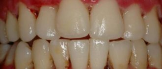
To easily identify pathology, you need to remember the norm. Let's remember what a normal, non-inflamed gum looks like.
Gums are normal
Normal gums have a characteristic appearance. It is convenient to describe it using several simple criteria:
| Sign | Norm |
| Color | Pale pink, coral, salmon (if we are talking about fair-skinned people) |
| Surface | Lumpy (“lemon peel”) |
| Circuit | Pointed or trapezoidal interdental papillae |
| Consistency | Dense |
| Bleeding | No |
A few words about each of the criteria:
Gum color
The color of the gums is noticeably different from the color of the mucous membrane of the alveolar process. The alveolar mucosa is more red, smooth and shiny. All due to the characteristics of its epithelium and connective tissue:
— the epithelium of the mucous membrane of the alveolar process is thin, non-keratinizing, there are no epithelial outgrowths;
- looser connective tissue contains more blood vessels.
(you can read more about the structure of the gums in the article “Structure of the periodontium”)
The black arrow indicates the gums, the white arrow indicates the mucous membrane of the alveolar process.
Gum surface
A lumpy surface is characteristic of the attached gingiva (the marginal gingiva is smooth). Microscopically, these are gingival depressions (arrows in the figure) and elevations due to the different arrangement of the connective tissue layer.
Gum contour
The contour, or shape of the edge, of the gum may be different. It can be more flat or, conversely, arched on the vestibular and lingual sides. This depends on the shape and inclination of the tooth crown: the more convex the crown, the greater its inclination, the more pronounced the curvature of the gums.
The gum contour is flatter in the upper central incisors, more curved in the canines.
The interdental papillae are also different in shape: the less space there is for them (the teeth are denser), the “sharper” they are; the more, the, on the contrary, they are wider:
Wide interdental papilla Pointed narrow interdental papilla
Gum consistency
Healthy gums are dense in consistency. This is all thanks to the abundance of strong collagen gingival fibers in its connective tissue.
Gingival fibers
Bleeding gums
There are also vessels there that are not pathologically changed. That is why there is no bleeding of the gums when brushing your teeth at home or probing them by a dentist.
| Gum probing |
Now let's figure out how the gums change when inflammation occurs - gingivitis. First of all, why does it arise?
Treatment with folk remedies
All “grandmother’s” advice for gingivitis is of an auxiliary nature. You cannot refuse traditional treatment, relying on “time-tested recipes.” This is not stated anywhere, but in ancient times, people, using exclusively natural raw materials, lost their teeth extremely early - by modern standards. It is worth taking advantage of the achievements of science to preserve a beautiful and healthy smile for many years.
As maintenance therapy, it is allowed to use rinses with tinctures and decoctions of herbs (excluding alcohol). Chamomile, St. John's wort, and eucalyptus are beneficial. A side effect of this treatment will be rapid darkening of tooth enamel. Therefore, special herbal remedies produced on an industrial scale will be a worthy alternative to home preparations.
Splinting teeth for periodontitis with fiberglass and tape
Laser gingivotomy - surgical treatment of complex forms of periodontitis
Catarrhal and hypertrophic gingivitis symptoms and treatment in adults
Treatment of generalized moderate and severe periodontitis
Open and closed curettage of periodontal pockets
The roots of the teeth are exposed - what to do and how to treat them
Causes of gingivitis
1) The main cause of gingivitis is microorganisms . “But there are always a lot of microbes in the oral cavity!” - you will say and you will be right. However, for gum inflammation to occur, more specific conditions are needed:
Condition once. So that these are certain microorganisms, here are these beauties:
- Streptococcus intermedius;
- Streptococcus sanguis;
- Actinomyces odontolythicus;
- Actinomyces naeslundii;
- Veilonella parvula.
Condition two. In the gingival sulcus:
| Gingival sulcus |
Both of these conditions are perfectly realized in dental plaque. It tends to accumulate in places that are difficult for a brush to reach (the gum area, for example) and creates an excellent environment in which there is little oxygen. For our microorganisms - facultative anaerobes - this is a resort.
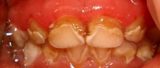
Colored plaque. The color is especially intense in the gingival area (where there is more plaque).
These microorganisms damage the gums by:
- The toxins they produce (exotoxins);
- Toxins that are released when bacteria die (endotoxins);
- Enzymes that destroy components of the connective tissue of the gums (hyaluronidase, collagenase);
- Products of their vital activity (amines, ammonia, organic acids).
There are other reasons why gum inflammation occurs. But these reasons are rather factors that accelerate the formation of plaque and the development of bacteria or help the plaque stay longer in the oral cavity. Their other effect is a decrease in immunity (both local and general).
2) Oral factors. In more detail, this factor could be:
- small amount of saliva, its high viscosity. Teeth are less easily cleared of plaque naturally. Local immunity suffers, because saliva contains important antibacterial substances (lysozyme, immunoglobulins).
- violation of the load on the periodontium aggravates inflammation.
Such a violation occurs with an incorrect bite.
The destruction or even absence of a tooth leads to the fact that the load falls on neighboring teeth. At the same time, chewing is also impaired. Due to pain or if there is, roughly speaking, nothing to chew on one side, it becomes one-sided. Then, on the chewing side, the teeth are overloaded, and on the opposite side they are not cleaned of plaque mechanically (with solid food).
Other reasons for increased load on the periodontium:
- overbite on the filling. As a result, so-called supercontacts arise in the area of this tooth and its antagonist;
- incorrect design of the prosthesis when the supporting teeth suffer;
- abrasion of teeth.
— factors that increase plaque retention, among them:
- malocclusion, when it is difficult to reach an incorrectly positioned tooth with a brush;
- carious cavities of class II, V (proximal, cervical areas);
- The fillings that exist after treatment of these cavities are of poor quality, poorly polished. A microbial plaque will linger better on a rough surface, and cleaning such a filling with a toothbrush will not give an ideal result.
- injury to periodontal tissue disrupts its blood supply and depletes its protective forces. Mechanical damage can occur as a result of:
- anomalies of attachment of soft tissues of the vestibule of the oral cavity. These are short frenulums of the lips and tongue, pronounced buccal cords, and a small vestibule (less than 5 mm).
A short wide frenulum of the lower lip injures the periodontium of tooth 41
- incorrect movements when brushing teeth, inaccurate cleaning of interdental spaces;
3) General factors reduce our resistance to gum attack by microorganisms.
These include immunity problems, general diseases (diabetes mellitus, hypothyroidism), and certain vulnerable periods in life (pregnancy, puberty). And the omnipresent stress that accompanies every step in the modern world.
We are what we eat. This is also true for gingivitis. On the one hand, if your diet is high in carbohydrates, plaque accumulates better. On the other hand, a lack of vitamins in food worsens the body’s protective functions. Vitamin C has a special role, because if there is not enough of it, the renewal of collagen fibers suffers. And this, for a minute, is as much as 60% of the composition of the connective tissue of the gums.
And also, importantly, bad habits. The most dangerous one is smoking. It harms not only general, but also local immunity in the oral cavity. The local inflammatory response, blood supply to the gums, and the composition of the gingival fluid are disrupted, and the number of bad, pathogenic microorganisms increases.
So, we figured out the reasons. Let's take a hypothetical patient who has one or more of these. Obviously, the development of inflammation in the gums is inevitable. How does it arise, and then develop and progress? Let's figure it out.
What happens if gingivitis is not treated?
Many forms of gingivitis do not seem dangerous at first glance, especially when the acute stage passes. A person gets used to mild pain and bleeding, so he does not go to the doctor. However, if gingivitis is not treated in time, it can lead to the following unpleasant consequences:
- Periodontitis is an inflammatory disease of the gums, in which teeth become loose and fall out, causing severe pain and bad breath.
- Frequent caries and throat diseases.
- The gums have receded to such an extent that the exposed roots of the teeth begin to cause severe discomfort.
- The spread of inflammation to the jaw bones and the body as a whole, a serious deterioration in overall health, in the worst case, even blood poisoning.
In addition, gingivitis is usually a sign of other unpleasant diseases or conditions, such as poor oral hygiene, which can cause serious dental problems.
All this means that at the first signs of gingivitis, you need to contact your dentist for treatment.
Pathogenesis of gingivitis
In the pathogenesis of gingivitis, there are several stages that transform into one another:
- initial stage;
- early stage;
- stage of stable damage;
- progressive stage.
The initial stage of gum inflammation develops in the first 2-4 days. Polymorphonuclear leukocytes begin to fight dental plaque microorganisms. Let me remind you that these include basophils, eosinophils and neutrophils. In our case, neutrophils predominate:
They penetrate from the blood vessels into the connective tissue of the gums, the epithelium of the gingival sulcus, and the attachment epithelium. They displace and compress the collagen fibers around the vessels, releasing collagenase, which destroys them. In response to the appearance of such a large number of immune cells, capillaries dilate and blood flow increases. And, as a result, more exudate and extravascular proteins are released from the gingival sulcus - the amount of gingival fluid increases.
This stage is also called subclinical gingivitis, because outwardly the gums look absolutely healthy. Pathological changes are visible at the microscopic level:
Neutrophils (NF) in blood vessels near the junctional epithelium (JE) and even between its cells. There are no neutrophils in the gingival crevicular epithelium (GED)
1 - healthy gums. Connective tissue (CT) contains fibroblasts, blood vessels, and dense collagen fibers. 2 – after a few days of plaque accumulation. Many immune cells in connective tissue, epithelium (CE). Dilated blood vessel (CV) in the center
The body's response determines how the initial stage will end. Recovery and return to a normal state or transition to the next stage - the stage of early damage.
The initial stage becomes early after about 1 week of plaque accumulation. Other immune cells - macrophages, lymphocytes - begin to migrate into the inflammation zone, forming an infiltrate. Around such infiltrates, as well as around vessels in the initial stage, collagen is destroyed:
The appearance of lymphocytes of small (ML) and large (BL) sizes. Light spaces around them are missing collagen
Moreover, its production by fibroblasts decreases. The worst conditions are for circular (4) and dentogingival fibers (1,2,3 in the figure below).
The connective epithelium and blood vessels grow: epithelial cords appear, between which there are capillary loops. Therefore, the color of the gums changes and erythema occurs. The first clinical sign also appears - bleeding gums.
Over time (according to various sources, from 2-3 weeks to even 1-2 months), the process enters the stage of stable damage (developed). This period can already be characterized as a chronic inflammatory process in the gums. Many immune cells of previous stages (neutrophils, macrophages, lymphocytes) are destroyed. From them there is only a trace in the form of expanded intercellular spaces, granular residues and lysosomes. They are replaced by others - B lymphocytes and plasma cells. The amount of collagen continues to decrease.
Plasma cell degeneration, cellular debris (debris)
Epithelium of the gingival sulcus with increased intercellular spaces and lymphocytes
The blood vessels change significantly: they are overloaded. As a result, their fragility increases, blood circulation in them slows down, and venous stagnation occurs. The color of the gums becomes darker and cyanotic. Plus, red blood cells leave the capillaries into the connective tissue, the hemoglobin in them breaks down, this enhances the staining.
Strands in the epithelial attachment can become very pronounced. So much so that, continuing to grow in the direction of the connective tissue of the gums, they can partially destroy the basal lamina (with the help of which the epithelium is attached to the connective tissue).
The stage of sustained damage can have several scenarios:
- With one of them, the process remains stable for several months or years.
- In the other case, it is more active and causes progressive destructive lesions.
- But the most favorable path is tissue restoration thanks to successful therapy. This happens due to the return of healthy periodontal microflora, which causes the inflammatory response of immune cells to disappear.
Some authors also distinguish stage 4 of gingivitis: progressive damage. But this is already periodontitis. This stage is left and considered, because gum inflammation with the involvement of other periodontal structures (ligament, bone) does not disappear anywhere. But this is a completely different story, so we’ll limit ourselves to three.
For clarity, we can summarize the results with a table:
| Stage | Time (days) | Blood vessels | Connective and sulcus epithelium | Predominant immune cells | Collagen | Clinical signs |
| Initial | 2-4 | Dilation, inflammation, increased permeability | Infiltration of polymorphonuclear leukocytes | PMN (mainly neutrophils) | Destruction around blood vessels | Increased amount of gingival fluid |
| Early | 4-7* | Proliferation | The same + epithelial strands, atrophy zones | Lymphocytes | Destruction around infiltrates | Erythema Bleeding (probe) |
| Persistent damage (developed) | 14-21* | Same + blood stagnation, fragility | Same + progression | Plasma cells | Progressive destruction | Changes in color, size, texture, surface, etc. |
* other authors note the duration of stage II is 1-3 weeks, stage III – 1-2 months.
I hope I was able to lift the veil of mystery about the occurrence and development of gingivitis. With this knowledge, it will be easier to understand where all the clinical signs, or symptoms, of gum inflammation come from.
The inflammation itself can occur and manifest itself in different ways. Due to these features, various forms of gingivitis are distinguished.
Forms of the disease
In dentistry, there are two main forms of the disease:
- acute catarrhal gingivitis. This is a pronounced one-time inflammation of the gums, which has a temporary limitation and can develop into a more serious form;
- chronic inflammation. It develops when the acute stage is ignored and self-medication; it is a low-grade inflammation. It recurs periodically and requires repeated treatment.
In terms of prevalence, gingivitis can be localized, when 1-3 gums become inflamed, and generalized, in which the entire tissue of the dentition or both jaws is affected.
Diagnostics
The differential diagnosis of catarrhal gingivitis makes it possible to distinguish it from periodontitis and prescribe appropriate treatment. Diagnostics involves examination by a dentist and laboratory tests to determine the degree of development of inflammatory processes, the amount of microbial plaque and the quality of hygiene procedures performed by the patient. During the examination, the dentist pays attention to the degree of hyperemia, the color of the gums, the amount of plaque and tartar.