Pemphigus is a very serious dermatological disease that can lead to serious consequences and complications. The development of pemphigus in newborns is especially dangerous; in this case, even death cannot be ruled out.
People of all ages are susceptible to this disease, however, in most cases it is observed in adults 40-60 years old. Children suffer from this disease extremely rarely. Since the disease is an autoimmune pathology, it is difficult to treat and is chronic.
The disease is expressed by the appearance of blisters filled with exudate on the skin and mucous membrane. New growths merge with each other and quickly spread throughout the body.
Causes of herpangina
Herpangina can be caused by about 70 serotypes of enteroviruses. Most often these are Coxsackie B, Coxsackie A17 viruses and enterovirus 711.
Since the only carrier of enteroviruses is humans, you can become infected through contact with a sick person or with a virus carrier who has no symptoms of the disease1. According to the literature, the number of virus carriers can be up to 46% of people2.
The virus is released into the external environment with feces and droplets of saliva. It is also contained in bubbles that appear in the patient’s throat. Enterovirus infections most often affect children, although the disease also occurs in adults5.
The patient or virus carrier excretes viruses from the upper respiratory tract within 3 weeks after infection, and with feces - up to 8 weeks. In the first two weeks, herpetic sore throat is most contagious1.
You can become infected in the following ways:
- through dirty hands, objects and food if they are exposed to the virus;
- drinking contaminated water from a reservoir;
- upon contact with a patient or virus carrier.
The herpangina virus is also transmitted transplacentally - from mother to fetus3.
Up to contents
Types of pemphigus
First of all, the pathological process of the disease is classified into:
- true pemphigus (acantholic);
- benign pemphigus (neacantholic).
True pemphigus, depending on the manifestation of the pathological process, is classified into the following subtypes:
- Vulgar (ordinary) pemphigus . The most common type. It is characterized by the appearance of blisters on the skin without signs of inflammation. If there is no proper treatment, there is a rapid spread of bubbles throughout the body. When the disease is advanced, death often occurs.
- Erythematous, also known as seborrheic pemphigus. Difficult to treat, characterized by the appearance of red spots with dense crusts.
- Leaf-shaped . Pemphigus is characterized by the appearance of flat blisters and many crusts overlapping each other, which create the effect of leaves (photo on the right).
- Brazilian . A similar pathology is common in Brazil. As a rule, all family members suffer. Both children and elderly people are susceptible.
Symptoms of herpetic sore throat
The disease begins acutely. From the moment of infection to the first symptoms, it takes from 2 to 14 days3. The temperature rises to 38-39°C. The patient feels weakness, headache, chills, less often nausea, possible vomiting and enlargement of the submandibular lymph nodes, 1,2,3.
Herpangina goes through several stages2:
- The day before the rash appears in the throat, the patient feels a mild pain. On examination, you may notice redness of the palatine arches and the back wall of the pharynx.
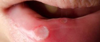
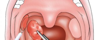
- Then, rashes appear on the mucous membrane of the soft palate, palatine arches, tonsils and uvula - small papules (nodules) up to 5 mm in diameter with a red rim.
- The nodules turn into vesicles, which open after 1-2 days.
- In their place, painful erosions with a gray-white coating form.
Up to contents
Diagnosis and severity of the disease
Since at the initial stage of the development of the disease the symptoms are similar to many other dermatological diseases, to accurately determine the diagnosis it is necessary to conduct a comprehensive examination, namely:
- examination of the patient - the nature of the lesion and location;
- studying the patient’s medical history and the presence of concomitant diseases;
- performing Nikolsky's test, which will distinguish pemphigus from similar pathological processes;
- carrying out histological and cytological studies;
- the use of immunological methods that confirm or refute the autoimmune nature of the disease.
As the blisters develop and spread, the disease is characterized by varying degrees of severity:
- Easy . The pathological process appears on the mucous membranes or skin gradually with a minimum number of foci.
- Average . With a moderate course of the disease, an increase in the spread of blisters on the skin and mucous membrane is observed.
- Heavy . Most of the skin and oral cavity are affected. The ulcers merge into large lesions. Complications and pathologies begin.
Herpetic sore throat in children
Children usually become infected at school or kindergartens2,3. Due to pain and fever, they are restless, tearful, and often refuse to eat and drink because food irritates erosions on the mucous membrane and causes discomfort. But due to refusal to drink water or juices, children often develop dehydration. At the same time, the child’s tongue becomes dry, and the elasticity of the skin decreases1. Convulsions may occur due to high temperature1.
Blistering rashes in children can appear not only on the mucous membrane of the throat, but also on the hands and feet, and even on the buttocks and forearms. This manifestation of enterovirus infection is called viral pemphigus of the oral cavity and extremities or mouth-hand-foot syndrome. The disease is contagious in 100% of cases, is often mild, and can affect nails3.
Up to contents
Pathohistology of pemphigus
At the onset of the disease, intercellular edema of the epithelial layer and destruction of intercellular bridges in the lower parts of the germ layer appear. As a result of the loss of communication between epidermocytes, cracks are formed first, and then blisters , predominantly of suprabasal localization. The basal cells, although they lose contact with each other, remain attached to the basement membrane. The cavity of the bladder, as a rule, contains round acantholytic cells with large hyperchromatic nuclei and pale cytoplasm. During the healing process, proliferation of the papillae and elongation of the epithelial outgrowths are noted. The infiltrate consists of eosinophilic granulocytes, plasma cells and lymphocytes.
In other words, the basis of the pathological process in pemphigus is acantholysis - a disruption of the connection between the cells of the spinous layer as a result of disruption of the desmosome - tonofilament complex, resulting in the formation of acantholytic cells, which are characterized by a large nucleus. The cytoplasm of the cells is strongly basophilic and pyroninophilic, which is due to the high content of RNA in it. In acantholytic cells there is a high level of protein metabolism and oxidative processes (exceeding the metabolism of spinous cells), which indicates their secretory activity. It is assumed that acantholytic cells originate from the basal and lower rows of spinous cells.
Course of herpetic sore throat
The diagnosis of herpetic sore throat can be made by an otolaryngologist, therapist or pediatrician after examining the patient and clarifying his complaints. To monitor changes characteristic of a viral infection, the doctor may prescribe a general blood test, and to confirm enteroviral sore throat, a specialist may prescribe a pharyngeal smear and a blood test for specific antibodies. The pathogen can also be detected in stool or inflammatory fluid that is released from vesicles1,4.
Manifestations of herpetic sore throat can go away on their own in less than 10 days. But in any case, at the first symptoms of the disease, you should definitely consult a doctor. You cannot self-medicate2,3.
In some cases, herpetic sore throat can cause complications from the nervous system. In this case, 1 appears:
- severe spasm of the neck muscles, due to which the child cannot bend his head;
- weakness of the muscles of the limbs;
- disturbance of consciousness.
A severe complication of herpetic sore throat is damage to the soft membranes of the brain, brain and spinal cord1,3.
Newborns are at highest risk of developing complications, so they need careful treatment and care3. It is important to maintain hydration and give your child enough fluids1.
Up to contents
Causes of the disease
Specific causes of the occurrence of vesicles have not been identified, but provoking factors are identified that influence the development of the disease, namely:
- hereditary predisposition;
- viral infections;
- harmful working conditions;
- allergic reaction to certain types of products, metals, chemicals;
- disruption of metabolic processes in the body;
- endocrine diseases;
- long-term use of certain types of medications.
An autoimmune cause of the disease is also being considered. In children, the cause of the disease can be viruses and bacteria, the main one being Staphylococcus aureus.
You can become infected through airborne droplets or by using the patient’s belongings.
Treatment of herpetic sore throat
Patients with complications require hospitalization in an infectious diseases hospital and treatment under the supervision of specialized specialists - a neurologist and a cardiologist. If the doctor has recommended treatment at home, it is necessary to closely monitor the patient's condition2.
The sick person should be isolated and stay in a clean, well-ventilated area so as not to infect other family members. Quarantine must be observed until symptoms subside1.
For herpangina you should 1,3,4:
- Wash your hands as often as possible, including after feeding and changing a sick child’s diaper.
- Disinfect surfaces and objects with which the patient has been in contact.
- Drink enough fluids to avoid dehydration. At the same time, pay attention to the temperature of the drink: hot, warm drinks irritate the mucous membranes and cause additional discomfort. You can drink cool drinks.
- Consume food in liquid or mushy form. Spicy, salty, sour foods, including fresh fruits even in the form of puree, are not suitable for a patient with herpetic sore throat.
- Rinse your mouth with a saline solution after every meal to maintain oral hygiene and prevent bacterial infections from erosions.
- Use a soft toothbrush to reduce trauma to the mucous membrane.
Currently, there is no proven antiviral drug to treat herpangina by acting on its causative agent. Sometimes a doctor may prescribe medications that support local immunity of the pharyngeal mucosa1. Antibiotics are not prescribed for herpangina6.
The goal of treatment for herpangina is to relieve the symptoms of the disease4.
If the body temperature is above 38.5°C, physical methods such as cold compresses and ice packs may be used. Your doctor may also recommend anti-inflammatory and antipyretic medications1. Local treatment includes agents with anti-inflammatory, analgesic, enveloping and antiseptic effects1.
For the symptomatic treatment of herpetic sore throat, the doctor may prescribe the drugs HEXORAL®7,8,9,10,11. It is convenient to use HEXORAL® spray to irrigate the pharyngeal mucosa. The active substance of the spray is hexethidine. It acts against the main bacteria found in the oral cavity and pharyngeal mucosa8. The drug is also active against some viruses and fungi of the genus Candida8. Thanks to the local anesthetic effect of hexethidine, HEXORAL ® spray helps reduce pain8. HEXORAL®7 solution is suitable for rinsing. The use of HEXORAL ® spray and solution is allowed in children over 3 years of age7,8.
If herpangina causes severe pain and discomfort, adolescents over 12 years of age and adults can benefit from HEXORAL ® TABS EXTRA lozenges, which contain the anesthetic lidocaine10. For children over 4 years of age, HEXORAL ® TABS lozenges may be suitable. The anesthetic benzocaine in their composition helps reduce pain in the throat and mouth9.
All medications for herpetic sore throat should be used only after consultation with a doctor. In case of severe erosions, HEXORAL ® solution and spray are contraindicated7,8, and lozenges can only be prescribed by a specialist after examining the pharynx9,10.
Up to contents
The information in this article is for reference only and does not replace professional advice from a doctor. To make a diagnosis and prescribe treatment, consult a qualified specialist.
Clinic for pemphigus
Of the 4 forms of acantholic pemphigus, of particular interest to dentists is pemphigus vulgaris , which affects the oral cavity most often (75% of patients with true pemphigus).
Pemphigus vulgaris is a severe bullous dermatosis that affects people aged 40-60 years, mainly women. The course of the disease is most often chronic or subacute, rarely acute.
The mucous membrane of the oral cavity with pemphigus vulgaris is affected in the majority of patients (about 70%), and these lesions for a long time, several months and even years, may be the only symptoms of the disease.
In the oral cavity, the process proceeds differently than on the skin, which is explained by the anatomical features of the oral epithelium.
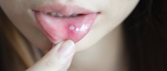
But in the oral cavity, pemphigus vulgaris occurs in different ways. In one case, initially the epithelium at the site of the lesion becomes cloudy, erosion occurs in the center of the lesion, quickly spreading to the periphery. If you run a swab over such clouded epithelium, the top layer is easily removed, exposing the erosive surface. Pemphigus erosions come in various sizes - from small abrasions to large bluish-red surfaces, often they are “bare”, without plaque, or covered with a fairly easily removable fibrinous coating. Rashes appear on the unchanged mucous membrane.
In other cases, damage to the mucous membrane in the form of blisters of various locations is often caused by an isomorphic reaction as a result of irritation in the oral cavity by microtraumas. Bubbles ranging in size from 1-2 to 30-40 mm (from lentil grains to pigeon eggs) initially have transparent contents, later they can take on a yellowish and cloudy tint. The covering of the blisters is initially tense, then becomes flabby, the opening of the bubble occurs as a result of rupture of the covering or resorption of the exudate (crusts form on the skin when the blisters dry out, and dense deposits form on the mucous membrane).
In the oral cavity, lesions are located throughout the mucous membrane, especially in easily injured folds. The mucous membranes of the nose, eyes, genitals, larynx, pharynx, and esophagus may also be affected.
On the skin, blisters are localized on the back, chest, arms, legs, neck, and less often on the face.
The first phase of the disease is characterized by the appearance of single or multiple small blisters on the mucous membrane, not accompanied by pronounced general reactive phenomena. When they are opened, erosions form. Nikolsky's symptom is not always positive .
The second phase is characterized by the development of large blisters, which, when opened, leave erosions. They often merge to form a bright red erosive surface. Erosions bleed easily when eating or touching with an instrument.
The tongue is swollen and there are tooth marks. Nikolsky's symptom is positive . Hypersalivation and severe pain in erosions are noted. When localized on the mucous membrane and red border of the lips, erosions quickly become covered with yellowish-brown or bloody crusts. There is an unpleasant odor from the mouth. Severe general condition has sometimes previously led to cachexia and death.
The third phase - predominant epithelization - is characterized by the subsidence of acute phenomena. Erosion heals, new blisters form less frequently. Nikolsky's symptom in the lesions is difficult to determine. Subjective sensations are accompanied by a slight burning sensation, or tingling, or paresthesia, which are often precursors of the disease.
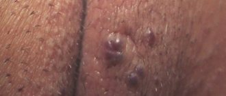
Pemphigus vegetans is much less common. The initial sign is often damage to the oral mucosa, localized on the cheeks, tongue, palate and corners of the mouth. The formation of bright red soft vegetation against the background of an erosive surface after the opening of the blisters is characteristic. Usually, the process also includes areas of the skin adjacent to the oral cavity, where the vegetations are covered with loose, dirty-brown crusts. Painful bleeding cracks form in the corners of the mouth. Blisters appear on normal or slightly hyperemic skin. They are smaller than in pemphigus vulgaris, have a thinner covering, and are located more superficially in the epidermis. The predominant localization is in the armpits, navel, inguinal folds, genitals and anus (in areas of maceration). After epithelialization of the rash, dark brown pigmentation remains. Nikolsky's symptom is positive .
Pemphigus foliaceus is rare and has a sudden onset with the patient generally feeling well. Blisters appear on the skin of the scalp and torso and can be localized for a long time, but rapid generalization of the process with damage to other areas of the skin is also possible. The blisters are located in the superficial layers of the epithelium, so when the blisters are opened, the exudate shrinks into thin crusts, reminiscent of sheets of puff pastry. Typically, the blisters merge with each other and, when opened, form large erosive surfaces similar to burns. Nikolsky's symptom is positive . As the process generalizes, the general condition of the patients worsens: weakness occurs and the temperature rises. The oral mucosa is rarely involved in the process.
Seborrheic pemphigus is a fairly rare disease. Erythematous lesions against the background of oily seborrhea form thin yellowish crusts that are easily rejected without the subsequent formation of atrophic areas. When the process spreads to the skin, diffuse hyperemia and peeling are noted, reminiscent of seborrheic eczema with symptoms of impetiginization. Cases of primary localization of blisters on the oral mucosa have been described.
Non-acantholytic pemphigus (benign) is characterized by the formation of subepithelial blisters, without acantholytic cells, Nikolsky's sign is negative . The prognosis of the disease is favorable if there are no secondary aggravating complications.
Non-acantholytic pemphigus itself is observed in older people and has a chronic course. The onset is characterized by the appearance of tense blisters on the mucous membrane of the oral cavity, and less often on the lips. Bubbles develop on a hyperimproved or unchanged base and may not open for a long time. When the blisters are opened, erosions heal without the formation of scars or atrophic areas.
Mucosynechial atrophying bullous dermatitis ( pemphigus of the eye ) is benign and affects the mucous membrane of the eyes or oral cavity with the formation of scars. The first blisters may appear on the oral mucosa. Unlike ordinary pemphigus, erosions do not have a fringe of exfoliating epithelium along the periphery and are not prone to peripheral growth. They do not bleed and are not painful. Chronic rhinitis, damage to the esophagus, adhesions or atrophy of the mucous membrane of the external genitalia are often observed. Blisters occur on the skin in about a third of patients.
Benign non-acantholytic pemphigus of the oral cavity only is not accompanied by a violation of the general condition of the patient’s body. Small tense blisters with transparent or hemorrhagic contents appear on the mucous membrane, when opened, low-painful and quickly epithelial erosions are formed. Nikolsky's symptom is negative . During the period of epithelization of erosions, there are no scars, adhesions, or atrophies. Bubbles usually form in fixed areas.
The severity of the process in pemphigus is determined by the appearance of blisters and erosions. In favorable cases, which happens infrequently, erosions epithelialize after 3-6 weeks, but new rashes appear to replace them or even during their existence. Sometimes, with this course of the disease, spontaneous remission occurs, which can last weeks or months. Usually, without adequate therapy, epithelization of erosions does not occur, and the disease progresses.
If the process began on the mucous membrane of the oral cavity, then in the absence of appropriate therapy, after 1-6 months, rashes begin to appear on the skin of the torso and limbs, and the patient’s general condition deteriorates sharply. In the absence of corticosteroid therapy, intoxication increases, cachexia develops, and, 1-2 years after the onset of the disease, patients die.
Sources
- Corsino CB, Ali R, Linklater DR. Herpangina. 2021 Jun 23. In: StatPearls [Internet]. Treasure Island (FL): StatPearls Publishing; 2020 Jan–. PMID: 29939569. https://www.ncbi.nlm.nih.gov/books/NBK507792/
- Ter-Baghdasaryan L.V., Ratnikova L.I., Stenko E.A. Clinical and epidemiological aspects of enterovirus infection // Infectious diseases: news, opinions, training. 2021. Vol. 9, No. 1. pp. 88-93. doi: 10.33029/2305-3496-2020-9-1-88-93 https://infect-dis-journal.ru/ru/jarticles_infection/672.html?SSr=2601343bdb01ffffffff27c__07e4040b011a36-9772
- Alacheva Z. A., Rybalka O. B., Kulichenko T. V. Should everyone escape from Coxsackie?! Or fear has big eyes. Issues of modern pediatrics. 2017; 16 (4): 286–290. doi: 10.15690/vsp.v16i4.1774) https://vsp.spr-journal.ru/jour/article/viewFile/1787/713
- Herpangina Brenda L. Tesini. University of Rochester School of Medicine and Dentistry // MSD Handbook - 2019 https://www.msdmanuals.com/ru/professional/infectious-diseases/enteroviruses/herpangina
- Kozlovskaya O.V., Katanakhova L.L., Kamka N.N., Evseeva A.N. Epidemiological, clinical and diagnostic features of enterovirus infection in children and adults. Bulletin of Surgu State University. Medicine. 2018;(2):56-60. https://surgumed.elpub.ru/jour/article/view/140/141
- Kuo KC, Yeh YC, Huang YH, Chen IL, Lee CH. Understanding physician antibiotic prescribing behavior for children with enterovirus infection. PLoS One. 2021 Sep 7;13(9):e0202316. doi: 10.1371/journal.pone.0202316. PMID: 30192893; PMCID: PMC6128467. https://pubmed.ncbi.nlm.nih.gov/30192893/
- Instructions for use of the drug HEXORAL® SOLUTION:
- Instructions for use of the drug HEXORAL® AEROSOL:
- Instructions for use of the drug HEXORAL® TABS:
- Instructions for use of the drug HEXORAL® TABS EXTRA:
Up to contents
Diagnosis of pemphigus
An important diagnostic sign of pemphigus vulgaris is a positive Nikolsky sign . There are 3 methods. One Nikolsky symptom does not confirm the diagnosis of pemphigus; it is also observed in medicinal stomatitis. In this regard, the diagnosis of pemphigus must be confirmed by cytological studies - detection of Tzanck cells. Acantholytic cells are round and smaller than normal cells of the spinous layer, the nucleus is large relative to the entire cell, its diameter is 1/3-1/2 or more than the diameter of the cell, the nucleus is loose with 1-6 lighter nucleoli; the cytoplasm is two-layered - light (perinuclear zone) and dark blue (peripheral).
With pemphigus vegetans, the cytological picture is identical to that with pemphigus vulgaris. However, in seborrheic and pemphigus foliaceus, multinucleated cells are not found, acantholytic cells are found in smaller numbers, are monomorphic, and often have an oval and irregularly triangular shape.
You should also use the immunofluorescent method, which allows you to detect in the blood serum of patients with pemphigus antibodies to the intercellular substance of the spinous layer of the epidermis (with indirect RIF) of the IgG type and IgG deposits in the area of the intercellular substance and the membrane of the cells of the spinous layer (with direct RIF).
Differential diagnosis of pemphigus and diseases that make up the group of blistering dermatoses is based on symptoms associated with the localization of blisters in relation to the epithelium. With pemphigoid, the blisters are located subepithelially, so they have a thicker cover than with pemphigus and last a longer time, and therefore, when examining the oral mucosa in such patients, blisters with transparent contents are observed, which is impossible with pemphigus.
Erosions formed at the site of blisters with pemphigoid are usually located on a slightly hyperemic base, their surface is often covered with fibrinous plaque.
With pemphigus vulgaris, the oral mucosa around the erosions is not externally changed, and the erosions themselves can be covered with a collapsed bladder cover, which is very easily removed with a spatula. Acantholytic cells and a positive Nikolsky sign distinguish pemphigus vulgaris from non-acantholytic pemphigus.
For pemphigus, RIF allows one to determine deposits of immune complexes containing IgG in the area of the membranes of spinous cells and the cementing substance between them. With indirect RIF, circulating IgG is determined.
In non-acantholytic pemphigus, these same immune complexes are also found in the basement membrane area.
A blistering rash as a manifestation of an allergy to drugs helps to distinguish between anamnesis (medication intake) and relatively rapid healing after discontinuation of the causative drug.
The blisters are located subepithelial. There is no acantholysis, Nikolsky's sign is negative, acantholytic cells are absent. Allergy tests clarify the diagnosis. Blisters in the bullous form of lichen planus also occur under the epithelium, but there is no acantholysis. Around the bladder or in other areas of the mucous membrane, typical papules of lichen planus can be observed.