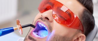
Radiovisiographic examination is a method of computer radiographic examination in dentistry. Its goal is to obtain an image of the tooth and tissues around it with minimal radiation exposure. It has proven itself well in diagnosing hidden carious processes, inflammation of the pulp or periodontium, and identifying foreign objects in the tooth canal. Thanks to him, the dentist has the opportunity to control the quality and effectiveness of the treatment. During the procedure, a detector is inserted into the oral cavity, which is aimed at the area under study: its image is displayed on the monitor.
You can undergo radiovisiography in Moscow at the dental department of CELT. Our clinic has been operating in the capital's paid medical services market for almost three decades. We have a modern radiovisiograph, which allows us to accurately and quickly make diagnoses, identifying diseases in the early stages of their development. Our surgeons, orthodontists, therapists, and orthopedists have decades of experience in scientific and practical work. They constantly improve their professional knowledge and skills and use effective and gentle treatment methods. You can find out the price of the study by going to the “Services and Prices” tab. To avoid misunderstandings, check the numbers with the information line operators or at your doctor’s appointment.
Consultation with a dentist-therapist - 1,000 rubles.
Radiovisiographic examination - 370 rubles.
At CELT you can get advice from a dental specialist.
- The cost of a consultation with a dentist-therapist is 1,000
Make an appointment
Radiovisiography in dentistry
Without radiovisiographic examination, diagnostics in dentistry would be impossible. This is computer radiography, which replaced the outdated film radiography. Thanks to it, you can get a clear image in just a few seconds, while the patient’s radiation exposure will be minimal - ten times less than when using a film device.
A digital visiograph is equipment that should be in any modern dental clinic, since today it is the only method that fully complies with international standards. Its operation is based on the same principle as a standard device, but the signal is sent not to the film, but to the CCD sensor (or, as it is also called, the matrix). The latter has greater light sensitivity than conventional film, so minimal exposure to radiation is sufficient to obtain a clear image.
The visiograph sensor is connected to a converter, which reads its signals, transforming them into a digital image. The latter is sent to the monitor where the doctor can see it. The digital format also has many advantages: it does not take up much space on your hard drive, where it can be stored for a long time, it can be transferred to a USB drive or sent by email.
Advantages and disadvantages of a visiograph
The main advantages of radiovisiography:
- compactness of the device;
- image accuracy;
- the ability to instantly view a diseased tooth on a computer screen with multiple magnification of the resulting image, storing the image on a PC and transmitting it via a local network and e-mail;
- safety of the procedure;
- speed of manipulation;
- editing a digital visiogram, using various filters to improve the visual perception of the picture.
Radiovisiography of the tooth is carried out in a fairly low spatial resolution. This is the main disadvantage of the procedure. The device's sensor does not clearly show the difference between different structures with unequal densities. But thanks to the ability to adjust the contrast and brightness of the image, details become more visible.
Despite the fact that doctors do not prohibit performing the procedure during pregnancy, the expectant mother should warn the attending physician about her situation.
Indications and contraindications for dental radiovisiography
The study can be used for targeted study of dental units, as well as for identifying hidden carious processes and inflammation of surrounding structures.
Indications
- Diagnosis of caries, inflammatory processes of pulp and periodontium;
- Monitoring their treatment (quality of endodontic treatment, in particular, filling of canals, correct placement of pins, tightness of seal to the walls of the tooth);
- Preparation for surgery to increase bone tissue to determine the degree of its atrophy and the characteristics of the process;
- Suspicions of neoplasms in the thickness of the soft tissues of the oral cavity;
- Cystic formations and granulomas at the roots of teeth;
- Determining the location of fragments of broken dental instruments in the root canal;
- Traumatic damage to a tooth (dislocation, bruise, fracture);
- Control of the implant after installation;
- Before sealing the fissures of teeth in order to ensure the absence of caries.
Contraindications
Absolute contraindications:
- The patient has serious mental disorders;
- Pathologies of a neurological nature;
- Depressive states.
Due to the minimal level of radiation, radiovisiography of the maxillofacial area can be performed on a child, provided that he can sit still for several seconds.
If indicated, the study can be performed on women during pregnancy and lactation. It is optimal to do this in the second trimester, after warning the diagnostician about pregnancy. This is necessary so that she is put on a special apron, which provides additional protection from x-rays.
What does a radiovisiographic image reveal?
A digital radiovisiograph allows you to identify any foci of infection, the location of which is difficult to determine visually. A visiogram is done in case of possible occurrence of the following diseases:
- Caries;
- Pulpitis, which causes inflammation of the tissues surrounding the tooth;
- Small neoplasms in the oral cavity;
- Periodontitis.
The image allows you to detect hidden foreign bodies that have entered the canal. Any foci of inflammation, granulomas and cysts are painted black, which stands out sharply against the gray background of bone and connective tissue. With the help of detailed visual images, the dentist will be able to make an accurate diagnosis and prescribe appropriate treatment.
Radiovisiography allows you to determine the depth of tissue damage and control the quality of filling.
Advantages of radiovisiographic examination
This research method is considered the best option both for a dental office and for a large clinic in which several specialists work at once. Its advantages:
- Efficiency and the ability to receive an image instantly, without waiting;
- High clarity and information content of the image, in which you can see the condition of the teeth, surrounding tissues, and implants;
- Minimum number of restrictions (possibility of use for children and pregnant patients);
- Convenient digital format, the ability to print the image on paper.
Do not forget that visiography is an environmentally friendly and harmless research method. It does not require the use of film or aggressive chemicals for development. The radiation dose received during filming of the lower jaw is 2 microsieverts, the upper – 5 microsieverts. For comparison, when using a film apparatus, it is equal to 30 and 80 microsieverts, respectively.
The resulting images clearly show:
- Carious lesions, cystic formations, granulomas and inflammatory processes. They are transmitted in black;
- Fillings, inlays, pins, dentures – white areas;
- Connective and bone tissue are gray areas.
Features of the orthopantomograph
Dental orthopantomograph is equipment for creating high-resolution panoramic images. Devices of various types have an overview imaging system, which makes it possible to visualize the entire jaw, the patient’s teeth, and jaw joints. The principle of operation is that the X-ray emitter and sensor in digital modifications rotate around the head, forming a clear three-dimensional image of the jaws.
Digital orthopantomographs make shooting as safe as possible for humans - the radiation level is 2-3 times less than with film units. Innovative technology does not require laboratories to develop images; the picture will be ready quickly and visualized on the computer screen without waiting for results.
If the orthopantomograph has a cephalostat, the doctor can take a frontal profile photograph of the skull, which is needed to build orthodontic structures.
Innovative technology works with different shooting programs:
- Standard overview.
- Separately upper and lower jaw.
- Photo of the maxillary sinuses.
- Children's photography.
- Image enlargement mode.
- Other programs for different clinical cases.
Safety, speed, high-resolution image quality, and information content are the characteristics of equipment used by clinics all over the world.
Reviews about our dental therapists
I am very grateful to Evgeniy Borisovich Antiukhin for removing my three eights.
Especially considering that the lower tooth was not the simplest (it was located in an embrace with a nerve). The removal took place in 2 stages, one tooth under local anesthesia, two under general anesthesia. I had no idea that wisdom teeth could be... Read full review Sofia
28.12.2020
Words cannot express my gratitude to Elena Nikolaevna Kiseleva. This is the best doctor in the world. I got an appointment after many years of being ignored by the dentist’s office and with a bitter experience of treatment in another paid clinic, the mistakes of which had to be corrected in the first visits. Thank you for this... Read full review
Roman Stanislavovich Sh
25.07.2020
Analysis of targeted dental images –
Analyzing X-rays is not difficult if they are of good quality.
Almost any patient will be able to see signs of periodontitis or cysts in the image, and will also be able to determine how well his root canals are filled. All you need here is skill. But it is worth remembering that absolutely everything cannot be diagnosed using X-rays, for example, inflammation of the nerve of a tooth. You can diagnose from the image -
- caries (but not in full),
- periodontitis (granulomas, cysts),
- quality of root canal filling after treatment of pulpitis and periodontitis,
- perforations and root cracks,
- fragments of instruments in root canals,
- presence of periodontal pockets...
1) A group of images indicating the development of inflammation (periodontitis) at the apexes of the roots of previously untreated teeth. In this case, you will always see a clear or vague darkening at the apex of the tooth root, which can be of different sizes and shapes.
2) A group of photographs taken after root canal filling. The first 2 pictures (Fig. 14-15) show what well-filled root canals look like. The following images show poor quality treatment and complications that arose (read the descriptions in each image).
How is dental radiovisiography performed?
The procedure is very simple and absolutely painless and does not require preparation. The patient is asked to take a sitting position on a chair near the device, after which a sensor is applied to the area under study and the patient is asked to gently bite the sensor with his teeth. This prevents its displacement. After this, the tube of the device is directed to a certain area of the jaw. The diagnostician starts it, and X-rays begin to illuminate the tissue. The signals are captured by the sensor, then sent to the converter and (already in the form of a graphic image) to the monitor.
What is a visiograph?
A visiograph is a part of a visiographic complex, working in tandem with an X-ray unit. It consists of three main elements:
- Sensor: a silicone chip that detects and transmits incoming signals to the device.
- Analog-to-digital converter: a monoblock that processes information and displays it on the screen.
- Connection cord: connects the sensor and the ADC.
The patient is seated next to the visiograph, then the doctor, directing the X-ray tube, leans the sensor against the tooth being examined. All you have to do is turn on the device and take a picture - it only takes a few moments.
Difference between radiovisiography, OPTG and computed tomography
Unlike visiography, OPTG - orthopantogram (or, as it is also called, panoramic image) covers not one or three teeth, but the entire dental system. An orthopantomograph is used to obtain images. Most often, such diagnostics are prescribed to young patients in order to assess the condition of the rudiments of permanent teeth. As for computed tomography, it provides not a two-dimensional, but a three-dimensional image of the patients’ jaw system. For this reason, it is carried out before implantation and bone augmentation operations that precede it, in order to plan them in detail.
You can make an appointment with dentists and undergo radiovisiography at CELT online or by contacting our information line operators: +7 (495) 788‑33‑88.
Make an appointment through the application or by calling +7 +7 We work every day:
- Monday—Friday: 8.00—20.00
- Saturday: 8.00–18.00
- Sunday is a day off
The nearest metro and MCC stations to the clinic:
- Highway of Enthusiasts or Perovo
- Partisan
- Enthusiast Highway
Driving directions
Is it possible to take a dental x-ray during pregnancy?
Is it possible to take a photograph of a tooth during pregnancy? This issue is regulated by the recommendations of SanPiN 2.6.1.1192-03. These recommendations do not prohibit taking dental x-rays during pregnancy, but it is strongly recommended to use x-rays only in truly necessary cases, for example, in case of acute pain and the provision of appropriate emergency care.
It should be noted that over the past 20 years, the radiation doses received by patients with 1 x-ray of the teeth have become tens of times less, thanks to the advent of radiovisiographs and ultra-sensitive photographic films, which require significantly lower x-ray radiation (24stoma.ru). Therefore, the risks of fetal pathologies have decreased significantly in recent years.
Of course, if possible, X-ray examination of teeth should be avoided, but there is nothing terrible about this today, because The radiation dose of 1 image on a radiovisiograph is approximately equal to the radiation dose of any person to natural background radiation in 1 day. In addition, the irradiation time on the visiograph will be only 0.05-0.3 seconds, which, if protective measures are observed (lead apron), can significantly increase the safety of the procedure.
Additionally, it is worth considering the following recommendations –
It is better not to do a dental x-ray during pregnancy in the early stages, because at this most important time for the laying of organs and tissues of the fetus. And if x-rays are taken, then it is in the second half of pregnancy, because the risks to the fetus during this period are significantly reduced. Please note that you can only take pictures using a modern digital radiovisiograph of the latest generation, because Their radiation dosages are significantly lower than those of outdated digital radiovisiographs and, even more so, film devices.