Almost every person on earth has experienced toothache at least once. It can be very difficult to tolerate, and sometimes simply unbearable. To avoid complications, one of which may be periodontitis, you should immediately consult a doctor.
Periodontitis is an inflammatory process of the apex of the tooth root, as well as in the tissues adjacent to it. This is a rather dangerous disease that can lead to even more serious problems, so you should not delay treatment. Periodontitis can occur as a result of advanced pulpitis or poor-quality canal filling.
Also, the causes of this disease can be an infection, injury, or the consequences of medication. According to statistics, this disease accounts for 45-50% of all dental diseases. People with weakened immune systems are most susceptible to rapid development of the disease.
- The occurrence of severe pain that is constant;
- The appearance of painful sensations when chewing or touching;
- A clear feeling of which tooth hurts, with a feeling of bursting from the inside;
- Discharge of a small amount of fluid from under the tooth, accumulation of pus in the gums;
- The pain may be throbbing and radiate to the temporal or ear region;
- Bad breath;
- Visible swelling and redness of the gums.
General characteristics of the disease
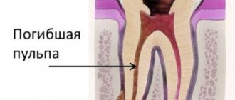
Pulpitis is an inflammation of the dental nerve (pulp), which is located in the dental cavity and root canals.
The pulp is extremely sensitive, and if a tooth is deeply damaged by caries, there is a crack in it, or a filling has fallen out of it, then the likelihood of developing pulpitis is quite high. Inflammation can also occur during treatment or preparation for dental surgery if the doctor is not careful enough. In rare cases, a previous infectious disease becomes a provoking factor. Pain can occur when the tooth is exposed to cold or heat, sour foods, alcohol, or sugar. If you see a doctor right away, the inflammation can be suppressed. However, patients often try to relieve pain with analgesics, wasting precious time. As a result, nerve damage becomes irreversible and the pulp must be removed. The danger of pulpitis is that without treatment, inflammation can spread to the tooth root, which increases the risk of tooth loss. Sometimes the pain goes away spontaneously, but this is not an indicator of recovery. This situation may indicate that the nerve is irreversibly damaged by harmful bacteria and a purulent mass begins to form in the tooth cavity. The latter, if it gets into the base of the tooth root, can cause gumboil - a purulent lesion of the jaw.
If the inflammatory process has been causing you discomfort for a long time, you should not delay going to the doctor: removing the nerve in time will help avoid complications. Of course, there is a psychological factor: many patients do not like going to the dentist, are afraid of pain, and do not want to waste time and money. It is important to make a sound decision here so as not to aggravate the situation, especially since modern dentistry is accessible and practically painless.
Inflammation of the nerve of the tooth is characterized by:
- Pain from temperature stimuli;
- Very severe pain that radiates to the ear, temple or larynx;
- Night pain.
In the first stages, pain appears rarely and quickly subside, but as pulpitis develops, pain appears more often and more severely. The pain becomes more severe and throbbing when pulpitis becomes purulent.
When the first symptoms appear, you should immediately consult a doctor, because pulpitis is dangerous due to its complications. Treatment of pulpitis consists of removing the dental pulp (nerve) under anesthesia or using arsenic paste, mechanical and medicinal treatment of the canals, filling them and placing a filling, if necessary, using pins.
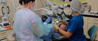
To perform high-quality work in root canals, qualified doctors at our center use modern treatment methods. The work is carried out with “four hands” (doctor-assistant), which improves the quality of the doctor’s work and the patient’s comfort.
Treatment of tooth pulpitis from 6,500 rubles.
Recommendations before visiting the dentist
A dentist treats diseases of the oral cavity. There are several sections in dentistry: surgical, therapeutic, periodontics, orthodontics, orthopedics. Before visiting the dentist, you should brush your teeth or at least rinse your mouth to remove food particles. It is advisable not to take painkillers so as not to change the clinical picture of the disease. During treatment, the doctor will administer local anesthesia, if necessary. If dental treatment was previously performed in another clinic, it is better to pick up the card and bring it with you. This will allow the doctor to assess the condition of the teeth over time. X-rays and tomograms are usually taken at the clinic when you visit as prescribed by a doctor, but if any pictures have already been taken, you need to take them with you.
Methods for filling root canals
There are several basic techniques used in the treatment of pulpitis and periodontitis:
- One-paste method
The channel is filled with a plastic, subsequently hardening material. The method is outdated and gives a large number of complications. - Single pin method
First, the root canal is filled with a special paste, and then a gutta-percha pin is inserted into it. The percentage of complications is lower, but this method is also gradually being phased out. - Filling with “thermophile”
A more modern method, also based on the use of an initially plastic material - heated gutta-percha. The fluid state allows the material to densely fill all cavities inside the canal, which significantly reduces the risk of complications. The disadvantages of the method include high requirements for the doctor’s qualifications and relatively high cost. - Gutta-percha condensation method
This method ensures even more dense filling of the canal lumen with filling material. Currently, the method is one of the most common. The procedure is performed in several stages.
Biological (conservative)
Conservative treatment of pulpitis is the most gentle way to eliminate pulp inflammation.
The biological method is used in cases where it is possible to stop the inflammatory process while preserving the nerve. Not all dental clinics offer services for biological treatment of pulpitis. This is explained by the fact that the doctor performing this type of therapeutic manipulation must be highly qualified and have extensive experience in performing such procedures. In addition, when using a conservative treatment method, cases of relapse of the disease are common; for this reason, dentists prefer to resort to nerve removal.
If your tooth hurts after filling
So, why does a tooth hurt after filling? Any installation of a filling is an intervention in the tissue, so pain during and after treatment may well occur. On the other hand, one pain is a natural stage in the recovery of the body after treatment, and the other is a sign of incipient complications or injury. So when should you sound the alarm? Experts distinguish different types of toothache: according to duration, nature of manifestation and correlation with various types of manipulations.
Indications and contraindications
The biological method of treating pulpitis has a number of indications. These include:
- chronic and focal inflammation of the pulp in the acute stage;
- fracture of the crown of a tooth with damage to the root;
- age up to 28 years;
- the initial stage of development of carious lesions.
This method of eliminating pulp inflammation is most often prescribed to children under 12 years of age, since the tops of their teeth are not yet fully formed. The use of a conservative method of treating pulpitis is contraindicated in:
- the patient has acute infectious diseases and a burdened allergy history;
- impossibility of preserving the dental nerve;
- treatment of a patient over 28 years of age.
Patients, as a rule, begin to take measures to eliminate the disease only when it has already become advanced. For this reason, this treatment method is used quite rarely.
Medicines
It is impossible to say exactly how long this treatment method takes.
As a rule, it is not possible to eliminate pulp inflammation in one visit; this will take several days. The specialist carefully selects the method of pain relief, removes damaged areas of dentin and forms a cavity for the affected tooth. All medications used in the treatment process are selected in strict accordance with the patient’s health condition and taking into account his individual characteristics. The stages of the biological method of treating pulpitis include the following procedures:
- administration of anesthetic;
- treatment of the carious cavity using warm antiseptics and enzymes (Chlorhexidine, Iodinol, Trypsin, Lysozyme, Dimexide);
- drying the work area with preliminary degreasing;
- applying a bandage impregnated with calcium hydroxide to the treated carious cavity;
- installation of a temporary filling;
- determining the condition of the pulp after 2–3 days, for which radiography is performed;
- carrying out unsealing in the absence of an inflammatory process and installing a permanent filling.
This treatment method involves applying a special paste directly or indirectly to the inflamed root. The first option is used in cases where it is impossible to clean the bottom of the carious cavity without damaging the pulp chamber. The second method of application is used when the pulp is completely or partially exposed.
After all medical procedures have been completed, the patient is prescribed non-steroidal anti-inflammatory drugs, such as Ibuprofen, Nise or Ketonal. As noted earlier, this treatment method is used quite rarely.
Due to the impossibility of ensuring absolute sterility during the procedure, there is a high probability of further development of the inflammatory process.
Typically, this method is used for inflammation of the pulp in children, when it is necessary to preserve the structure and function of baby teeth.
Physiotherapeutic procedures
In parallel with the described method of treating pulpitis, physiotherapy is prescribed. It includes the following activities:
- ultra-high-frequency inductothermy, which helps eliminate the source of inflammation in the pulp and reduce pain;
- infrared laser therapy, which has a beneficial effect on the regeneration of damaged tissues;
- diathermocoagulation, which prevents infection from penetrating into periodontal tissues;
- transcanal electrophoresis, which stimulates the restoration of pulp and periodontal tissue, which helps prevent the development of apical periodontitis.
In most cases, the doctor prescribes a course of physiotherapeutic procedures. Already after the first implementation of these manipulations, the patient feels a significant improvement in his condition.
Types of cysts according to their causes:
- Eruption cyst – most often occurs in children aged 7 – 10 years.
- Paradental (retromolar) cyst - appears when there is difficulty in the eruption of a wisdom tooth and its chronic inflammation.
- A follicular (tooth-containing) cyst is formed due to infection of a tooth germ or an unerupted or supernumerary tooth.
- Primary cyst - is formed when the development of a tooth is disrupted from the remains of dental tissue.
- Radicular cyst - a cyst that forms on the root of a tooth and usually develops due to chronic periodontitis
- A residual cyst appears in the bone after tooth extraction.
Surgical treatment of pulpitis
A traditional method that has different implementation options depending on the specific case and the age of the patient.
Consists of complete or partial extraction of pulp from the tooth cavity. • Extirpation. Used in the vast majority of cases. Represents complete removal of the pulp.
- Vital. Under anesthesia, living pulp tissue is removed. First, the dental tissue affected by caries is removed, treated with an antiseptic, then the infected nerve is removed and the cavity is filled. The procedure requires just one visit to the dentist; it is universal for any form of pulpitis, but is not applicable for patients with allergies to anesthetics.
- Devital. If it is not possible to carry out vital extirpation, the pulp is exposed to paste-like toxic substances (for example, arsenic) so that it can then be painlessly removed. The paste is applied for 24–48 hours, if the patient can come for a second appointment quickly enough, or for up to 14 days - in this case, mildly acting formulations are used. The cavity is closed with a temporary filling until the next procedure. At the second appointment, the doctor removes the dead pulp, cleans the canals and places a permanent filling. For purulent pulpitis or tissue necrosis, the method is not used.
• Amputation. In this case, only the coronal part of the pulp is removed, the root part remains in place. For teeth with one root, amputation is not suitable, since in this case it is almost impossible to isolate individual elements of the pulp. Amputation is usually prescribed for acute pulpitis or accidental mechanical damage to a tooth.
- Vital. The necessary part of the nerve is amputated under anesthesia in one procedure. The method is indicated for patients under the age of 45 with healthy periodontium.
- Devital. After exposing the pulp to a toxic paste, the dead area is removed, and the healthy one is deliberately mummified, for example, with an antiseptic paste based on zinc oxide-eugenol, so that the infection cannot develop again. Devital amputation is used in difficult cases when it is impossible to reach the desired area of the pulp. The modern arsenal of dentists includes flexible nickel and titanium instruments. With their help, you can treat even the most curved canals, which eliminates the need for complex techniques.
The use of arsenic for the treatment of pulpitis was proposed only at the end of the 19th century. Before this, the inflamed pulp was destroyed with hot oil or burned with a hot iron. These methods were used by ancient Greek and ancient Egyptian dentists.
Treatment of dental cyst
A dental cyst is the last stage in the development of chronic periodontitis. It is a cavity of various sizes, which is formed in the thickness of the jaw and is connected to the root of the tooth. This cavity is lined with a membrane and filled with liquid. When microbes enter, the contents of the cyst suppurate. Usually the cyst grows for a long time without any subjective sensations or clinical manifestations. The first symptom that attracts attention is the deformation of the jaw in the form of a protrusion at the site of the growing cyst.
Tooth cyst
If the cyst does not suppurate, then it can reach a significant size without causing any reaction from the general condition of the patient and without causing him pain.
What to do after treating pulpitis?
Pulpitis occurs when the soft tissue of the pulp (dental nerve), which is located in the oral cavity, becomes inflamed.
Recommendations after treatment of pulpitis:
- There may be discomfort after anesthesia. On the upper jaw, numbness can last for 1-2 hours, on the lower jaw much longer - for 2-4 hours, depending on your anatomical features. Try not to eat at this time, so as not to damage (bite) the soft tissues of the cheek, tongue, lips. After anesthesia, it is possible: swelling of soft tissues, hemorrhage at the injection site. In this situation, we recommend applying cold for 15 minutes.
- After administration of anesthesia, decreased attention may occur. We recommend: do not drive or be attentive to the situation on the road.
- Delayed allergic reactions to the anesthetic may occur in the form of swelling of the face, tongue, and rash. We recommend: take a suprastin tablet and be sure to call your doctor; if swelling increases, call an ambulance.
- If, after anesthesia, sensitivity to any area is not completely restored, then you should definitely seek help from a doctor.
- After the anesthesia has worn off, you may feel a slight aching pain in the tooth and discomfort when biting - this is normal, since the tooth canals were treated with mechanical instruments and antiseptic solutions. We recommend taking 1 tablet of an analgesic (ketorol) or a non-steroidal anti-inflammatory drug (nimestil - 1 sachet).
- The temporary filling hardens within 2 hours, please do not eat during this period. It may crumble, but should not fall out completely. Look carefully, if there is no filling, then you need to put it in as quickly as possible, since the prognosis and result of treatment for this tooth depends on this.
- Trauma to the gums is possible, since for the treatment of pulpitis a special insulation is used, which is put on the diseased tooth and protects the oral cavity from the aggressive effects of the antiseptics used. When restoring the correct anatomical shape of a tooth, matrices and special threads are used, which are inserted under the gum. After placing the filling, it must be polished, which can also injure the gingival margin. If the gums are damaged, we recommend: brushing your teeth, rinsing your mouth with water and lubricating the gums near the tooth with Actovegin gel for mucous membranes for 3-7 days after each meal.
- After filling the canals, there may be painful sensations when biting on a tooth for 7 days, so that the discomfort goes away faster, we recommend taking: nimesil 1 sachet 2 times a day after meals.
- There may be discomfort after placing a filling: the filling may interfere with the bite. In this case, be sure to consult a doctor.
There should NOT be: swelling, fever, severe pain. In these cases, we recommend that you consult a doctor immediately. You cannot warm your cheek in the area of a sore tooth, rinse your mouth with alcohol-containing solutions and take them orally, or pick out a temporary filling. DO NOT: walk with a temporary filling for more than 7 days, as the seal of the temporary filling is broken, and microorganisms from the oral cavity penetrate into the canals, resulting in the need for re-treatment of the canals, and the favorable prognosis of treatment is reduced.
Types of periodontitis
Depending on the nature of the processes occurring in the area of the tooth root, periodontitis has three forms.
- Chronic fibrous. A type of chronic periodontitis. This disease most often affects older people. The disease is practically asymptomatic, but during an exacerbation, pain may occur when chewing food. This form of periodontitis is manifested by fibrous changes in the periodontal tissue, that is, the tissue around the tooth becomes pale pink, and the tooth itself may become gray.
- Chronic granulating. It is also a chronic form of periodontitis. It is often the cause of complicated caries, and is characterized by inflammation in periodontal tissues. In this case, granulation tissue appears, which grows and can have devastating consequences for the bone.
- Granulomatous. Chronic stage of periodontitis with an inflammatory nature. Granulomas appear in the tissues at the apex of the tooth root, that is, formations that separate the infectious focus from part of the healthy tooth. Gradually, the tissue surrounding the tooth is destroyed.
In addition, according to the speed of the disease, periodontitis can be of the following types.
- Spicy. The disease acquires this form at the final stage. This form is characterized by rapid development, accompanied by acute pain (especially when eating hot food), redness and swelling of the gums, and the appearance of a fistula.
- Chronic. This form is characterized by sluggish processes due to unresolved inflammation. Pathogenic microorganisms constantly penetrate inside the tooth and contribute to its death, as well as damage to the tissues surrounding the tooth and bone destruction.
- Chronic in the acute stage. When the disease is in the acute stage, a person may experience a rise in temperature, a headache, a feeling of weakness, swelling of the gums and face, enlarged lymph nodes, and mobility of a damaged tooth.
Complications of pulpitis without treatment
If pulpitis is not treated, the inflammation progresses, extends beyond the root canal, and affects adjacent periodontal and periodontal tissues.
Most often this leads to periodontitis. Acute periodontitis is manifested by sharp pain, which is localized in the area of the affected tooth. The intensity of the pain gradually increases, it becomes throbbing when purulent inflammation begins. If treatment is not started at this stage, purulent exudate begins to separate, and the periodontal collagen fibers are destroyed. Soft tissues swell, temperature rises, and general condition worsens. With periodontitis, the ligaments that hold the tooth in the alveolus are destroyed, and the destruction of bone tissue begins. The tooth becomes loose and falls out. Large cysts may appear.
Another possible complication of pulpitis is periostitis. With periostitis, inflammation spreads to the periosteum (tissue surrounding the bone), and a flux is formed. The gums swell and the jaw becomes painful. Periostitis can appear after pulpitis or accompany periodontitis. Without treatment, the swelling of the gums increases, the pain intensifies, the cheek and lip near the site of inflammation swell. The pain becomes shooting and can affect the temple, eye, ear. A fistula (passage) may appear in the swollen gum, through which pus comes out. After the fistula tract appears, the inflammation becomes less intense, but the tissues remain infected, and the infection continues to spread, and periostitis from an acute form becomes chronic.
Pulpitis can provoke the development of periodontitis - an inflammatory disease of the gums, dangerous by the destruction of bone tissue, gradual recession of the gums, and adentia. Against the background of acute inflammation and tissue infection, severe systemic diseases may appear: general sepsis (blood poisoning), phlegmon (breakthrough of pus into the soft tissues of the face, requiring surgical intervention), amyloidosis (occurs due to the constant toxic effect of pus).
Symptoms and causes of pulpitis
Depending on the clinical picture, pulpitis can be acute or chronic. The acute form usually develops into a chronic form, and then the sick person simply does not pay attention to periodic pain. This condition is dangerous because periodontitis can begin, in which periodontal inflammation occurs. The reasons for the formation of pulpitis are the following factors:
Caries
The disease leads to the gradual destruction of hard dental tissues and pathogenic microorganisms enter the pulp chamber, leading to pulpitis
Trauma or bruise of a tooth
Trauma or bruise of a tooth can also lead to the development of pulpitis
Complications of certain infectious diseases
When pulpitis forms in the pulp chamber. In such a situation, pathogenic microorganisms enter the pulp from a cyst or periodontal pocket
Due to incorrect previous medical procedures
In particular, pulpitis often appears after incorrectly performed dental canal filling technology.
Complications of pulpitis due to treatment errors
The instrument broke off inside the root canal.
The problem arises when passing through narrow, curved channels, with insufficient access to them, or with the wrong choice of tool. If the instrument breaks off in the channel, they try to remove the fragment. In some cases, it is possible not to remove the fragment and continue filling the canal. If a fragment remains in the root canal, the quality of the filling must be monitored using radiography. Perforation of the cavity bottom or tooth walls. Perforation is an artificial through hole in the hard tissues of the tooth. This is a dangerous complication that may require tooth extraction. Perforation can occur due to medical error or due to severe demineralization of hard tissues. If the hard tissues are demineralized, the doctor may perforate the cavity even with normal probing. This is not a medical error and is associated with the low strength of hard tissues. During treatment, the cavity may be perforated due to the inclination of the teeth, curvature or insufficient width of the canals. At the Dentospas clinic, before treating pulpitis, radiography and its analysis are performed to exclude such complications - an x-ray allows you to assess in advance the location of the root canals.
The filling material extends beyond the root tip. If a little filling material extends beyond the root apex, this is not a complication (it may be required in the treatment of complicated pulpitis accompanied by periodontitis). With a significant amount of filling material, if it comes out into the maxillary sinus, sinusitis (inflammation of the mucous membrane) may develop. In case of such a complication, the tooth is removed and the sinus is cleared of infection.
After filling, pain persists. In almost all cases, after treatment of pulpitis, pain persists when biting on a tooth. It may not go away for two weeks after filling the canal. If the pain gradually subsides, this is a normal response to treatment. The doctor may prescribe pain medication and physical therapy to relieve it. If the pain increases and becomes throbbing, this indicates ongoing inflammation, and the tooth needs to be re-treated.
Change in crown color. After depulpation, the hard tissues of the tooth may darken and become brittle. This may occur due to the preservation of the nerve during treatment (it was not completely removed), due to poor-quality filling of the root canals. If the darkening of the crown is accompanied by pain and swelling, the tooth will have to be re-treated. To remove the aesthetic defect, restoration or installation of an artificial crown will be required.
You can eliminate the complications of pulpitis by performing quality treatment in a timely manner. Dentospas dentistry performs endodontic treatment, dental retreatment, and treatment of complications of pulpitis.
Treatment of chronic periodontitis
Before starting treatment for chronic periodontitis, it is necessary to conduct a thorough examination of the patient. To do this, probe and x-ray of the affected area of the jaw are performed. Based on the results, an effective treatment method is prescribed.
Periodontitis is treated in two ways.
- Treatment of the disease with a therapeutic method involves the use of antibacterial medications that have a wide spectrum of action, localize inflammation and relieve symptoms. Periodontal canals inside the tooth are treated with antiseptics, and general strengthening drugs are also used to accelerate tissue regeneration. It is also possible to use laser processing. Treatment lasts up to six months. The dynamics are monitored using x-rays.
- The surgical method is used if therapeutic treatment does not produce the desired results. It consists of removing the affected tissue, resection of the apex of the tooth, or its complete removal. If there is a large amount of pus, installation of elastic drainage is required. After the procedure, the patient must take medications prescribed by the doctor to avoid infection and to accelerate tissue regeneration.
When not to worry
Dental depulpation surgery may not seem like such a serious procedure, however, like any other surgical intervention, it can lead to pain.
Soreness in the root of the treated tooth, especially when biting or pressing on the crown, is normal for up to 3-5 days after completion of treatment. In this case, there is no need to panic, you just need to carefully monitor your condition. However, sometimes the pain experienced is very severe or other alarming symptoms are added, for example, swelling of the gums, the appearance of ulcers on the mucous membrane and elevated body temperature that does not subside for several days. In such cases, you should immediately contact your dentist in order to make treatment adjustments or receive qualified recommendations for pain relief. Sources:
- https://www.pravda.ru/navigator/kak-lechat-pul-pit.html
- https://DocDoc.ru/treatment/lechenie-pulpita
- https://MikDent.ru/stomatolog/bolezni/lechenie-pulpita-u-stomatologa.html
- https://fdclinic.ru/aboutfd/forp/posle-lecheniya-pulpita/
- https://dentospas.ru/inf/bolezni-zubov/oslozhneniya-pulpita/
- https://ZubNeBoley.ru/lechenie/karies-i-pulpit/prichiny-zubnoj-boli-posle-lecheniya-pulpita/
How to restore a tooth if the tooth tissue is severely damaged
When is an inlay used to restore a tooth? It is in case of severe destruction of the tooth crown that a pin tab or crown is used
The choice of material depends on the doctor’s recommendations and the patient’s capabilities. It can be a zirconium insert, it can be metal, made of precious metals or ordinary ones.
Here it is important to understand that this must be done, since it is important to close the entire structure made - the sealed canals and the filling in the crown of the tooth, to “seal” it.
There is an opinion that with good sealing, mediocre endodontics last longer, and vice versa, when the filling and the crown above it have microcracks, microleakages, even perfectly sealed canals will gradually begin to collapse.