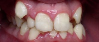
Dentofacial anomalies can be associated not only with a deviation in their position, but also with their irregular shape. One of the pathologies of the shape of teeth is their narrowing: conical and awl-shaped (in the form of a screwdriver). Teeth of such a sharp shape are extremely rare - one case per thousand people.
Contrary to the erroneous opinion of some Patients, this is not only an aesthetic problem, but also a serious pathology that can seriously damage the soft tissues of the oral cavity. In addition, this form of teeth can lead to disruption of chewing function, as well as cause increased abrasion of the enamel and sensitivity of the teeth.
This anomaly is characterized by several signs:
- sharpening of the teeth to the cutting part, their conical or barrel-shaped shape;
- the presence of the same slope on all sides;
- smaller size compared to other teeth;
- Predominant location on the upper jaw.
Reasons for development
All causes of the development of dental anomalies are usually divided into internal (endogenous) and external (exogenous).
Endogenous factors
The root cause of the development of pathology may be a genetic predisposition. Hereditary anomalies of the dental system include: diastemia (gap between the front teeth), adentia (congenital absence of a dental unit), changes in the shape of the dental crown, craniofacial dysostosis (Treacher Collins syndrome), macro- and microgantium (hypertrophied or, on the contrary, underdeveloped jaw ) and others.
Diastoma on the left, adentia on the right
In this case, a child can inherit from parents who do not have pronounced anomalies, different structural features of the dental system: for example, from the father - large teeth, and from the mother - a narrow upper jaw. Such a child has a high probability of developing a congenital anomaly of the dental system - crowded teeth.
Exogenous factors
The formation of the human jaw system begins during intrauterine development, and is completely completed by the age of 13-15 years. An important role in the development of anomalies of the dental system in adults is played by poor ecology, insufficient content of inorganic salts, vitamins and minerals in water and food.
After the birth of a child, the causes of anomalies of the dental system can be:
- lack of vitamins and minerals supplied with food and water;
- diseases associated with insufficient bone mineralization (rickets);
- frequent diseases of the upper respiratory tract, accompanied by difficulty in nasal breathing;
- artificial feeding, long-term use of a pacifier;
- bad habits (thumb sucking, sleeping with your hand under your cheek).
Crooked teeth are not a death sentence
With the onset of a certain age, people often come to the understanding that it is time to change something. Including in appearance. Modern plastic surgery can correct any mistakes of nature, erase traces of years and fatigue from the face. It's sad when all your efforts are ruined by an ugly smile.
Along with cosmetic manipulations, bite correction in adults becomes an important step on the path to transformation and gaining self-confidence. A good orthodontist can help straighten teeth for patients of any age.
Types of dental anomalies
There are different classifications of HFA. The most popular and the only international one is considered to be proposed by the American orthodontist E. G. Engle. According to Engle's classification, based on physiological and functional characteristics, disorders of the structure of the dentofacial apparatus are divided into several types.
Dental pathologies occur both isolated and in combination with each other. Thus, anomalies in the position of teeth often develop against the background of disturbances in their shape or the shape of the dentition, both of which lead to the formation of malocclusion.
Anomalies of teeth position
This type of pathology includes displacement of individual dental units:
- Distal, oral - back, towards the palate or tongue. In this situation, when there is a shortage of space in the dentition, the central incisors and premolars most often find themselves. Mesial, vestibular - forward along the dental arch. The most common causes of this pathology are a narrow jaw or incorrect direction of the alveolar process during eruption.
- Rotation is a position in which the dental unit is in its place, but rotated relative to its own axis. This anomaly is characteristic of incisors.
Anomalies of the dentition
With normal development of the jaw system, the dental arch of the upper jaw has the shape of a semi-ellipse, the lower one is less rounded, and looks like a parabola. In some people, the shape of the jaw arches is changed; they may be longer or shorter, wider or narrower than normal, or located asymmetrically relative to each other. The consequence of such anatomical disorders (most often congenital) is the development of various types of anomalies and deformations of the dental system.
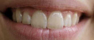
Crowding of teeth . This pathology is diagnosed quite often: about 34% in mixed dentition and 68% in permanent dentition. Due to the narrow arches, there is not enough space for the teeth to be in a normal position, therefore, when they erupt, they can turn around their axis, tilt, move outward (vestibular), inward (oral), and grow outside the dentition.
Violation of the distance between teeth. The reason for this anomaly in the dentition is the opposite of the reason for the development of crowding, it is too wide a jaw. The gaps between the front incisors are called tremata, and between the lateral premolars and molars - diastemas.
Abnormal bite. The bite is the relative position of the upper and lower teeth at the moment of closing the jaws (occlusion position). If the jaws are narrowed, widened, shortened, lengthened or asymmetrically positioned, the bite will be incorrect:
- protruding upper jaw - distal (prognathic);
- protruding lower jaw - mesial;
- the upper jaw is wider than the lower jaw and its teeth completely overlap the lower dentition
- The upper and lower jaws are asymmetrical - cross.
- The jaws do not match in size, which is why the rows of teeth do not overlap each other, but only close at the ends - open.
Anomalies in the shape and size of teeth
The most common types of this type of pathology:
- Macrodentia - giant teeth. This anomaly of tooth shape is typical for the upper central incisors or lower premolars, and is most often diagnosed in mixed dentition, at the age of 7–9 years.
- Microdentia - small teeth. This pathology is rare and usually appears symmetrically, on both sides of the jaw.
- Hutchinson's teeth, Fournier. With this pathology, the central incisors have the shape of a screwdriver with a rounded notch at the end and flat edges.
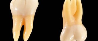
- Pflueger's teeth. Large molars, in which the chewing surface area is narrower than the neck of the dental crown, are more often susceptible to this pathology.
- Spiked teeth. The coronal part of the lateral and central incisors has the shape of a cone.
The reason for the development of malformed teeth is considered to be a violation of mineral and protein metabolism in the prenatal period, when the formation of dental buds and alveolar processes of the jaw occurs in the fetus.
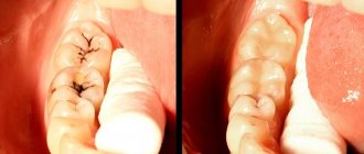
Let's consider a clinical example of transposition correction using orthodontics
Patient N., 14 years old, came with her mother to the dental clinic. Complaints were made about the incorrect location of the canine on the upper jaw on the right. We already had a panoramic radiograph in hand, which complemented the analysis of the clinical situation in the oral cavity. To clarify the diagnosis and determine the optimal treatment plan, the patient was referred for a computed tomography scan of the jaws
After analyzing the diagnostic data, it was found that the root of the first premolar 1.4 is inclined towards the persistent primary canine 5.3, and the root of the permanent canine 1.3 is projected into the area between the roots of the premolars. Consequently, the situation was complicated by the fact that it was necessary not only to move the crowns, but also to “separate” the roots of the canine and premolar in their places.
When discussing the treatment plan with the patient and parents, a clear decision was made: to put the teeth in their places, despite the duration and complexity of the correction. The treatment method involved fixing a metal brace system initially only on the upper jaw, and after moving the crowns and roots, installing a brace system on the lower jaw for final correction and detailing the position of the teeth.
Treatment
In most cases, it is possible to partially or completely achieve normal functioning of the dental system and restore its anatomical shape using methods of orthodontics, physiotherapy or surgery.
The most popular method of correcting PCA today is the use of various types of orthodontic appliances. With their help, it is possible to correct the position of the dentition or change the shape and size of the jaw. The principle of operation of such devices is based on the pressure force applied to a certain area of the dentition and directed in the desired direction.
Of all types of orthodontic structures, braces are considered the most effective for correcting anomalies and deformations of the dental system. These are non-removable dental systems consisting of small metal, plastic or ceramic plates connected by an arch (the arch, trying to take a given position, is the source of force). Vestibular braces are placed on the outside of the teeth, lingual braces are placed on the inside of the teeth.
The following removable intraoral devices are used:
- Orthodontic plates. These are rigid plastic structures consisting of a base adjacent to the palate and a screw located in its central part that regulates the force of pressure on the jaw.
- Trainers are soft silicone or polyurethane splints that are worn on the jaw for several hours during the day and all night.
- Aligners . Rigid structures made of transparent plastics. They are made individually, using dental impressions. The course of treatment for CFA involves the sequential use of several aligners.
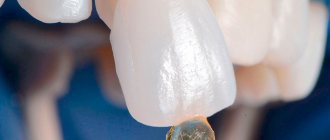
To eliminate minor defects (slightly curvature of teeth, irregularities in their color or shape, gaps between them), veneers can be used to improve aesthetics. These are thin plates made of porcelain or composite material that are attached to the outer surface of the teeth with a special glue.
When correcting dental anomalies, physiotherapeutic procedures are also indicated in combination with hardware methods; they can significantly speed up treatment. Applicable:
- myogymnastics - exercises for certain groups of jaw muscles;
- massage of the alveolar process to stimulate its growth;
- vacuum, ultrasound, vibration therapy;
- electrical stimulation;
- electrophoresis.
Surgical methods for eliminating anomalies of the jaw are used when a serious reconstruction of the jaw is required, a significant change in its shape, if it cannot be achieved with the help of orthodontic devices.
Prevention
A set of measures aimed at preventing the development of dental anomalies in children includes:
- balanced nutrition of a woman during pregnancy in order to provide the fetus with the substances necessary for its skeletal system;
- inclusion in the diet of a growing child of vitamins and microelements necessary for the proper formation and mineralization of teeth;
- preventing the development of bad habits in children that cause occlusion disorders (prolonged use of a pacifier, finger sucking);
- timely treatment of dental, infectious, endocrine and other somatic diseases;
- regular monitoring of the condition of the dental system.
What symptoms of a wedge-shaped defect should you pay attention to?
Painful sensations during caries appear when destroyed dental tissues (the so-called carious cavity) are no longer able to protect the neurovascular bundle located in the pulp chamber. With a non-carious defect, the affected area and decaying dentin are covered by the gum, and the walls of the formed wedge-shaped cavity remain smooth and hard. Therefore, the patient does not begin to experience unpleasant sensations immediately, but only in the third or fourth stages. Treatment of advanced pathology turns out to be more complex, costly, and in some cases ineffective.
Therefore, you should definitely see your doctor if you have:
- individual teeth have become hypersensitive and react to external irritations with sharp, piercing pain;
- the enamel has become cloudy, pale or more pigmented;
- The gum line has changed and the necks of the teeth are exposed.