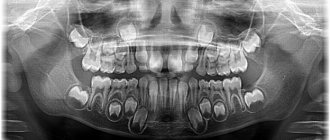
Milk teeth are bone formations that are designed for mechanical processing of food in order to prepare it for subsequent digestion. They also influence diction and make the child’s speech correct and clear. In terms of their anatomical structure, they are in many ways similar to permanent ones, but they all have some differences. Among them:
- the coronal part is smaller;
- the thickness of enamel and dentin is less (from 0.6 to 1 mm);
- reduced degree of mineralization;
- no immune zones;
- the pulp volume is large;
- dental canals are shorter;
- poorly developed tubercles in the closure zone.
As for the number of root canals, their number remains unchanged.
The roots of baby teeth are slightly inclined. This is explained by the fact that the rudiments of permanent units are located above them.
It is a mistake to believe that children's teeth do not have roots. But this is what many adults think when they look at their children’s fallen teeth. In fact, by the time of prolapse, the formations located in the alveoli are almost completely resolved.
How do the roots of baby teeth dissolve?
The process of replacing children’s teeth with “adult” ones normally occurs at the age of 5-7 years. It is during this period of life that a person first becomes partially toothless. But before the first incisor falls out, serious physiological changes will occur in the structure of its roots. We are talking about the process of resorption, better known under the term “resorption”. Let's look at how this happens.
First, the hard plate that separates the germ of a permanent tooth and the roots of a milk tooth begins to collapse. Small pits with osteoclasts form inside the roots. The roots located near the bud are the first to disappear. The process always starts from the apex and moves towards the gum.
Soon, the loose fibrous connective tissue located inside the crown is replaced by granulation tissue. The latter is also important for timely resorption. After this, very little time passes before the tooth begins to become very loose and one day falls out. A permanent unit is soon shown in the vacated space.
In most cases, resorption proceeds in balance with the eruption of permanent teeth, but sometimes failures occur. Then the roots of children's baby teeth are preserved for a long time or, conversely, are destroyed much earlier than expected. Such phenomena are undesirable.
Forced resorption occurs due to traumatic injuries, tumor growths, strong pressure from “neighbors,” deep bite, and orthodontic disorders. A slowdown in the process is possible if there are no rudiments at all.
Thus, children have roots of baby teeth. They are simply subject to resorption. That is why they cannot be seen by the time they fall out.
Lower premolars
Two in each quadrant, they erupt between 10 and 12 years of age, with full root formation after about 3 years. They replace primary molars. In contrast, the upper premolars are distinct from each other (Fig. 1.32).
Fig 1.32 Lower premolars: occlusal view
Lower first premolar
The lower premolar erupts sometimes several months before and sometimes after the lower canine and is the smallest of all premolars. The crown has two cusps, one very large and similar to the cusp of a canine tooth. The other is shorter by about 2 mm, similar to the lingual marginal ridge of the canine (Fig. 1.33).
Figure 1.33 Lower premolars: graphical representation of the access cavity
Length is about 21-22 mm, usually has one root with one canal (73-74%). Sometimes one root with two canals is possible (19%) (Fig. 1.34a, b), two roots with two canals (6%) (Fig. 1.35a, b) or three canals (1-2%).
Rice. 1.34 (a, b) First lower premolar: occlusal and proximal view
Rice. 1.35 (a, b) Second lower premolar: (a) coronal view; (b) Proximal view showing the presence of a single root
The root is often distally curved in 35% of cases and with a sharp curve in 7% of cases. The shape of the access cavity to the pulp chamber is oval, extending from the main tubercle to the tip of the minor lingual tubercle (Fig. 1.36).
Figure 1.36 Anatomical pictures from Hess and Keller: a mandibular premolar with a complex canal system that has two main canals connected by several fins and two separate exits
Lower second premolar
The second lower premolar erupts at the age of 11-12 years and is larger than the first premolar by 1 or 2 mm. The crown has two cusps (a larger buccal and a smaller lingual), but the lingual cusp is often divided into two parts (Fig. 1.37).
Rice. 1.37 Anatomical pictures from Hess and Keller: lower premolar with two roots and two canals; several short lateral canals present in the apical third
It has only one root with one canal in 85% of cases, but we can also find two separate canals in one root (11.5%) or two canals that merge into one apical foramen (1.5%) (Fig. 1.37 ). Rarely three channels are possible (0.5-1%). In approximately 40% of cases the root is straight, while in approximately 40% the apical third is distally curved. Severe curvature (7%) and vestibular curvature (10%) are possible.
The access cavity has an oval shape, also in the buccal-lingual direction, located in the center of the occlusal surface (Fig. 1.36).
Root canal treatment in children
The roots of children's baby teeth are filled with a special paste. It has the ability to dissolve in parallel with the process of resorption of hard tissues. This is the only way to ensure that the order of changing teeth is not disrupted and that there are no obstacles to the resorption of milk roots.
Once the root canals are treated, they are closed with a filling. Sometimes the doctor decides to use temporary filling material. This measure is justified if the likelihood of complications is high and you need to make sure that the inflammation has subsided. Only when the dentist is sure that everything is in order will he install a permanent filling.
My baby is teething - how can I help him?
The child's profuse salivation will tell you that the first tooth is on the way. 1-2 months before the first tooth erupts, the baby’s saliva begins to flow so actively that it is already difficult to do without aprons and bibs.
All your older relatives will probably tell you about the unpleasant side effects of teething. However, there are many misconceptions here.
The very first teeth usually come out painlessly. What most often happens is this: while spoon-feeding a child, the mother hears the sound of metal on the edge of a tooth - that is, she detects an event that has already happened, without even noticing anything unusual in the child’s behavior.
The emergence of canines and molars may be more difficult. The baby may be capricious, refuse to eat, sleep poorly, and put everything in his mouth. You should take care of inflamed gums - regularly treat them with special gels, offer your child to chew a cooling ring (cold relieves pain well).
However, do not believe that an increase in temperature is associated with teething. Fever and catarrhal symptoms are caused by an infection that is “caught” by the child’s body, weakened by malnutrition and lack of sleep. That is why, during the period of teething, it is better to protect the baby from communicating with strangers. Digestive disorders, an upset stomach of a child at the time of teething, is associated with his desire to chew and suck everything he can reach, just to relieve discomfort in the gums. This is how pathogenic microbes enter his mouth. Try to surround your child with clean objects, wash his hands and toys more often. Regularly let your baby chew small pieces of solid food - dried bread, bagel, apple slice, etc. This will help the teething of those teeth that are already “on the way”, improve blood supply, and therefore nutrition of the gums, develop the chewing reflex, and help the formation of the speech apparatus.
Why is it important to treat baby teeth?
Now that it is clear that the structure of baby teeth is very similar to permanent teeth, the question “To treat them or not to treat them” should not arise for parents at all. Advanced caries often leads to destruction and damage to the pulp. Then the roots become involved in the pathological process. Removing dental nerves is not a pleasant procedure. It is important to prevent its occurrence.
Moreover, refusal of dental treatment can result in a number of other problems. This means:
- Incorrect bite formation. If, due to the destruction of the crown, the doctor has to remove it ahead of time, voids appear in the row, which “neighbors” tend to occupy. Then, by the time the “adult” unit erupts, there may not be a place for it - it will begin to grow somewhere on the side and ruin the smile.
- Damage to future teeth even before they erupt. With deep caries and periodontitis, cysts often form. They affect the rudiments of permanent teeth. Then the child is faced with the fact that his new tooth turns out to be sick and requires urgent treatment. Needless to say, its service life will be significantly reduced because of this.
- Diseases of the gastrointestinal tract. If parents believe that it is better to remove baby teeth rather than treat them, then by the age of nine their child (especially if he is prone to developing dental diseases) may lose half of his teeth. Is it possible to fully chew food in such a situation? No. The child regularly swallows poorly chewed foods. Because of this, the load on the intestines increases, which can result in frequent abdominal pain, stool disorders, nausea, gastritis and more serious gastrointestinal diseases.
- Psychological complexes. At 9-10 years old, a child evaluates his appearance and sees that his smile is very different from the smiles of his peers - it does not have a large number of teeth or they are dark in color, half destroyed. Because of this, he begins to be embarrassed to smile, laugh, and tries to talk less. All this negatively affects his self-esteem and does not allow him to quickly adapt to new situations and successfully go through the process of socialization in elementary school.
Don't waste your children's teeth. Teach your child from childhood to undergo preventive examinations in the pediatric dentistry department. At the same time, choose a specialist for him who you can trust. Then the child will not perceive another visit to the dentist as something terrible. He will be happy to go to the appointment, knowing that he will not be hurt.
Upper premolars
Two per quadrant, premolars erupt between 10 and 12 years of age, and root formation is completed after 3 years. They replace temporary molars.
The first and second premolars have a similar crown but different morphology.
Upper first premolar
The first upper premolar erupts when the child is about 10-11 years old. It has a length of about 21-22 mm, and its buccal-palatal dimensions are 9 mm and mesial-distal dimensions are 7 mm. It has two cusps, buccal and palatine, which are slightly shorter (about 1 mm). The pulp space is determined by the shape and size of the outer crown (Fig. 1.24). In approximately 72% of cases, two roots with two different apical foramina are present (Fig. 1.25). Premolars may also have only one root with two canals (13%) (Fig. 1.26a, b), and in some cases three roots (6%) (Fig. 1.27). In addition, in 37% of cases we found a distal root bend in the apical third.
Rice. 1.24 First upper premolar: occlusal view
Rice. 1.25 Upper premolars: graphical representation of the access cavity; note the mesial position of the second premolar access cavity (right in the figure) to facilitate passage of the distally curved canal
Rice. 1.26 (a, b) Upper single-root premolar: buccal and proximal view
Rice. 1.27 Anatomical pictures from Hess and Keller: complex pulp space of an upper single-rooted premolar; two different channels have multiple messages along the root and two separate outputs
The access cavity to the pulp chamber should have an oval shape in the buccal-palatal direction, from one tubercle to another Fig. 1.28.
Rice. 1.28 Anatomical pictures from Hess and Keller: an upper premolar with three roots, two buccal and one longer palatal
Upper second premolar
The second upper premolar erupts between the ages of 10 and 12 years. It is very similar to the upper first premolar, both in size and crown shape. But there are some main differences between the roots, presented in three versions:
One root with one canal in 75% of cases (Fig. 1.29a, b)
Rice. 1.29 (a, b) Upper first premolar: mesial and distal view
One root with two canals and one or two separate apical foramina (12%) (Fig. 1.30 and 1.31)
Rice. 1.30. Anatomical pictures from Hess and Keller: an upper single-rooted premolar having two canals merging into one exit; lateral canals are also present in the apical and middle third of the canal
Rice. 1.31 Anatomical pictures from Hess and Keller: upper single-rooted premolars have two canals with separate exits, apical and lateral
Two separate roots and canals (12%)
Three separate roots (usually two of them buccal) with three canals (1%)
On the buccal side, the roots have a distal curvature in 27% of cases, and a vestibular curvature in 12% of cases, and in 20% of cases they have two sharp curvatures.
The endodontic access cavity to the pulp chamber has an oval shape in the buccal-palatal direction. If there is significant distal apical curvature, the approach should be moved closer to the mesial marginal ridge, maintaining an oval shape (Fig. 1.28).