Problem: a woman came to the Family Dental Clinic with a complaint of aching pain in the area of the upper tooth, a feeling of fullness above the tooth, heaviness, nasal congestion on this side, and recently there was pain when biting on the tooth. The tooth was treated a long time ago in another clinic. A comprehensive diagnosis showed periodontitis (inflammation around the roots of the tooth), an abscess at the root of the tooth (acute purulent inflammation of the jaw bone), and right-sided sinusitis (inflammation in the maxillary sinus).
Solution: the patient was treated jointly by a dentist-endodontist (treatment of tooth canals under a microscope) and an ENT specialist (treatment of sinusitis). The treatment brought relief, eliminating all unpleasant symptoms. Long-term results after 3.5 years - complete restoration of bone tissue around the roots of the tooth, no signs of inflammation in the maxillary sinus, excellent health of the patient.
How does an infection in a tooth cause the lining of the maxillary sinus to thicken?
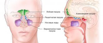
The apices of the roots of the teeth are located under the Schneiderian membrane, that is, under the mucous membrane that lines the bottom of the maxillary sinus.
This is the normal anatomical structure of the sinus - when the tops of the teeth are located in it. Some people have the tops of the fourth, fifth, sixth, seventh there. Some people only have the sixth. Some have sixth and fifth. But, one way or another, they are almost always there.
When a tooth is affected by caries, the infection penetrates through the dentinal tubules into the pulp chamber. Through it, the infection slowly progresses further through the blood and lymphatic vessels into the periodontium of the tooth.
Pulp microvessels emerge from the periodontium directly under the mucous membrane of the sinus, and over time it begins to react with inflammation.
Immune system cells responsible for protecting against infection initiate chronic inflammation. And the mucous membrane gradually thickens. This irritation happens all the time.
Infection from the carious process, which is thus restrained by immunocytes, causes a gradual degeneration of the structure of the sinus lining in the form of polyps and cysts.
Diagnostics
When teeth hurt due to sinusitis, the patient should be examined not only by an otolaryngologist, but also by a dentist. In case of inflammation of the maxillary sinuses, which are combined with damage to the molars (dental sinusitis), treatment is prescribed after the following examination:
- X-ray of the oral cavity and maxillary sinuses;
- diaphanoscopy (examination of the paranasal sinuses with a narrow beam of light);
- MRI or CT;
- puncture of the maxillary sinus.
In addition, to obtain the maximum clinical picture, endoscopy is used. To carry it out, a microscopic endoscope is used, which is inserted through pre-extended small holes connecting the paranasal sinuses and its cavity. This allows you to expand the viewing angle and enlarge the image of the pathological focus.
Can a dentist diagnose sinusitis?
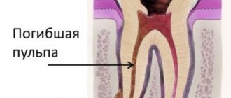
In case of pulpitis, the patient finally comes to the dentist. The doctor fills the canals, removes the nerve, but part of the infection still remains in the apical delta - after all, it has penetrated there a long time ago!
The apical delta is a branching at the tips of the roots that can never be adequately sterilized. And if a tooth has many roots, then, naturally, the entire system of root canals is one, like the root of one tree.
Therefore, if an infection gets into a tooth, it is already inside.
And it’s normal that the body fights it, and the sinus mucosa begins to thicken. In the process, some people develop granulomas, others - cysts. But cysts are changes in the periodontium, and the mucous membrane itself begins to thicken and degenerate.
Sometimes it happens that polyps are formed - spherical thickenings on a stalk. Hypertrophy of the columnar epithelium may occur. That is, as a result of chronic inflammation, many, many layers are formed.
The dentist can conduct a computed tomography scan, but it can only identify changes in the mucous membrane in volume - simply establish the presence of this fact.
And only an otolaryngologist can say what exactly happened there. To do this, you need to conduct an endoscopic examination.
But in most cases, it is the dentist who is the first doctor to detect sinusitis on a CT scan.
When a person realizes from various symptoms that he has inflammation in the sinus, he also learns about this from the dentist.
Treatment of periodontitis
The endodontist explained to the patient that in order to stop the inflammation, it is necessary to eliminate its cause - that is, eliminate the infection in the tooth canals. Retreatment of the tooth canals will be carried out under a microscope, which allows you to better clean the canals and see the branches along the canals.
Commentary by endodontist T.I. Matienko: “When periodontitis with an abscess develops, it is most often suggested to remove the tooth, but at the clinic we try to save the tooth, which is why treatment is carried out under a microscope, which improves the quality of treatment and helps to see many problems (narrow canals, canal branches, organic residues on the canal walls etc.) that are not visible to the naked eye. The decision on the choice of treatment tactics is made very carefully, after a thorough analysis of the computed tomogram. Not all teeth can be saved by retreatment. Sometimes I recommend removing a tooth because, based on some signs visible on a CT scan, its treatment will not be effective.”
To treat periodontitis, the canals were unfilled, cleaned with special endodontic instruments, washed with medicinal solutions and dried. A medicinal paste is placed in the canals to completely suppress the infection inside the tooth and surrounding tissues. After 3 weeks, the process of washing, drying and adding the medicine was repeated. After another 3 weeks, the patient did not experience any unpleasant sensations either in the tooth area or in the maxillary sinus area, so the canals were again washed, dried and hermetically sealed with hot gutta-percha, which penetrates well into all branches and bends of the canals.
The duration of each visit is about 1.5-2 hours, since root canal treatment requires careful adherence to the treatment protocol (use of a rubber dam, adherence to the time of exposure to medicinal substances) and attentiveness from the doctor.
After canal treatment, a ceramic inlay is installed in the tooth (E.max ceramics). An inlay in a tooth increases the strength of the tooth and its resistance to chewing loads. In this case, the tooth was badly damaged, and a large filling would have been unreliable.
Sinusitis or tooth – what to treat first?
In this case, many otolaryngologists prescribe tooth extraction.
In our clinic, we collaborate with specialists who treat odontogenic sinusitis or help the patient prepare the sinus mucosa for sinus lifting.
Of course, it is not always possible to prosthetize and treat a tooth with a source of infection in the periodontium in the upper jaw. Therefore, such teeth often still need to be removed.
Tooth extraction surgery is a major source of irritation for the body and the immune system. After it, you need to wait 4-6 months until the porous bone tissue of the alveoli is restored.
In this case, conservative anti-inflammatory therapy, antibiotics, and active copious rinsing of the nasal sinuses are prescribed. Treatment lasts about a month after tooth extraction.
If CT and diagnostics revealed a source of infection (cyst or granuloma) on the tooth, then treatment for odontogenic chronic sinusitis begins immediately after removal. Then the chance to reduce inflammatory changes in the mucosa is quite high.
Otherwise, the patient risks getting large growths, which sometimes occupy more than half the height of the sinus.
This is manifested by nasal congestion, because when we have ARVI, the nasal mucosa swells greatly. And our nasal mucosa is exactly the same as in the sinus: the same columnar epithelium. It swells very much when initially, as a result of constant irritation, the infection has already grown by more than half.
In this case, the half of the nose that is more blocked will be an indicator to check what is wrong with the teeth on the same half of the jaw.
Treatment result
A follow-up visit was scheduled 6 months after treatment, but since nothing bothered her, the patient refused the visit and came to the dentist 3.5 years later with another problem.
The CT scan shows:
- calm state of the maxillary sinus mucosa;
- complete restoration of bone tissue around the roots of the tooth;
- high-quality sealed canals;
- hermetic tooth restoration with an inlay to replace the old filling;
- complete restoration of bone tissue in the area where there was an abscess.
Analysis of computed tomography confirms that the treatment was very high quality and effective.
The cost of retreatment of a tooth with 3-4 canals as of September 2019 is 27,500 rubles, the cost of a ceramic inlay per tooth is 45,000 rubles, the cost of a dental CT scan is 2,900 rubles.
Implant into a modified sinus - is it possible?
Some time after tooth extraction, the patient may decide to install an implant. If sinus treatment was not carried out in a timely manner after tooth extraction, it remains to treat sinusitis before sinus lift surgery and implant installation.
It is advisable to do this together with an otolaryngologist.
But at the same time, the patient must understand that during a sinus lift, the altered sinus may rupture because it has an altered structure. And if this does happen, the sinus lift may not take place. Therefore, odontogenic sinusitis still needs to be treated during tooth extraction. And prepare the sinus together with the otolaryngologist.
Specialists who took part in the treatment:
- Dentist-endodontist – diagnosis of periodontitis, treatment of tooth canals under a microscope, tooth restoration with a ceramic inlay.
- ENT A.V. Arkhandeev – diagnosis and treatment of sinusitis.
- Dental assistant A. Antoshkina.
- ENT assistant E. Shilova.
See other examples of the work of specialists from the Dial-Dent clinic here.
Make an appointment for a consultation by phone +7-499-110-18-04 or through the form on the website. You can ask questions about dental treatment to the chief doctor of the clinic, Sergei Vladimirovich Tsukor, at
Types of odontogenic inflammation
The disease begins with a serous form. It is characterized by swelling of the mucous membrane, nasal congestion, dilation of capillaries, and increased secretion production. Swelling of the tissues leads to a narrowing of the anastomosis, the mucous masses do not find a way out and mucus with pathogenic contents accumulates in the sinus. This is how serous inflammation turns into purulent inflammation.
There are also acute and chronic forms of the disease. Chronic odontogenic sinusitis occurs due to improper treatment of acute sinusitis. Chronic odontogenic sinusitis lasts for years, worsening during a decrease in immunity or when infected.
Odontogenic sinusitis is diagnosed by an otorhinolaryngologist, and treatment is carried out by two specialists: a dentist and an otorhinolaryngologist.
Symptoms of the disease
The symptoms of the disease are similar to classic inflammation of the maxillary sinuses. Only a highly qualified otolaryngologist will be able to distinguish odontogenic sinusitis from ordinary sinusitis.
Signs of odontogenic sinusitis are:
- constant nasal congestion;
- purulent nasal discharge with a characteristic odor;
- the appearance of odor from the patient’s mouth;
- pain in the cheek area under the eyes;
- acute toothache in the upper jaw;
- elevated body temperature.
Symptoms also include a general deterioration in well-being, a persistent feeling of fatigue, and problems sleeping.
Specific symptoms of purulent odontogenic sinusitis are pain when lightly pressing on one of the cheeks or when lightly tapping on the teeth located in the area of the inflamed sinus.
Chronic odontogenic sinusitis is manifested by discomfort in the affected sinus area. In general, the patient feels satisfactory until a period of exacerbation occurs.