05.11.2019
The maxillary sinus in humans is located directly above the gum. Sinusitis after tooth extraction is caused by the penetration of an infectious agent into the sinus. This complication occurs infrequently, but is quite severe.
Classification and manifestations of pathology
According to the duration of the disease:
- acute - no more than three weeks;
- subacute - duration of illness up to 6 weeks;
- chronic - the disease lasts more than 6 weeks.
Depending on the location of the inflammation:
- pathology on the left;
- pathology on the right;
- bilateral pathology.
When pathology develops due to an extracted tooth, unilateral sinusitis most often develops, but if left untreated, the infection can spread to both sides.
In any case, the disease manifests itself with the same symptoms, but in acute pathology they are more pronounced:
- nasal congestion;
- profuse mucous or purulent runny nose;
- pain from the extracted tooth (localized in the cheek or under the eye);
- throbbing headache;
- pain when tapping on the cheekbones;
- fever;
- general intoxication syndrome - fever, chills, weakness
A distinctive feature of odontogenic sinusitis is its connection with previous dental procedures.
How does maxillary sinusitis manifest?
When visiting an ENT doctor, patients usually complain of heaviness, sometimes pain or swelling of the soft tissues in the area of one maxillary sinus, prolonged runny nose, hyposmia (decreased sense of smell) or, conversely, a constant unpleasant odor in the nose. Often such patients report frequent and painful sinusitis, multiple punctures, numerous courses of antibiotics, which brought only a temporary improvement in their condition or did not help at all. And only with a detailed collection of the medical history is it possible to find out about the recent or long-standing treatment of the “causal” teeth by the dentist, namely “7s”, “6s” or “8s” of the upper jaw of the corresponding side. But it happens the other way around: the patient does not make any complaints, but when preparing for dental prosthetics or sinus lifting, the dentist identifies pathology of the maxillary sinus on a computer tomogram and refers it to an ENT specialist. The reason for such careful preparation and alertness of our colleagues is the golden rule of planned surgery - the absence of inflammatory changes in the surgical area, because otherwise there is a high risk of implant rejection, scarring and a generally worse outcome of the operation.
Diagnostics
Diagnosis is carried out by an ENT doctor, and, if necessary, by a dentist. It is based on an analysis of the medical history, examination data, instrumental and laboratory examination.
- Anamnesis collection. The doctor listens to the patient’s complaints, asks about the dental procedures performed, and finds out how long ago the manifestations of the disease began.
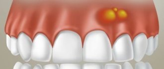
- Inspection. Swelling of the cheek, upper lip, and redness of the skin over the source of inflammation are detected. Palpation reveals pain in the area of the maxillary sinuses. During rhinoscopy, swollen and reddened mucous membrane is noticeable.
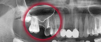
- X-ray of the maxillary sinuses. The image reveals a darkening of the cavity, and fragments of the tooth roots are visible.
- CT. Determines perforations and the presence of root fragments in the sinus as accurately as possible.
- Sinus puncture. A diagnostic and therapeutic procedure that allows you to both identify the contents of the sinuses and rinse them with antiseptic solutions.
- General blood analysis. An increase in leukocytes and an increase in ESR are detected, which confirms the presence of an active focus of infection.
Differential diagnosis with allergic and rhinogenic sinusitis is also carried out.
What is odontogenic sinusitis?
- Sinusitis is an inflammation of the maxillary sinus, the causes of which can be different.
- If the cause is a diseased tooth, then it is odontogenic sinusitis.
- Causes of odontogenic sinusitis: massive caries, purulent-inflammatory pulpitis, improper dental treatment, filling material getting into the sinus.
- Today, such sinusitis is not uncommon, but eliminating it completely is very difficult.
- Typically, patients with odontogenic sinusitis turn to either ENT doctors or dentists, although joint management of such a patient by an ENT doctor and an oral and maxillofacial surgeon is mandatory.
! A patient with odontogenic sinusitis must be observed by two specialists - an otolaryngologist and an oral and maxillofacial surgeon.
Treatment methods
Therapeutic tactics depend on the course of the disease. Treatment of the chronic form is carried out in a clinic, while acute purulent sinusitis requires hospitalization.
Treatment of odontogenic sinusitis after tooth extraction begins with removing the source of infection and establishing drainage to remove pus. The cavity is washed with an antiseptic solution and hypertonic saline solutions.
An oral antibiotic must be prescribed. The course of treatment is 7-10 days. After this, the patient is advised to carefully observe the hygiene of the nasal and oral cavity and not to become overcooled.
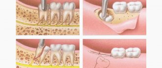
If perforation of the maxillary sinus occurs during tooth extraction, treatment depends on the duration and extent of the injury. A small fresh defect is covered with a plastic plate.
Old injuries, with the formation of fistulas and purulent discharge, first require careful sanitation of the sinus. Then part of the gum is removed and bone grafting is performed.
Treatment Options
A feature of the treatment of sinus perforation is its urgency, since infection occurs literally in a matter of hours.
If a “pure” perforation occurs, not complicated by a foreign body, pus, etc. remaining in the cavity, then the first aid will be the formation of a blood clot that will close the hole. To do this, a small swab containing iodoform is carefully inserted into the lower part of the socket. If it is not fixed tightly enough, silk thread sutures are placed on the gums, and sometimes mouthguards are used. After a few hours, a blood clot forms and is held in place by a tampon for five to seven days. During this time, the sinus mucosa grows together and a scar forms. At the same time, a course of anti-inflammatory drugs and antibiotics is prescribed to avoid complications.
In the case when a foreign body remains in the maxillary sinus, an inpatient operation is performed, during which the sinus is opened and the object is removed, dead tissue is cleaned out and perforation plastic surgery is performed. The use of anti-inflammatory drugs is indicated for two weeks.
Perforation of the sinus can occur not only during the removal of upper teeth, but also during implantation, prosthetics, endodontic dental treatment, and during the insertion of a pin. In such cases, perforation is always a medical error caused by the incompetence of the dentist.
Features of the maxillary sinus
The maxillary sinus (its other name is the maxillary sinus) is located in the thickness of the bone tissue of the upper jaw. It is separated from the oral cavity by the alveolar process of the upper jaw, which forms its bottom. The volume of such a sinus is quite large, and in adults it can reach 10 cubic centimeters.
This sinus, or sinus, is not airtight. It communicates with the nasal cavity through a narrow slit.
Typically, perforation of the maxillary sinus occurs in the area of its bottom. Some of its features contribute to this:
- Close proximity of the roots of molars and premolars. In some cases, the thickness of the bone layer between the tooth roots and the bottom of the maxillary sinus can be relatively large - up to 1 cm, but in some people the bone border between these formations is very thin - no more than 1 mm.
- Sometimes the roots of the first and second molars are located in the sinus cavity itself, separated from it by just a layer of mucous membrane.
- Rapid thinning of the bone layer in the presence of acute or chronic inflammatory diseases: periodontitis, periodontitis, cysts.
- Relatively thin bony trabeculae in the tissue of the upper jaw.
All this predisposes to the occurrence of perforation during dental procedures, even if the treatment technique was not violated and the doctor did not apply significant traumatic force.
Navigation
In ENT practice, there are cases when a patient comes to an otolaryngologist with complaints of severe nasal congestion and suppuration from the nasal cavity. At the same time, the patient has an elevated body temperature and headaches. But the ENT doctor, having examined the patient, sends him to see a dentist. “Why?”, you will be surprised. Because in this case we are dealing with odontogenic sinusitis.Sinusitis is a type of sinusitis. The development of sinusitis of odontogenic nature is not associated with colds or acute respiratory viral infections. The main cause of the disease is bad teeth. First, the development of inflammatory tooth disease occurs, after which the inflammatory process from the upper jaw extends beyond the oral cavity and is localized in the maxillary sinus.
Such a state cannot be tolerated. Timely detection of symptoms and diagnosis carried out by a competent ENT doctor will allow timely diagnosis of dental sinusitis and avoid serious complications.
What are the maxillary sinuses
The maxillary sinuses (also called the maxillary sinuses) are special cavities on both sides of the nose that are filled with air.
Each cavity is connected to the nasal passage by small openings called anastomoses. The cavities are covered with mucous membrane. The function of mucus is to trap bacteria and harmful particles in it, and then remove them from the body through those same anastomoses. When edema occurs, the excretory opening becomes very narrow, as a result of which mucus, along with harmful particles and bacteria, cannot come out and stagnates. At this time, the patient begins to experience bursting pain in the cheek area - this is how inflammation of the maxillary sinus begins. Treatment of the maxillary sinus should not be neglected, since inaction can provoke serious consequences, including sepsis and meningitis. Classic sinusitis can be bilateral, when both sinuses are affected. In the odontogenic form, the inflammatory process starts in the sinus on which side the diseased tooth is located.
Types of odontogenic inflammation
The disease begins with a serous form.
It is characterized by swelling of the mucous membrane, nasal congestion, dilation of capillaries, and increased secretion production. Swelling of the tissues leads to a narrowing of the anastomosis, the mucous masses do not find a way out and mucus with pathogenic contents accumulates in the sinus. This is how serous inflammation turns into purulent inflammation. There are also acute and chronic forms of the disease. Chronic odontogenic sinusitis occurs due to improper treatment of acute sinusitis. Chronic odontogenic sinusitis lasts for years, worsening during a decrease in immunity or when infected.
Odontogenic sinusitis is diagnosed by an otorhinolaryngologist, and treatment is carried out by two specialists: a dentist and an otorhinolaryngologist.
Causes of odontogenic sinusitis
It is very easy to understand why acute odontogenic sinusitis occurs - just look at the anatomy. If you look at the structure of the skull, you can see: the 5th, 6th and 7th teeth of the upper jaw are located very close to the maxillary sinus. There are cases that these teeth even go into her cavity. An infection in a tooth easily thins the already small distance between the tooth and the wall of the sinus and easily penetrates inside, thus starting the inflammatory process.
Development in acute form occurs due to the following reasons:
• Improper oral hygiene. The most common cause of the disease is neglect of basic rules of oral care: improper and irregular brushing of teeth, delaying a visit to the dentist when there are problems with the teeth. At particular risk are those patients who have been diagnosed with an advanced form of caries with developing necrosis of the dental nerve. • After tooth extraction. Infection after tooth extraction is associated with the formation of a fistula - a small channel through which the infection enters the maxillary sinus. This occurs when the roots of a diseased tooth enter the maxillary cavity. • Incorrectly installed seal. Unfortunately, there are cases when the development of inflammation is facilitated by the inept actions of insufficiently qualified dentists. Part of the filling can get into the sinus, and it will immediately be perceived by the body as a foreign particle, and the inflammatory process will start. If after visiting the dentist you have a runny nose, you need to consult an otolaryngologist. • Oral diseases. Caries, gum disease, periodontal disease, the presence of cysts are all diseases in which there is a source of infection on the oral mucosa. • Reduced immunity. Decreased immunity leads to the activity of bacteria that are constantly in the oral cavity.
Symptoms of the disease
The symptoms of the disease are similar to classic inflammation of the maxillary sinuses. Only a highly qualified otolaryngologist will be able to distinguish odontogenic sinusitis from ordinary sinusitis.
Signs of odontogenic sinusitis are:
• constant nasal congestion;
• purulent nasal discharge with a characteristic odor; • the appearance of odor from the patient’s mouth; • pain in the cheek area under the eyes; • acute toothache in the upper jaw; • increased body temperature. Symptoms also include a general deterioration in well-being, a persistent feeling of fatigue, and problems sleeping. Specific symptoms of purulent odontogenic sinusitis are pain when lightly pressing on one of the cheeks or when lightly tapping on the teeth located in the area of the inflamed sinus.
Chronic odontogenic sinusitis is manifested by discomfort in the affected sinus area. In general, the patient feels satisfactory until a period of exacerbation occurs.
Diagnosis and treatment
Treatment of an inflamed maxillary sinus, like any other disease, begins with diagnosis and determination of the causes that led to the inflammatory process.
Odontogenic sinusitis is an insidious disease that must be treated under the supervision of an otolaryngologist. He will identify the exact cause of the disease and tell you how to treat the disease. Diagnosis begins with examination of the sinuses. To identify and treat a diseased tooth that has caused inflammation, the patient is sent for an X-ray examination, and in especially severe cases, a computed tomography scan. Effective treatment consists of several stages. Traditionally, the first stage of treatment is to eliminate the cause of suppuration. If the root cause of the disease is a problematic tooth, then at the initial stage the disease is treated by tooth extraction. Extraction is carried out in dentistry - before removing a tooth, you need to approach the choice of clinic with all responsibility, since an inexperienced doctor can only aggravate the situation.
If we are dealing with perforation of the maxillary sinus and penetration into it, for example, of part of the material for filling a tooth, then surgical treatment is indicated. The operation is performed to remove a foreign body from the sinus and subsequently eliminate the defect site.
If sinusitis without perforation, in order to release the purulent mass from the sinus, a puncture (puncture) of the maxillary sinus is performed.
Elimination of symptoms involves taking antibacterial drugs, rinsing the nasal cavity, using vasoconstrictor drops, and also performing physical procedures.
A highly qualified ENT doctor, modern equipment and a flexible pricing policy are the three components of the success of our clinic.
If you are faced with the problem of sinusitis, do not delay visiting the doctor!