When removing upper molars and premolars, the doctor must act with extreme precision and accuracy. After all, incorrect actions threaten that fragments of the root may get into the cavity of the maxillary sinus. Perforation is also possible when installing an implant used in implantology and in root canal treatment.
Perforation of the bottom of the maxillary sinus in the upper jaw is one of the complications that occurs after tooth extraction in the upper jaw. This is not a separate disease, but during dental exposure it occurs quite often. From the article you will learn what kind of pathological condition this is, how it can be eliminated surgically and therapeutically, and what it means for the patient.
The maxillary sinus is located in the thickest bone tissue located in the upper jaw. It is separated from the oral cavity by the so-called alveolar process, which is required for the formation of the floor. According to physiological indicators, the volume of the nasal sinus in adults can reach sizes of up to ten cubic centimeters.
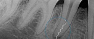
The location of the maxillary sinus poses a danger when performing certain surgical operations on teeth in the upper jaw. It's all about the roots of the teeth, which can reach the bottom of the nasal sinus or even penetrate the alveolar process. This can only be seen on a targeted photograph, which must be taken by the dentist before surgery.
Molars and premolars are located closest to the maxillary sinuses. It is worth saying that perforation can be predisposed not only by the particularly anatomical location of the upper teeth, but also by the presence of cystic neoplasms, periodontitis, and periodontitis as the main secondary factor.
Causes of perforation
The physiological location of the upper teeth and associated pathologies are a risk factor that can cause perforation of the maxillary sinus. Complications can be characterized by dental operations. In particular, this is the sanitation of dental tissues in case of deep caries, the removal of part of the tooth or its root. It is important for the dentist to carry out surgical intervention extremely carefully without fragmenting or causing fractures of the tooth roots, which is why mandatory x-ray monitoring is required.
When carrying out dental treatment associated with the removal of an upper wisdom tooth, perforation can occur when manipulating the root of the tooth, which is often removed in fragments. Pathology also occurs during manipulations in the root of the tooth during the treatment of caries and when intensive canal filling is done.
Perforation may occur during root resection and installation of implants in the upper jaw. The dentist must act with extreme caution when removing the upper tooth. First, an image of the upper jaw is taken, which allows one to assess the location of the roots relative to the maxillary sinus, and then the doctor determines the subsequent tactics of therapeutic or surgical treatment.
Article on the topic: Open and closed sinus lift surgery
What factors are taken into account by the dentist when performing an intervention?
The likelihood of sinus perforation increases when the tooth root is located in close proximity to it at the time of extraction. Therefore, surgical treatment should be carried out with extreme care and subsequent X-ray monitoring of the tissue condition after surgery. The dentist also monitors the condition of tissues when using pins for implantation or when treating root canals for filling purposes, since there is a high risk of filling material and root fragments penetrating into the maxillary sinus.
If the process occurs at the time of implantation into bone tissue or when filling canals, then this is a direct mistake that is made by the doctor during his therapeutic tactics. The only way to control manipulations is X-ray control and the experience of the dentist, who, when working, must take into account the anatomical structure of the patient’s upper jaw. In this case, damage to the bottom of the sinus is fraught with serious complications that are difficult to eliminate. Especially if this happens during the implantation of artificial roots. The bone tissue in the upper jaw quickly undergoes degenerative changes, and this leads to a decrease in the height of the alveolar process.
When performing root resection to eliminate cystic formations, as in the above cases, the dentist must fully examine the patient. If the history is insufficient, perforation may occur in a situation where the doctor does not know the exact size of the bone plate that separates the bottom of the sinus from the wall of the cyst itself. In this case, the prognosis worsens if it is necessary to remove a large volume of bone tissue.
Symptoms of perforation of the maxillary sinus
The symptoms of the disease are specific, especially if they occur after tooth root removal. In this case, the patient can note it virtually immediately after the intervention. It is as follows:
- The appearance of air bubbles in the blood that is released from the tooth socket. The number of bubbles increases with a sharp exhalation of air through the nose;
- The appearance of bloody discharge from the nose is noted. They can be scanty in nature and are detected from the side of the maxillary sinus perforated by the dentist;
- Sore throat, presence of nasal sound, the patient additionally notes a state of nasal congestion;
- Patients may also complain that they feel air passing through the hole. There may be a feeling of heaviness in the projection of the upper jaw. The oppressive feeling does not disappear, but only intensifies over time.
If such symptoms appear, you should immediately seek help from a doctor, who will examine the patient and do an X-ray examination, based on which it will be possible to judge the degree of perforation.
The dentist may suspect perforation of the maxillary sinus during implantation based on certain signs. The main one is the failure of the pin, which it is simply no longer able to strengthen at the base of the bone tissue. The doctor may also suspect the presence of pathology based on the following characteristic signs:
The appearance of small air bubbles in the hole;
- Infection of the cavity. As a rule, this happens because the diagnosis of perforation was not carried out completely or was not done completely;
- The patient complains of sharp, unpleasant and aching pain in the sinus area;
- Hypertrophy and swelling of the mucous membrane;
- The patient has difficulty breathing, with the development of an infectious process the appearance of purulent discharge.
General symptoms include fever, weakness, chills, headaches, which may accompany the inflammatory process.
Prevention of tooth sensitivity
A few decades ago, it was possible to escape from increased sensitivity of teeth only with folk remedies, but today there are a lot of special drugs that help in the prevention and treatment of this pathological condition - these are toothpastes, brushes and rinses, which, when used regularly, help cope with increased sensitivity. Based on the fact that the composition of all drugs is different, experts recommend changing the medicinal paste or rinse from time to time. But it is better to avoid using whitening pastes.
In addition, there are a number of standard recommendations, the implementation of which will protect the enamel from damage:
- Regular, thorough oral hygiene (2 times a day - morning and evening, during the day it is recommended to rinse your mouth after each meal). It is advisable to use a brush of medium hardness.
- The diet should be well balanced - vitamins, minerals, nutrients. Be sure to include dairy products, fish, and meat in your menu. But it is better to limit sweets and spicy foods as much as possible. Also, to preserve the integrity of the enamel, try to avoid dry solid foods - crackers, seeds, etc., as well as carbonated drinks.
- Consult your doctor and choose the appropriate course of vitamins and minerals for yourself.
- Don't forget to visit your dentist regularly - for a routine examination you need to make an appointment at least once every six months.
If, despite all preventive measures, your teeth react to cold or hot, be sure to consult a doctor - perhaps the problem lies deeper and only an experienced specialist can identify its cause.
Author: Elena Grunina Dentist-therapist, endodontist. Work experience more than 9 years. The information is for reference only. Before treatment, consultation with a doctor is necessary.
Methods for diagnosing perforation of the maxillary sinus floor
During diagnosis, a comprehensive study and analysis of the typical clinical picture observed with perforation of the maxillary sinus is used. The condition of the tooth socket, the presence of air bubbles, nosebleeds, and pain are taken into account. X-ray diagnostics are also performed, which show the area of perforation with high accuracy.
The dentist can perform a probing procedure for a perforated canal or an extracted tooth using a thin medical probe. This helps to reliably understand that there is no bone bottom in the wound. The instrument itself must pass freely through soft tissue without encountering obstacles along the way.
X-rays of the sinuses can detect characteristic darkening in the images, which are formed as a result of the accumulation of blood clots. Among other things, X-rays can show filling material, implants, and fragments of dental roots. X-rays with a contrast agent are often taken to help clearly visualize the condition of the bone tissue and surrounding tissues.
In such situations, a contrast agent is introduced into the cavity through a perforation fistula. An additional method of instrumental diagnostics is computed tomography, which determines the degree of perforation and the presence of foreign bodies, such as tooth fragments or residual filling material. If perforation is suspected, general clinical tests are required, which can show the presence of a focal infection in the patient’s body.
Classification and manifestations of pathology
According to the duration of the disease:
- acute - no more than three weeks;
- subacute - duration of illness up to 6 weeks;
- chronic - the disease lasts more than 6 weeks.
Depending on the location of the inflammation:
- pathology on the left;
- pathology on the right;
- bilateral pathology.
When pathology develops due to an extracted tooth, unilateral sinusitis most often develops, but if left untreated, the infection can spread to both sides.
In any case, the disease manifests itself with the same symptoms, but in acute pathology they are more pronounced:
- nasal congestion;
- profuse mucous or purulent runny nose;
- pain from the extracted tooth (localized in the cheek or under the eye);
- throbbing headache;
- pain when tapping on the cheekbones;
- fever;
- general intoxication syndrome - fever, chills, weakness
A distinctive feature of odontogenic sinusitis is its connection with previous dental procedures.
Treatment of the pathological process
Treatment depends on the severity of the observed pathological process. Typically, surgical intervention is not required in situations where the perforation was carried out during the extraction of a diseased tooth and was detected at the same moment. In this case, X-ray data should show the absence of an infectious process in the maxillary sinus.
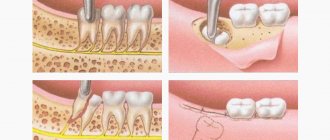
If there are no fragments, filling material or infection in the wound, then in this situation the dentist tries to form a blood clot in the hole after tooth extraction and make sure that the resulting cavity is not susceptible to infection. For such purposes, a small gauze swab can be used, which is soaked in iodine solution. Iodine itself has good bactericidal properties, allowing you to quickly eliminate all pathogenic microorganisms.
Most often, such a tampon can self-fix in the resulting wound cavity after tooth extraction. Sometimes stitches are required on the gums, which are done directly by the dentist. Treatment with an iodine solution is carried out for six to seven days until granulation tissue forms, which allows the perforation to close.
The dentist can temporarily close the defect using a special hypoallergenic plate made of plastic. The plate itself is securely fixed to adjacent teeth using clasps. It carefully separates the cavities of the maxillary sinus and mouth, which in turn promotes rapid healing and regeneration of healthy tissue in the perforation area.
It is important to carry out preventive measures aimed at preventing the development of any inflammatory complications. After all, after tooth extraction during perforation, pathogenic microorganisms that can cause a serious inflammatory process can enter the wound surface.
Prevention lies in the mandatory use of anti-inflammatory drugs and antibiotics prescribed by the doctor; agents that have a vasoconstrictor effect are also used to stop bleeding.
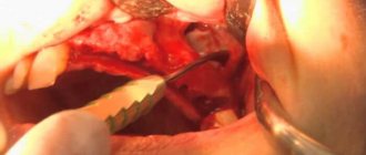
If foreign bodies penetrate into the maxillary sinus during perforation (fragments of tooth roots, filling materials, implant perforation), treatment is carried out in a hospital setting. In severe cases, surgical interventions can be performed on the maxillary sinus with its further opening, during which tissue is cleansed and foreign bodies are removed. After sanitation, plastic closure of the resulting perforation is performed.
Old perforations that occur after tooth extraction or therapeutic treatment
If perforation was not detected after tooth extraction, the patient may attribute the discomfort to the consequences of surgery. It is worth saying that after a couple of weeks the stage of acute pain passes, and in the area of the resulting defect in the maxillary sinus, a so-called fistula appears, which connects the surface of the gum to the sinus.
This process is accompanied by symptoms characteristic of chronic sinusitis; among other things, patients complain of pain, purulent discharge from the nasal cavity, and swelling of the cheek. Treatment consists of the use of therapy aimed at stopping the inflammatory process and surgical methods that eliminate foreign bodies in the maxillary sinus.
If we summarize the above, we can say that the removal of teeth in the upper jaw, as well as their treatment, should be carried out as competently as possible. Perforation of the maxillary sinus can have quite serious consequences for the patient’s body. In some cases, long-term hospital treatment is required. The addition of an infectious process is especially dangerous.
Such a complication can be avoided if the dentist is sufficiently competent, who must take into account the anatomical position of the teeth and the maxillary sinus before performing any complex dental procedures related to the upper jaw. Based on this, the dentist is obliged to highlight all the anatomical and topographical features of the patient and conduct qualified treatment.
Before the procedure for removing, implanting or treating upper teeth, the doctor must take a targeted photo, which will help visualize the area with which he will be working. If these conditions are met, such a serious complication as perforation of the maxillary sinus can be avoided.
Preventive actions
Prevention of perforations of the floor of the maxillary sinus consists of:
- in a full examination of the patient before complex dental procedures;
- in the correct assessment of the anatomical and topographical characteristics of each person;
- in strict adherence to the technology of therapeutic manipulations.
Timely detection of signs of perforation and its adequate treatment is the key to a favorable outcome for the patient. Incorrect therapeutic tactics or self-medication can aggravate the course of such a complication and cause the development of severe negative consequences.