Perforation of the maxillary sinus during the treatment of upper teeth is damage to its shell when trying to treat root canals without prior examination. The penetration of infection provokes inflammation with symptoms of sinusitis, which cannot be treated with medication.
For more than 20 years, Doctor Levin has specialized in eliminating complications after unsuccessful dental manipulations in the area of the maxillary sinuses. Surgical treatment is performed by maxillofacial surgeons with ENT training.
Causes of sinus perforation during dental treatment
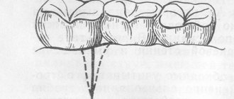
The maxillary sinuses or maxillary sinuses are located inside the upper jaw. They are separated from the oral cavity by a septum and the alveolar process, which contains the roots of the teeth. Treatment of the canals of such teeth is complicated by the risk of sinus perforation for a number of reasons:
- Anatomically thin septum Sometimes the height is only 1 mm. It happens that the teeth penetrate the cavity with their roots and only the sinus mucosa separates.
- Treatment of teeth with inflammation on the roots If there is inflammation on the roots of the teeth (periodontitis, cyst), the surrounding bone melts and becomes thinner.
- Excessive efforts by the doctor If the dentist does not calculate the efforts when passing the canals, the canal may rupture, damage the root, or perforate the bottom of the sinus.
Without a preliminary diagnosis, the endodontist, having no idea about the size of the sinus septum and the location of the roots, may not calculate the effort and damage the tooth and the thin bone layer that lies between the maxillary sinus and the root. As a result, infection penetrates into the sinus cavity, and fragments of instruments and filling material may fail. In our practice, there are cases when a foreign body in the sinus attracts secondary infections with the development of neoplasms - fungal colonies, cysts, polyps.
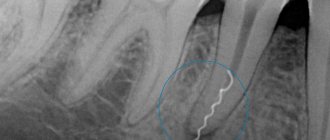
A fragment of an endodontic instrument in a tooth canal
Clinical picture
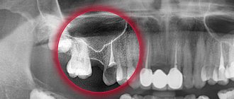
Anatomically, the maxillary sinuses are very close to the roots of the upper teeth.
Damage is likely in the maxillary sinus, where the roots of the following teeth are located:
- first molars or second;
- second premolars.
Risk factors:
- acute periodontal inflammation of the maxillary cavity above the upper teeth;
- osteomyelitis;
- formation and swelling of cysts in the upper jaw;
- traumatic damage to teeth;
- building up bone material inside the gums during dental treatment;
- procedure for installing prostheses;
- improper tooth extraction, resulting in swelling of the upper jaw;
- caries, which causes inflammation of the roots of the tooth.
Entering as a foreign body, it causes tissue irritation, and when a hole is formed, oral microflora can also penetrate through it into the cavity, which can cause the formation of ichor.
How to prevent perforation
If patients with anatomical features of the location of the roots in the sinus are treated according to the standard protocol, the threat of rupture of the membrane is very high
The doctor must have the necessary information about the anatomical and topographical features of the patient and be prepared for an emergency situation.
- To prevent perforation of the sinus membrane, computed tomography is required. After studying the CT image, the doctor selects the optimal treatment tactics. But not every equipment allows you to identify all the nuances. In our Center, diagnostics are carried out using a modern 3D tomograph Sirona Galileos with ENT mode settings. Provides high quality images, allows you to evaluate the structural features of the upper jaw and sinus, calculate the height of the septum, find out the location of the roots, their anatomy.
- When working with the canals of teeth located in the sinus, all manipulations must be extremely careful. The dental canals are very thin, sometimes tortuous, the doctor must control every action. Endodontic dental treatment in our Center is carried out using a Seiler dental microscope. Multiple magnification expands the view, allows you to examine the entire length of the dental canal in great detail, and prevent the filling material from leaving the root.
- The experience of a specialist is of great importance so that he can give an adequate assessment from a CT image, choose tactics, and calculate efforts. Our Center employs highly qualified endodontists, with excellent clinical thinking and honed manual skills. Qualifications and skills allow us to prevent complications and act promptly in the event of an emergency situation.
We have minimized the likelihood of complications after dental treatment
. Advanced CT diagnostics in ENT mode and work using a dental microscope allow us to select competent tactics to prevent perforation of the bottom of the maxillary sinus.
Levin Dmitry Valerievich Chief physician and founder of the Doctor Levin center
After endodontic treatment, control CT diagnostics is mandatory! This preventive measure allows you to timely identify perforation (even if the doctor did not notice it during treatment) and take measures to eliminate the consequences.
If a piece of instrument or filling material gets into the sinus, removal of the foreign body must be prompt and urgent ! Otherwise, there is a high risk of infection, development of inflammatory processes and neoplasms.
Preventive actions
The patient is unable to prevent a perforation of the maxillary sinus, and if this happens, he will not be able to overcome what happened without the help of a doctor. But an experienced dentist has the right to take measures to prevent an undesirable scenario. To do this, you will need to conduct a high-quality preliminary examination of the anatomical features of the person applying to the clinic and strictly follow the method of medical manipulation.
Having caused perforation, the doctor is obliged to draw up a treatment plan and bring the patient to a favorable outcome. And a person who experiences disturbing sensations after tooth extraction, treatment of pulpitis, or in other cases, should not endure and remain silent. A timely visit to the clinic will prevent the development of serious complications.
Signs if perforation was left without the attention of a doctor
Symptoms of inflammation after unsuccessful treatment occur in different ways - after a few days, weeks or even years. Depends on the sterility of the foreign body in the sinus, its ability to cause inflammatory processes, and the characteristics of the patient’s immune system. The following manifestations are possible:
- feeling of heaviness;
- pain when chewing;
- purulent and serous discharge from one nostril;
- elevated temperature;
- impaired sense of smell;
- pain when lightly tapped under the eye and in the nose area.
These are typical signs of odontogenic sinusitis caused by poor quality dental treatment. The key difference from rhinogenic sinusitis (when sinus infection occurs from the side of the nasal passages as a result of ARVI or influenza) is that signs of the inflammatory process are detected only on one side, where treatment was carried out.
What are the dangers of sinus injury?
Since the complication does not always manifest itself immediately, symptoms of sinusitis that arise after a few years force the patient to consult a regular ENT doctor. Treatment, as a rule, does not bring results - in most cases the cause is not found. You start going from one doctor to another, and precious time is lost. The longer a foreign body is in the sinus, the higher the risk of tumor formation. If sinus perforation is left unattended, it can lead to:
- chronic sinusitis and sinusitis
- inflammation of the roots of adjacent teeth
- osteomyelitis of the jaw
- encephalitis and meningitis
Why you should entrust your treatment to the ENT Department of Dentistry
ENT dentistry is a comprehensive approach to the treatment of complications in the maxillary sinuses after dental treatment
The symbiosis of two areas - dentistry and otolaryngology - makes it possible to identify the cause, assess the situation, and select competent treatment tactics.
The ENT department of the Doctor Levin Center has been providing assistance for many years when problems arise after treatment and removal of teeth located on the border with the maxillary sinus. Surgical treatment is carried out by candidates of medical sciences, maxillofacial surgeons with otolaryngological training .
Patients come to the Center after a painful search for a solution to the problem. Repeated treatment by an ENT doctor in the hospital does not bring results. But a thinking patient should understand that if there is concern on the side of the sinuses where endodontic treatment once took place, you need to contact an oral and maxillofacial surgeon with ENT training. Only in this case can a comprehensive assessment of the situation be made and an adequate treatment plan drawn up.
How can a complication be identified?
External signs of dental materials getting into the maxillary cavities and patient interview data may not be enough to make a correct diagnosis, and additional studies are carried out to confirm complications of the dental procedure:
- x-ray of the maxillary sinus;
- MRI;
- CT;
- puncture.
The diagnostic plan may include several procedures. Their number is determined by each doctor individually.
Diagnosis of perforation
Having an accurate understanding of the situation in the sinus, the maxillofacial surgeon chooses the correct tactics and method of eliminating the problem
As part of diagnostic activities, our Center carries out:
- X-ray allows you to assess the condition of the causative tooth and the quality of canal treatment
- Computed tomography in ENT mode allows you to get a complete picture of the condition of the maxillary sinuses
The modern Sirona Gallileos CT scanner guarantees high quality images , in which you can determine the location and size of the perforation, identify the presence and localization of foreign bodies, cysts and polyps (if they form), and assess the size of the inflammatory process.
Diagnostics and its features
The most effective way to make a correct diagnosis is computed tomography, but this does not exclude the use of other, more traditional methods:
- conducting a patient interview;
- use of rhinotherapy to study the maxillary sinus;
- X-ray examination;
- MRI;
- puncture of the maxillary sinus.
An experienced doctor can reliably assume the presence of such a disease, but it can be confirmed using computed tomography.
How is the treatment carried out?
We respect the patient’s personal time and strive to carry out all activities comprehensively, in one day :
- Professional hygiene Preparation of the oral cavity to ensure sterility during surgery to avoid secondary sinus infection
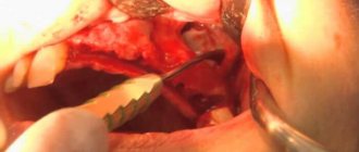
- Surgery to eliminate inflammation Performed while you sleep; the method of access to the sinus is selected depending on the location of the foreign body and the presence of tumors
- Temporary prosthetics If the causative tooth had to be removed, a temporary crown or immediate prosthesis is installed to mask the defect
Surgical operations in our Center are performed under sedation. The patient does not feel anything, fears and worries are excluded. Sedatives put you into a controlled drug-induced sleep without falling into unconsciousness - this is not general anesthesia! They do not contain toxic components, act gently, preserving reflexes. The artificial lung ventilation device is not connected, hospitalization is not required.
The surgical intervention is completed by a mandatory CT examination , which is necessary to assess the quality of the work performed.
After 10-14 days, the patient is invited to the clinic for suture removal and a control CT image. A schedule of professional inspections is drawn up.
How is a foreign body removed from the maxillary sinus?
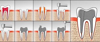
To prevent the development of complications in the presence of foreign objects in the maxillary sinus, minimally invasive surgical intervention is performed. Its main purpose is to remove filling material. The operation can be performed using optical instruments such as an endoscope or laparoscope. When carrying out such interventions, significant trauma to bone tissue and incisions is not required. These minimally invasive operations are pain-relieved with local anesthesia and take no more than half an hour. Traditionally, endoscopic techniques are used to remove foreign bodies from the maxillary sinuses. To perform the operation, a small incision is made above the upper lip through which an endoscope is inserted. This optical device helps the surgeon detect a foreign body. The dental material is then removed. When performing laparoscopic surgery, tissue puncture is performed in the place where the bone tissue is thinnest. A special instrument is inserted into the laparoscope, which captures and removes the foreign particle. This technique is used in cases where endoscopic intervention cannot be performed for some reason. Sometimes operations are supplemented by such manipulation as curettage. This method is necessary in cases where, due to the filling material, a granuloma has formed on the tissues of the cavity. After surgical removal of a foreign particle, the patient is prescribed anti-inflammatory drugs to prevent inflammatory and purulent processes. To achieve the desired result, antibiotics, immunostimulants and other drugs may be prescribed. In addition, the affected sinus is washed with antibacterial, antiseptic and anti-inflammatory solutions. To increase the protective functions of the body, the patient is prescribed vitamin and mineral complexes. The best prevention of such complications is regular visits to the dentist. In addition, it is necessary to exclude facial injuries, try to avoid stress, eat right and lead an active lifestyle.
No hospitalization required
Operations in our Center are performed by operating teams of experienced maxillofacial surgeons with ENT training in a sterile operating room. The treatment is as gentle and minimally traumatic as possible; a 24-hour hospital stay is not required; you will go home the same day .
The intervention lasts about an hour, under sedation. After completion, the patient quickly returns to clear consciousness without unpleasant consequences or risk of complications. After 30-40 minutes you can safely go home. For patients with concomitant cardiovascular diseases, a day hospital is provided . You can lie down for the time necessary for recovery under the supervision of our anesthesiologist-resuscitator.
Accelerated rehabilitation
After the operation, you will receive free medications and instructions with rules of behavior during the rehabilitation period. We have collected the necessary set of drugs to improve your well-being at home. Please take them as prescribed by your doctor and follow the recommendations to avoid unpleasant consequences.
For quick recovery after surgery, our Center offers its own method of rehabilitation in 1-2 days. The procedures allow us to minimize the formation of possible hematomas, swelling, and eliminate pain. The program uses:
Microcurrent therapy
A weak pulse current normalizes metabolic processes, activates ATP production, and improves tissue nutrition. Regeneration starts, healing accelerates, swelling decreases. Muscle spasm goes away.
PRP plasma therapy
Injections of purified and platelet-enriched plasma from one’s own blood trigger cellular regeneration processes using the body’s internal reserves. The manifestation of edema and hematomas is reduced.
Biostimulation of the face
Lymphatic drainage drugs D-NUCLEO and MesoSculpt C71 help reduce swelling. Active anti-inflammatory components improve blood flow, increase tissue nutrition, and improve healing.