The treatment of “fluxes” is carried out by a dental surgeon.
Do you feel afraid of the dentist?
Flux is the common name for any swelling in the cheek area and partly the neck - periodontitis, periostitis, complications of pulpitis and other inflammations. If you have inflammation of any origin or form, localized on the gums and in the oral cavity in general, you should definitely see a dentist.
Swelling in the cheek area, causes
The most common dental causes of cheek swelling are:
- advanced caries;
- complication of pulpitis;
- complication of gingivitis;
- periostitis, periodontitis and other diseases of teeth and gums.
If you suspect flux, call: 8 (495) 558-88-77
Flux is inflammation of tissues of various origins, for example, periostitis, an inflammatory process in the periodontal gap - periodontium. Such inflammation can occur in acute and chronic forms; the chronic form can periodically enter an exacerbation phase and then subside again.
By origin, periostitis is as follows:
- infectious;
- non-infectious;
- spicy;
- chronic;
The main role in the development of flux of infectious origin belongs to microorganisms, as well as their toxins. Microbes penetrate into the periodontium through the root canal, periodontal pocket, as well as through the blood or lymph flow.
Most often, flux is caused by an infection that enters through the root canal and is a consequence of acute diffuse and chronic gangrenous pulpitis, as well as pulp necrosis.
Microorganisms and their toxins, penetrating into periodontal tissue, cause dangerous acute inflammation
Non-infectious periostitis/periodontitis (fluxes) can develop as a result of trauma (blow, bruise, periodontal trauma after pulp extirpation, sharp, uncomfortable biting on a tooth; cracking nuts, gnawing bones) or chronic microtrauma (smoking pipe, brass band instruments, biting threads, pressing on the tooth with a pencil, pen, etc.); also as a result of the influence of medications - the ingress of potent substances into the periodontium during the expansion of root canals (trilon B, aqua regia), their sterilization (formalin, silver nitrate, etc.) and in the ingestion of arsenic paste.
Flux symptoms
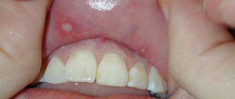
Clinical manifestations of acute periodontitis/periostitis are quite characteristic. Flux manifests itself as sharp pain in the tooth area, intensifying even with light pressure on it. Swelling and redness of the lips, cheeks, enlarged gums, and the tooth is often mobile.
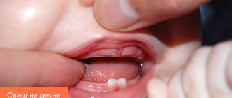
There is an unpleasant odor from the mouth, and sometimes fistulas on the gums. A characteristic sign of gumboil is the appearance of independent pain, weak at first, then intensifying, becoming pulsating and tearing. Its difference from pain with pulpitis is that it is strictly localized and becomes sharp when pressing on the diseased tooth, especially in the form of tapping. The closing of teeth is so painful that many people refuse to eat even liquid food.
Clinical manifestations of acute periodontitis/periostitis are quite characteristic. Flux manifests itself as sharp pain in the tooth area, intensifying even with light pressure on it. Swelling and redness of the lips, cheeks, enlarged gums, and the tooth is often mobile.
There is an unpleasant odor from the mouth, and sometimes fistulas on the gums. A characteristic sign of gumboil is the appearance of independent pain, weak at first, then intensifying, becoming pulsating and tearing. Its difference from pain with pulpitis is that it is strictly localized and becomes sharp when pressing on the diseased tooth, especially in the form of tapping. The closing of teeth is so painful that many people refuse to eat even liquid food.
It is also possible to increase body temperature to 37.5-38.0°C. Severe pain and difficulty eating food force the patient to see a dentist within the next few hours from the moment of illness.
Chronic form of periodontitis
In chronic forms of periodontitis, a different picture is observed. The most formidable and insidious in its course and possible complications is chronic granulating periodontitis and exacerbation of its course. The pain reaction is mild, but often in the vestibule of the oral cavity, in the projection of the apex of the root of the affected tooth, a fistula periodically opens with small purulent discharge.
When the fistula is closed during periostitis, swelling and hyperemia of the mucous membrane in the area of the flux and pain when pressing on the diseased tooth are possible. Chronic granulating periodontitis is diagnosed based on a comparison of clinical manifestations with X-ray data, which shows destruction of the periodontal gap and changes in the jaw bone in the form of “tongues of flame” in the area of the apex of the tooth root. The purulent process in the medullary spaces of the jaw adjacent to the periodontium leads to the spread of infection over a considerable distance.
The vital activity of pathogenic microflora during the long-term existence of a chronic pathogenic focus ultimately leads to odontogenic chronic sepsis. This provokes the emergence, formation or exacerbation of chronic infectious diseases of the heart, liver, kidneys and other organs.
The presence of such infectious foci can dangerously affect the course of pregnancy at any stage; complications such as infection of the female genital area, miscarriage, and disruption of the formation of fetal tissue occur.
Ulcers as local manifestations of general diseases on the oral mucosa
- Oral tuberculosis
is usually a secondary manifestation of pulmonary tuberculosis. It occurs as a result of penetration of tuberculosis bacteria into the oral mucosa through damaged epithelium. The membranes of the cheeks, tongue, and floor of the mouth are affected. First, typical tuberculous tubercles are formed, and then, after their disintegration, small ulcers are formed, which increase in size over time. The ulcer itself is not deep, with a loose bottom, which is covered with easily bleeding granulations (young tissue), uneven edges are observed, soft to the touch. With this disease, there is a sharp pain in the ulcer. In addition to the local manifestation in the form of an ulcer, there is a general deterioration in the well-being of patients: emaciation is noted, the amount of plaque on the tongue increases, sweating and body temperature increase. General treatment of tuberculosis of the oral mucosa is carried out in specialized anti-tuberculosis institutions. As for local treatment, in this case, sanitation of the oral cavity is carried out during the period of remission (weakening of the disease), treatment of the mucous membrane with antiseptic and anti-inflammatory agents. - Syphilis
is a chronic infectious disease caused by the so-called Treponema pallidum. All periods of development of this disease (in addition to the incubation period, which lasts 21 - 24 days) are characterized by the presence of ulcers in the oral cavity. At the initial stage of development of the disease, the presence of a painless ulcer is observed, which has a round or oval shape with raised, smooth edges and a cartilage-like specific infiltrate. The bottom of the ulcer is bright red, shiny or covered with a grayish-dark coating. The ulcers heal in 3 to 12 weeks with or without scar formation. Even with tertiary syphilis, there is no sharp pain, as, for example, with a tuberculous ulcer. The ulcer is surrounded by a powerful infiltrate, which is a dense bluish-red ridge that rises above the level of the mucosa. Its edges are smooth, bright red, covered with granulations, and bleed easily. After the ulcer heals, a retracted star-shaped scar forms. This process lasts 3 - 4 months. After the ulcers heal, scars remain, which are a sign of previous syphilis. General treatment of syphilis is carried out in a venereology hospital, local treatment is carried out during the period of remission or recovery (sanitation of the oral cavity, elimination of local traumatic factors). - Acute necrotizing gingivostomatitis
is a viral infectious disease. Most often, ulcers are localized on the mucous membranes of the gums, cheeks, soft palate, arches and tonsils. Favorable factors for the development of this disease are a decrease in the overall resistance of the body, a violation of the integrity of the oral mucosa, and a deficiency of vitamins in the body. There are also cases of this disease occurring against the background of cooling or overwork. It can also be a complication of viral infections, as well as allergic stomatitis. Usually young people (under 30 years of age) are affected, more often men.
Among the symptoms it is necessary to highlight: pain when eating; extremely unpleasant odor from the mouth; increased salivation; elevated body temperature. The gum mucosa becomes swollen, painful, and bleeds when touched. The epithelium of the gingival margin and gingival papillae becomes cloudy. The surface of the gingival margin is covered with a grayish-yellow coating, which is easily removed. The ulcers have soft, uneven edges and are covered with a dirty green coating (with a foul odor), which is easily removed. In this case, a loose bleeding bottom is detected. The surrounding tissues are swollen.
Treatment of necrotizing gingivostomatitis is carried out in accordance with the general condition of the body, taking into account its location and severity of the lesion. In moderate and severe stages of the disease, broad-spectrum antibiotics are prescribed, as well as drugs that prevent or reduce the manifestation of allergies. At any stage of development of the disease, vitamins C and P, high-calorie foods, juices are prescribed, and in some cases, according to indications, taking cardiac medications.
Local treatment is carried out under anesthesia (removal of necrotic tissue). The oral cavity is treated with warm solutions of antiseptics and anti-inflammatory drugs. The ulcer is also sprinkled with white clay powder. After acute inflammation has been relieved, professional oral hygiene is carried out.
Mouth ulcers can also form due to HIV (gum ulceration occurs in approximately 30% of people infected with HIV). How to cure mouth ulcers in this case? Treatment is specific and carried out by infectious disease doctors. Dental care is provided in all dental institutions with careful adherence to safety precautions.
Prevention of all of the above oral diseases consists of eliminating the causes of their occurrence. For example, to prevent infectious diseases that manifest themselves on the oral mucosa, such as syphilis, measures are necessary that prevent infection from entering the body. In other cases, wellness measures are very important, which include systematic independent and professional oral hygiene, which is offered by almost all dental clinics in Moscow.
The dangers of flux and the dentist's tasks
It is the identification of such foci and their elimination, especially in persons suffering from inflammatory processes of internal organs and in pregnant women, that is one of the essential tasks of a dentist. Untimely sanitation of the oral cavity or its absence in the presence of teeth affected by chronic periodontitis can lead to the development of a number of more severe inflammatory processes, the first of which is acute purulent periostitis.
Purulent periostitis
This disease is an acute purulent inflammation of the periosteum of the alveolar process of the jaw and accounts for up to 40% of complications of odontogenic infection, mainly chronic periodontitis. The cause of the development of this disease is most often large molars, which are primarily affected by the carious process.
The clinical picture of acute purulent periostitis is diverse and depends on the nature of the microflora, localization and extent of the inflammatory process. When the upper jaw is affected, the external manifestations of acute inflammation, in particular hyperemia, infiltration and swelling of soft tissues, are usually more pronounced than when the flux is localized in the lower jaw. However, the phenomena of general intoxication are more pronounced when the alveolar process of the lower jaw is affected, which is due to the difference in the anatomical and topographic relationships of soft tissues and jaw bones.
When examining the oral cavity, smoothness or swelling of the transitional fold of the mucous membrane of the vestibule of the oral cavity, its hyperemia, and sharp pain when touched by the dentist’s hands or instruments are noticeable.
It should be borne in mind that the pain in the “causal” tooth due to the release of exudate (liquid released into the tissue or cavity of the body from small blood vessels during inflammation) beyond the periodontal gap subsides to a certain extent, but slight mobility of the tooth appears. This circumstance sometimes serves as a justification for delaying visiting a doctor and as a cause of aggravated course of the inflammatory process in the future.
The diagnosis of “acute purulent periostitis” is an absolute indication for surgical intervention in the form of opening and drainage of the periosteal purulent focus and removal of the “causal” tooth.
Options for non-drug treatment (physiotherapy) depend on the patient’s general condition, age, and concomitant chronic diseases. In the latter case, the volume and nature of drug treatment is determined by the dentist of the appropriate profile. It is especially important to carry out adequate treatment in patients suffering from rheumomyocarditis, diabetes mellitus, nephritis and some other chronic diseases.
Another complication of chronic granulating periodontitis is the development of acute odontogenic osteomyelitis , accompanied by the formation of phlegmons and abscesses of the cellular spaces of the face and neck.
These complications of flux are the most severe and dangerous result of the progression of odontogenic infection, and depending on the location and nature of the microflora, they can lead to severe complications including the development of sinus thrombosis and mediastinitis.
It is their timely diagnosis in somatic patients that largely determines the outcome of the disease.
The clinical manifestations of phlegmon are different and depend on the location of the flux (deep, superficial), the nature of the pathogen (aerobic, anaerobic flora), body resistance, concomitant diseases, age, etc.
Today, persistent, sluggish chronic necrotic processes in the bone and soft tissues of the face often occur against the background of drug addiction.
Deep phlegmons of the pterygomaxillary, infratemporal, peripharyngeal spaces are manifested by sharp pain of the corresponding localization, severe manifestations of general intoxication up to acute intoxication psychosis, severe inflammatory contracture of the masticatory muscles, pain when swallowing, with an almost complete absence of external manifestations: hyperemia, infiltration, fluctuations, etc. .
Underestimation of certain symptoms of gumboil can lead to a delay in seeking dental care, ineffective therapy and progression of the inflammatory process.
Complications of flux
Complications with these localizations of phlegmon can be much more severe than with superficially located purulent foci with clear local manifestations. It is especially important to emphasize the danger of developing phlegmon of the floor of the mouth, taking into account the probable anaerobic component of the microflora.
Therefore, if there are complaints about difficulty swallowing, an increase in the volume of the tongue, its pushing towards the hard palate, a forced position of the head, you should urgently seek help from a dentist in order to avoid the development of the most common complication with this localization of phlegmon - anterior mediastinitis.
The occurrence of ulcers due to diseases/damage to the soft tissues of the mouth and tongue
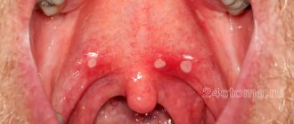
1. Recurrent
aphthous stomatitis is a chronic inflammatory disease that is characterized by periodic eruptions of aphthae (small ulcerations) on the oral mucosa. Aphthae can be localized on the tongue, buccal mucosa, hard and soft palate, and also on the mucous membrane of the lips. They are painful. In some cases, with constant injury, aphtha can turn into a long-term non-healing ulcer, after epithelization of which a scar is formed.
Patients with recurrent aphthous stomatitis usually suffer from colitis. Nervous strain, minor injuries to the mucous membrane, for example, when brushing teeth, as well as menstruation, can also predispose to the onset of the disease. The aphtha heals within 7 - 10 days. In case of complications of the disease, the number of ulcerations may increase, then the healing period is extended by 2 - 4 weeks.
2. Herpetiform stomatitis
characterized by the appearance of numerous small ulcers. In appearance they resemble the sores of herpes simplex. As a rule, they occur in women under 30 years of age. They are localized mainly on the lower surface of the tongue and in the area of the floor of the mouth. They do not have clear boundaries, the base is gray. The healing process is completed largely without scarring within 7 to 10 days. In simple forms of stomatitis, white ulcers in the mouth are observed (or rather, the center of the ulcer is covered with a thin, loosely fitting white or grayish film). Also, a white ulcer in the mouth can form in children in cases where there is candidal, or fungal, stomatitis.
As for the treatment of stomatitis, in this case the form of the disease and the severity of the lesion are taken into account. General and local treatment depends on this. But for almost all forms of stomatitis, vitamin C is prescribed to improve immunity.
3. Recurrent necrotizing peryadenitis
(Setton's aphthae) is characterized by the formation of a compaction in the submucosa, then painful ulcers with raised and compacted edges form in this place, as well as the presence of an inflammatory infiltrate (accumulation of cellular elements mixed with blood and lymph). Ulcers are localized on the upper and lower lips, cheeks, and lateral surfaces of the tongue. Eating food becomes extremely difficult for many patients, even to the point of giving it up. The same severe pain can be observed when talking. The ulcers do not heal until several months, and the disease lasts for years.
4. Afty Bednar
occur exclusively in children and are defined as traumatic erosions (ulcerations). The cause is poor oral hygiene or rough mechanical rubbing of the mucous membrane of the palate (this is where they are located). Covered with a whitish-yellow coating.
5. Traumatic ulcer
in the mouth is most often the result of physical impact, hence its name. As a rule, such an ulcer occurs as a result of an accidental or intentional bite of the mucous membrane, damage by a toothbrush. The presence of a traumatic ulcer in the oral cavity can provoke dental treatment (with careless use of dental instruments or, for example, with pointed temporary crowns).
Also among traumatic ulcers are the so-called prosthetic ulcers, which arise from exposure to complete or partial removable dentures, the dimensions of which are larger than necessary, or their surface is poorly processed and has sharp edges. Such ulcers can be located directly under the prosthetic structure. As a rule, healing takes place within 10 - 14 days, provided that the traumatic factor is eliminated. Treatment may not be necessary, since traumatic ulcers are often painless and small in size. In the case of the opposite, antimicrobial and anti-inflammatory drugs are prescribed, and the use of an ultrasonic brush is recommended, which not only cleans, but also has an antibacterial effect.
Also, the presence of ulcers can be caused by the effects of alkalis, acids, and certain medications on the oral mucosa.