Quite often in their practice, ENT doctors are faced with such a problem as paranasal sinus cyst . Despite the fact that the disease occurs frequently, in almost every tenth person, it is mainly discovered by chance, for example, during an X-ray examination for a completely different reason. This is explained by the fact that a cyst in the nose may not bother the patient for a long time and may not manifest any symptoms. Therefore, it is important to understand that timely treatment of cysts is necessary to avoid possible complications and to improve the patient’s quality of life.
What is a sinus cyst?
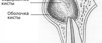
This is a large formation in which mucus or pus accumulates. The cyst is not dangerous, it does not degenerate into cancer, but over time it can grow and put pressure on the walls of the sinus. Because of this, breathing problems appear, the nasal mucosa swells, a runny nose almost never goes away, and recurrent bronchitis and pneumonia may occur. The cyst is sometimes located in such a way that there are no symptoms, and it is discovered by chance during another examination - for example, before dental prosthetics.
What causes cysts to form in the sinuses?
To ensure that the mucous membrane of the respiratory organs does not dry out, is not overcooled and is protected from dust, it contains glands that constantly secrete moisturizing mucus. This mucus flows to the surface through the ducts of the glands. If at least one of them is blocked, mucus continues to be produced, but will accumulate inside the gland, filling it and stretching the walls. As a result, a cyst appears.
The formation of cysts can occur due to:
- frequent infections;
- severe allergies;
- structural features of the nasopharynx;
- injuries and curvature of the nasal septum;
- inflammatory processes in the upper jaw (usually dental diseases).
The most common symptoms of a cyst are headaches, difficulty in nasal breathing due to swelling of the mucous membrane, discomfort or pain over the upper jaw.
Surgical methods for treating hypertrophy
Hyperplasia of the mucous membrane of the maxillary sinuses can be cured by certain surgical methods.
Surgical treatment methods:
| Method name | Main tool | How to do it |
| Galvanocaustics | Electrode | It is performed under local anesthesia. The pathological area is cauterized. After the procedure there is a recovery period, the formed scar resolves. Nasal breathing is completely restored. |
| Conchotomy | Wire loop | The technique involves resection of the enlarged mucosa. |
Upon completion of the procedure, the doctor should give detailed instructions about the features of the postoperative period.
In addition to surgical methods, drug support is prescribed:
- antihistamines;
- drugs to strengthen blood vessels;
- rinsing the sinuses and nasal turbinates with medicinal solutions;
- performing inhalations.
For successful treatment, the main thing is to see a doctor on time
Thickening and swelling of the mucous membrane of the maxillary sinuses causes pathological formation of mucus and pus. Pathogenic microorganisms successfully live in such an environment. This proximity is especially dangerous for the membranes of the human brain, which are located near the maxillary sinuses.
This disease cannot be determined independently, which means that even with a simple runny nose you need to consult a specialist. Self-medication in this case gives false hope, wastes precious time, the cost of which is very high, and is harmful to health.
Symptoms of a sinus cyst
The cysts and polyps themselves are soft and lack sensitivity, so if they are small, many people may not notice them at all. However, multiple growths or a large cyst can block the nasal passages and sinuses.
Common signs and symptoms of nasal cysts include:
- runny nose
- reduced or absent sense of smell
- loss of sense of taste
- facial or headache
- pain in upper teeth
- feeling of pressure on the forehead and face
- snore
- frequent nosebleeds
Indications for sinus cyst removal
It will not be possible to remove the cyst simply by relieving the inflammation; surgery is not necessary. Surgery is necessary if:
- pus accumulates inside the cyst;
- it is growing rapidly or its diameter is already more than 6 cm;
- pressure on the lower wall of the orbit increases (because of this, vision may deteriorate and double vision may appear);
- worried about frequent or constant runny nose and nasal congestion.
Important! Surgery to remove a sinus cyst is also recommended if the cyst is causing any discomfort. However, the size of the cyst in this case does not matter.
How to treat
In most cases, cysts cannot be treated without surgery. Only in unadvanced cases is it possible to get rid of the tumor by opening it and draining it.
The operations performed for the disease are called cystectomy and cystotomy. They both involve removing the formation. But in the first case, it is removed completely, and in the second, only the front wall is excised.
The main goal of a dental surgeon is to preserve the tooth growing in the area where the tumor is located. Unfortunately, this is not always possible. So, if the tooth root is immersed in the cyst cavity by more than 60% of its length, emergency removal is indicated. Such a unit can no longer be functional. Soon after excision of the tumor, it will still become loose and fall out.
Therefore, it is very important not to start the disease. Considering that it may not manifest itself for a long time, it is important to undergo a specialized examination at the dental office every year. This will increase the chances of successful early diagnosis.
Contraindications
The operations for removing sinus cysts are quite simple. Patients recover quickly after the intervention, and complications are rare. Contraindications are only general for any surgical interventions:
- acute infection;
- diabetes mellitus (severe);
- increased bleeding (blood does not clot well);
- pregnancy;
- epilepsy;
- oncology.
Before surgery to remove a cyst from the sinus, you need to take tests (for HIV, syphilis, hepatitis B and C, general blood test).
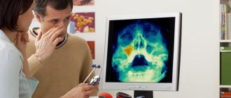
Diagnostics
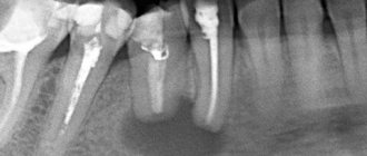
Figure 1. Cyst in the maxillary sinus.
Source: Contemporary clinical dentistry / Open-i (Attribution-NonCommercial-ShareAlike 3.0 Unported) Before surgery to remove a sinus cyst, you need to take tests (for HIV, syphilis, hepatitis B and C, general blood test).
Usually, to make a diagnosis, the doctor only needs to collect an anamnesis and a general physical examination, including the nose.
Other diagnostic tests include:
Endoscopy of the nose. The doctor performs a detailed examination of the nose and sinuses using a narrow tube with a lighted magnifying lens or small camera.
Visualization methods. Images obtained from a computed tomography (CT) scan can help the doctor accurately determine the size and location of cysts in the deeper areas of the sinuses, and assess the degree of swelling and inflammation.
Test for cystic fibrosis. If a child needs diagnosis, the doctor may suggest testing for cystic fibrosis. This is an inherited disease that affects the glands that produce nasal mucus, tears, sweat, saliva and digestive juices. This is usually a non-invasive sweat test that can determine whether your sweat is more or less salty than most people.
Types of operations to remove maxillary sinus cysts
The method of removing a cyst in the nose is chosen by the doctor. He takes into account its location and size. There are several types of operations.
Source: Symbolbild/Pixabay
Radical operation
This is a maxillary sinusotomy, in which an incision is made in the soft tissue (behind the upper lip) to gain access to the inside of the sinus in the oral cavity. They are peeled off, exposing the bone - the front wall of the sinus. Part of the bone fragment of the wall is removed to gain access to the sinus cavity. At the main stage, the surgeon cleans the sinus cavity from cysts, polyps and accumulated fluid, and rinses the sinus. The anastomosis with the nasal passage is widened, a drain is installed and taken out. The incision on the gum is sutured. The tampon is left in place for two days and then removed under anesthesia.
Figure 2. Stages of radical maxillary sinusotomy. Source: CC0 Public Domain
Important! Radical surgery is used relatively rarely, only if it is necessary to remove a large cyst or there are complications (separation of pus, inflammation, proliferation of polyps). Such an intervention allows you to completely scrape out and wash out the sinus, but it is dangerous due to complications (neuritis, the appearance of a fistula, bleeding). It is relatively difficult to tolerate and may require anesthesia and hospitalization.
Such an intervention allows you to completely clean and rinse the sinus, but is dangerous due to complications (neuritis, the appearance of a fistula, bleeding). It is relatively difficult to tolerate and requires hospitalization and anesthesia.
Endoscopic surgery
This is a minimally invasive procedure in which an endoscope is used to remove the cyst. The entire procedure takes about half an hour and is performed under local anesthesia.
To remove the cyst, an endoscope is inserted into the sinus through the nasal passage. Sometimes it is inserted through the tubercle on the upper jaw or the alveolar socket (through an additional channel with a diameter of 4–5 mm). The technique of access to the sinus depends on the location, shape and size of the cyst, as well as the individual characteristics of the patient’s nasopharynx. The path to the sinus through the nose is less traumatic and does not require an additional puncture. Access through the alveolar socket is used if the patient has had at least one molar removed (in adults, these are the sixth, seventh and eighth teeth on the left and right sides) on the upper jaw.
After entering the sinus under video control, the doctor pierces the cystic cavity and excises its walls, performs sanitation of the maxillary sinus, cleans it and rinses it with an antiseptic solution.
Endoscopic surgery has several advantages:
- there is no need to make an incision, the tissue is not injured;
- a video endoscope allows you to combine diagnostics with surgery (examine the sinus and immediately remove the tumor);
- The tissues of the mucous membrane are not damaged and scarring does not occur.
- Complications rarely occur;
- patients tolerate the intervention more easily and recover from it faster.
Therefore, the endoscopic technique is used more often than the radical one.
Figure 3. Endonasal removal of nasal cysts. Source: CC0 Public Domain
Important! Endoscopic removal is used for certain anatomical features of the structure of the nasopharynx, which can be diagnosed by a doctor. The technique is safe, almost does not injure the nasopharynx, and long-term rehabilitation is not required after its use.
Microsinusrotomy
In this case, a trocar (something like a surgical awl) and an endoscope are used. The procedure is also considered gentle. It is performed if the tumor can be removed through an opening in the anterior wall of the maxillary sinus.
At the first stage of the operation, access to the sinus is provided. To do this, a puncture with a diameter of 7 mm is made in the upper jaw. The puncture is performed with a trocar and an endoscope is inserted into the sinus cavity through it. Under video control, the cyst is punctured, its walls are excised, and the contents are removed.
In almost a third of cases, after maxillary sinusotomy, trigeminal neuropathy develops, which usually regresses without special treatment. Advantages: quick recovery, low swelling. Healthy tissues are almost not damaged.
Laser therapy
The laser is rarely used and only in combination with other surgical methods, provided that the cyst is small (otherwise it will take too long to remove it).
After surgery to remove a cyst, swelling of the mucous membrane, a feeling of dryness or profuse discharge from the nose, and pain in the area through which the sinus was accessed may appear. These symptoms are not complications of surgery and usually go away within a few days. If they persist and intensify, you should consult a doctor.
The nature of complications after surgery depends on the technique used:
- if access to the sinus was carried out through the alveolus of the tooth, an incision or puncture in the upper jaw, a fistula tract may form;
- with micromaxillary and radical maxillary sinus there is a risk of damage to the infraorbital or trigeminal nerve with the development of neuropathy. In this case, areas of the skin, nasal mucosa and mouth become numb, which may require treatment;
- inflammation and/or suppuration of the wound develops if it becomes infected (with poor quality oral care, in cases where the patient does not follow the doctor’s orders).
Hospitalization
The day hospital at our Center is a post-operative support service for patients. Necessary if the patient has concomitant cardiac issues, such as hypertension, arrhythmia, AOS, etc.
Gentle ultrasound surgical protocols for low-impact PiezoSurgery operations in combination with microscopic surgery performed by operating teams consisting of pairs of the most experienced surgeons at our Center allow you to carry out treatment so delicately that you will not need mandatory hospitalization, which is so popular in Moscow hospitals.
All operations are performed only in medicated sleep, without pain or nervous overload.
The treatment will be carried out with respect for your personal time, in a short time and at a comfortable time, the operating teams and anesthesiology department work seven days a week and holidays.
Possible complications
After surgery to remove a cyst from the maxillary sinus, swelling of the mucous membrane, a feeling of dryness or profuse nasal discharge, and pain in the area through which the sinus was accessed may appear. These symptoms are not complications of surgery and usually resolve within a few days. If they persist and intensify, you should consult a doctor.
The nature of complications after surgery depends on the technique used:
- if access to the sinus was carried out through the alveolus of the tooth, an incision or puncture in the upper jaw, a fistulous tract may appear;
- with micromaxillary and radical maxillary sinus there is a risk of damage to the infraorbital or trigeminal nerve with the development of neuritis. In this case, areas of the skin, nasal mucosa and mouth become numb, which requires additional treatment;
- inflammation and/or suppuration of the wound develops if it becomes infected (with poor quality oral care, in cases where the patient does not follow the doctor’s orders).
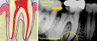
Establishing diagnosis
With the help of rhinoscopy, a preliminary conclusion can be made
The following methods are used to diagnose the disease:
- X-ray examination;
- MRI;
- Rhinoscopy.
The research method is prescribed by the attending physician. Often you have to resort to more than one method. It is especially important during the examination to determine the exact location of mucosal hypertrophy.
Important: it is not possible to cure this disease at home, since a diagnosis can only be made after a series of procedures and consultation with a specialist.
In case of a serious condition of the patient, surgical intervention will be indicated. The video in this article and photos will explain the features of x-ray diagnostics.
X-ray is the main diagnostic method when there is a suspicion of thickening of the mucous membrane of the maxillary sinuses
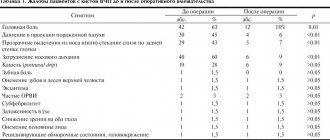
Rehabilitation period
Figure 4. A cyst removed from the maxillary sinus with a tooth in it.
Source: Indian journal of ophthalmology / Open-i (Attribution 2.0 Generic) After surgery to remove a sinus cyst, no special recovery or treatment is usually required. Full recovery takes only a few days. With a small amount of intervention, patients feel well within a few hours after the intervention.
For the first time after surgery, it is advisable to avoid physical activity. You cannot visit the pool and sauna, you must not allow injuries. To avoid inflammation and other complications, you need to ensure high-quality oral hygiene and follow your doctor’s prescriptions.
Causes of the disease
Already based on the classification, it becomes clear that tumors appear in the tissues of the oral cavity for a variety of reasons. Among the main provoking factors:
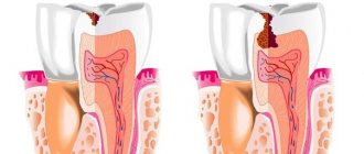
- Damage to hard dental tissues. Untreated caries, periodontitis, pulpitis are all diseases in which pathogenic agents penetrate the periodontium through unsealed root canals.
- Inflammation of the area adjacent to the area where the cyst is located. Quite often it turns out that a benign tumor formed after sinusitis or inflammation of the gums. This happens because the infection penetrates through the bloodstream into the bone tissue from the maxillary and nasal sinuses.
- Injuries, bruises, blows. The first time after them, a person may not notice anything. But later a nagging pain appears, indicating a problem.
Very rarely do doctors have to treat congenital cysts. It has not been established why they occur.